Abstract
High-intensity circuit training (HICT) using body weight as resistance is a popular fitness trend and an ideal exercise modality in terms of functionality and economy. Given the popularity of HICT, evidence suggests that sex may elicit unique physiological and perceptual responses to this mode of exercise and there is a need for further work in this area. The purpose of this study was to examine physiological and perceptual responses of females and males to HICT using body weight resistance exercise. Forty-five participants (23 females and 22 males) completed baseline testing and a 15-minute HICT exercise bout wearing a portable metabolic analyzer. %VO2max, %HRmax, and RER were monitored during exercise and analyzed at 3 different 5-minute time segments during the HICT (beginning, middle, end). RPE was assessed half-way through the circuit (7.5), immediately upon cessation of exercise (15), and 15-minutes post-exercise (Session RPE). There was a significant (p<0.01) time effect on %VO2max, %HRmax, RER, and RPE. At all three time points, %VO2max was significantly (p<0.02) higher among females compared to males. RER values were significantly (p=0.02) higher among females during the last 5-minute segment (i.e. the end) of the exercise bout. However there were no differences in %HRmax (p>0.20). Males reported a higher RPE immediately post-exercise compared to females (p=0.01). Taken together, these data suggest that there are distinct, sex-specific physiological and perceptual responses to HICT; thus, sex-specific exercise prescription considerations are warranted.
Keywords: %VO2max, %HRmax, RER, RPE
INTRODUCTION
Body weight resistance training and high-intensity interval training are two fitness trends gaining momentum among the health and fitness community (23). High-intensity circuit training (HICT) using body weight as resistance is an ideal exercise modality in terms of functionality and economy (11). Several body-weight exercises utilized in HICT mimic movements used in activities of daily living (e.g. squats, step-ups, etc.); these exercises provide functional fitness benefits. Additionally, HICT requires little to no equipment and can be done in a variety of environments (e.g. outside, in the home, in small spaces)(11). Despite the recent popularity, there are few studies investigating HICT using body weight resistance exercise. The physiological and cardiometabolic responses to HICT using body weight resistance exercise have not been studied.
This is an important area of study as the physiological and cardiometabolic responses to HICT using body weight resistance exercise can influence a variety of training prescription variables and outcomes relative to both competitive and recreational athletes. For example, these responses have implications on self-selected exercise intensity and perceived exertion during and after the exercise bout (12, 17). Perceived exertion is associated with exercise enjoyment and adherence (15, 19, 26). A thorough understanding of the physiological and cardiometabolic responses to HICT using body weight resistance exercise can also improve the effectiveness of the exercise training and minimize injury (6, 9). For example, exercising at an intensity that is too high could result in non-functional overreaching. On the contrary, exercising at an intensity that is too low also can have undesirable outcomes such as a lack of beneficial physiologic adaptations related to improved performance.
Further, previous studies have reported sex-specific differences in physiological measures and perceptual responses in response to high-intensity interval training (10, 14, 18), suggesting that sex-specific considerations when prescribing high-intensity exercise prescriptions are warranted. For example, a recent study by Panissa et al. reported sex-specific differences in cardiometabolic responses to high-intensity exercise (18). The authors reported sex-specific differences in anaerobic power reserve and respiratory exchange ratio during all out exercise. They conclude that males self-selected a higher exercise intensity and maintained a higher anaerobic power reserve compared to females. Interestingly, they reported no differences in oxygen uptake, which suggests sex-specific metabolic adjustments are taking place during high-intensity interval training. A different study by Laurent et al. (2014) stated that sex-specific differences in recovery during high-intensity interval training may translate into sex-specific differences in perceptions of effort during exercise (14). The authors suggest that females demonstrate improved recovery during high-intensity interval training, which supports higher self-selected exercise intensity among females, leading to greater cardiovascular strain. The authors also noted that females typically reported higher perceptual strain during a bout of high-intensity exercise, but lower values of perceptual strain post-exercise. However, there are mixed findings in the literature regarding perceptual strain, as another study reported little-to-no sex-specific differences in perceptual responses during all out cycling and treadmill exercise bouts (10).
To our knowledge no study has investigated potential sex-specific differences in response to a HICT using body weight resistance exercise. Given the popularity of this exercise modality and evidence suggesting that sex may elicit unique physiological and perceptual responses to HICT, there is a need for further work in this area. Therefore, the purpose of this study was to examine physiological and perceptual responses of females and males to HICT using body weight resistance exercise.
METHODS
Participants
Fifty participants were recruited from a medium-size, Southern/Midwestern university. The study was advertised using flyers posted on campus. Forty-five participants completed both testing sessions. The study was approved by the University’s Institutional Review Board (IRB 16–067). Following a comprehensive explanation of procedures, all participants signed written informed consent. To be eligible, participants had to be free of injury and healthy enough to participate in vigorous-intensity exercise (i.e. have no signs, symptoms, or known history of metabolic, cardiovascular, or renal disease)(21). Subjects were excluded if: they had a medical contraindication to exercise or if they were taking any medications known to alter metabolism, indicated by the health history questionnaire.
Protocol
Participants reported to the lab for two different study visits (Session 1 and Session 2).
During Session 1, eligibility was confirmed through health screening questionnaires. Each participant completed a Physical Activity Readiness Questionnaire (PARQ) as well as a health history questionnaire to determine ACSM risk-factor assessment. Physical activity levels were assessed using the International Physical Activity Questionnaire (IPAQ). Baseline measures were taken including resting heart rate, age-predicted max heart rate using the equation (220-age), resting blood pressure, height, weight, and maximal aerobic capacity (VO2max). VO2max was obtained using the Bruce treadmill protocol. Participants were deemed successful in achieving VO2max if they accumulated at least 2 of the following criteria: an elevated respiratory exchange ratio (RER) ≥ 1.10, a rating of perceived exertion ≥ 17 on the 6–20 Borg scale, and a HR within ± 12 beats per minute of their age-predicted maximum. Age-predicted HR maximum (HRmax) was estimated by subtracting the person’s age from 220.
Following baseline testing procedures, each participant was introduced to the body weight resistance exercise in the HICT protocol (Table 1). The rational for the exercise selection and protocol was based on previously published work (11). Participants were familiarized with each of the seven exercises (and/or the modified version of the exercise) in the circuit. After participants were comfortable with the seven exercises, they were informed not to consume any caffeine and/or alcohol and to avoid heavy exercise within the 24 hours preceding Session 2.
Table 1.
Experimental high-intensity circuit training (HICT) using body weight as resistance exercise protocol.
| Complete as many rounds as possible in 15 minutes |
|---|
| 12 air squats |
| 12 butterfly sit-ups |
| 12 push-ups (modified = 18 in incline push-ups) |
| 12 forward alternating lunges |
| 12 pull-ups (modified = ring row) |
| 12 step ups (20″ box) |
| 12 high knees |
Participants reported to the lab for the HICT exercise session between 24 hours and 7 days after Session 1. Upon arrival to the lab, the participant was fitted with a Polar heart rate monitor (Polar Electro Ltd., Warwick, UK) and was instructed to complete a five minute walking treadmill warm-up at a self-selected pace. After the warm-up participants reviewed the body weight resistance exercise and potential modifications to ensure they remembered how to correctly and safely perform each exercise. Participants were fitted with the K4 COSMED (Concord, California) portable metabolic analyzer to measure oxygen consumption, carbon dioxide production, and heart rate throughout the bout. Participants were instructed to complete as many rounds of the body weight resistance exercise protocol as possible during the 15-minute time allotment. During the exercise bout, participants were constantly monitored by a research team member to ensure safety. If the participant could no longer properly complete the standard (unmodified) version of an exercise, they were instructed to continue the workout using the modified version. The Omni-Scale (0–10) was used to assess RPE at minute 7.5 and minute 15. Upon completion of the exercise bout, overall Session RPE was assessed by asking the participant to report an RPE for the entirety of the exercise bout.
VO2 (ml/kg/min), HR (beats/min), and RER were the physiological responses related to cardiometabolic demand assessed throughout the exercise session. To calculate %VO2max and %HRmax values, the measured VO2 and HR values assessed during the exercise bout were divided by VO2max and HRmax values obtained in Session 1 (respectively). The 15-minute exercise bout was divided into three 5-minute segments in order to investigate these measures at the beginning (minutes 0:00–4:59), middle (minutes 5:00–9:59), and end (minutes 10:00–15:00) of the HICT session. Thus, %VO2max, %HRmax, and RER were calculated for each participant during the “beginning,” “middle,” and “end” of the exercise session. Additionally, the “overall” value was averaged for the entire exercise session.
The rating of perceived exertion (RPE) is a widely used validated scale to assess exercise intensity. The session RPE has been deemed an accurate reflection of the overall intensity of bout of exercise ranging from low to high-intensity modalities (4). The Omni-RPE (0–10) scale (24) was used to assess perceived exertion at minute 7.5, immediately post-exercise, and 15 minutes post-exercise (Session RPE). The Omni-RPE scale, as opposed to the Borg (6–20 scale), was selected because it was easier for participants to report as they were able to hold up zero to 10 fingers to indicate their RPE while exercising at high-intensity with their faces covered by the metabolic analyzer mask.
Statistical Analysis
All analyses were performed using SPSS for Windows software (version 25.0; SPSS, Inc., Chicago, IL, USA). Descriptive statistics (mean ± SD) were generated to show group characteristics. Independent samples t-tests were applied to detect differences in group means between males and females. Sex-specific physiological (%VO2max, %HRmax, RER) and perceptual (RPE) responses were assessed using repeated-measures ANOVA with emphasis on a sex (female, male) X time (beginning, middle, end) interaction, to determine if male and female participants responded differently during the HICT session. Sex-specific perceptual (RPE) responses also were assessed using repeated-measures ANOVA with emphasis on a sex (male, female) X time (minute 7.5, immediately post-exercise, 15 minutes post-exercise) interaction. Post hoc comparisons were performed with contrast analyses. Data are presented as mean ± SD or means ± SD. All data were assessed for normality and equal variance. Statistical significance for all comparisons was set at p<0.05.
RESULTS
Participants’ baseline measurements are presented in Table 2. There were no age or baseline blood pressure differences between males and females; however, males had a significantly (p<0.05) higher body mass index (BMI) and VO2max values compared to females (Table 2).
Table 2.
Participant characteristics (mean ± SD).
| Combined (n = 45) | Men (n = 22) | Women (n = 25) | |
|---|---|---|---|
| Age (y) | 28.0±10.9 | 30.2±11.7 | 25.8±9.7 |
| BMI (kg/m2) | 22.9±3.3 | 24.0±3.6* | 21.8±2.6 |
| VO2max (mL/kg/min) | 48.6±8.6 | 55.1±6.8** | 42.4±4.6 |
| SBP | 117.5±9.0 | 120.1±9.3 | 115.0±8.3 |
| DBP | 75.2±8.2 | 75.8±8.2 | 74.6±8.4 |
BMI = Body Mass Index; VO2max = maximal oxygen consumption; SBP = Systolic Blood Pressure; DBP =
Diastolic Blood Pressure
Significant difference (p < 0.05) between males and females.
Significant difference (p < 0.001) between males and females.
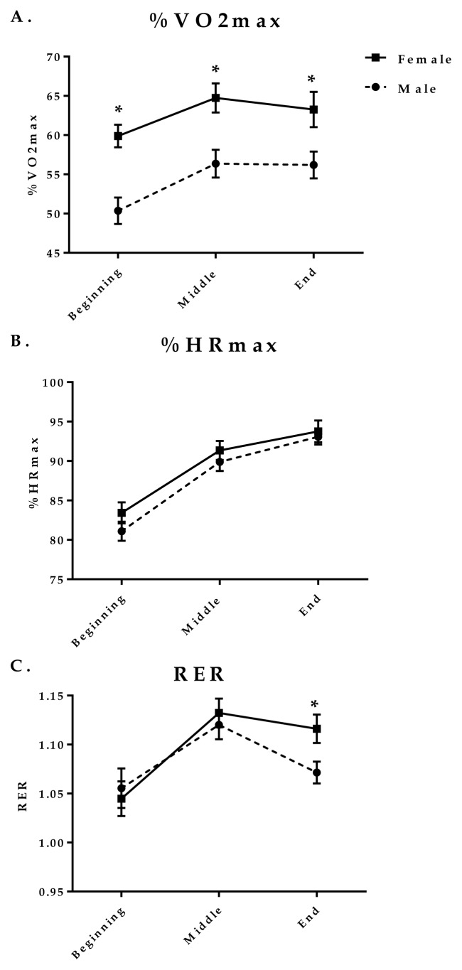
The overall %VO2max, %HRmax, and RER values for all participants (averaged from the entire HICT session) were 58.7±9.2%, 88.9±5.0%, and 1.09±0.07% respectively. There was a significant (p<0.01) time effect for all three measures. As a whole, participants worked out at a significantly (p<0.01) greater %VO2max during the middle and end of the HICT session compared to the beginning. The beginning %VO2max (55.2±8.8%) was approximately 5% lower compared to the middle %VO2max (60.6±9.5%) and the end %VO2max (59.8±10.0%)(See Figure 1A). %HRmax increased significantly (p<0.01) throughout the HICT session (beginning 82.3±6.0% vs. middle 90.6±5.4% vs. end 93.4±5.5%) (See Figure 1B). RER significantly (p<0.01) increased from the beginning to the middle of the workout and then significantly (p<0.01) decreased from the middle to the end of the workout (beginning 1.05±0.09 vs. middle 1.13±0.07 vs. end 1.09±0.06) (See Figure 1C).
Figure 1. Sex-specific changes in response to HICT body weight resistance exercise.
%VO2max, %HRmax, and RER at three five-minute time segments (“beginning” = minutes 0:00–4:59, “middle” = minutes 5:00–9:59, and “end” = minutes 10:00–15:00) during a 15 minute bout of HICT. To illustrate sex-specific responses, the solid line represents females and dashed line represents males. Data are presented as means ± SEM.
*Significant difference between females and males (p<0.05).
There were sex-specific differences in %VO2max at all three time points. %VO2max was significantly higher among females compared to males at the beginning (females 59.0±7.5% vs. males 52.2±6.3%; p<0.01), middle (females 63.3±9.3% vs. males 60.0±7.0%; p<0.01), and end (females 62.9±10.0% vs. males 59.7±5.9%; p=0.02) (See Figure 1A). Interestingly, there were no sex-specific differences in %HRmax at any of the three time points (beginning, middle, end) (See Figure 1B). However, there was a significant (p=0.02) interaction effect (sex X time) for RER where females had a significantly higher RER compared to males at the end of the exercise bout (females 1.12±0.07 vs. males 1.07±0.05; p=0.02) (See Figure 1C).
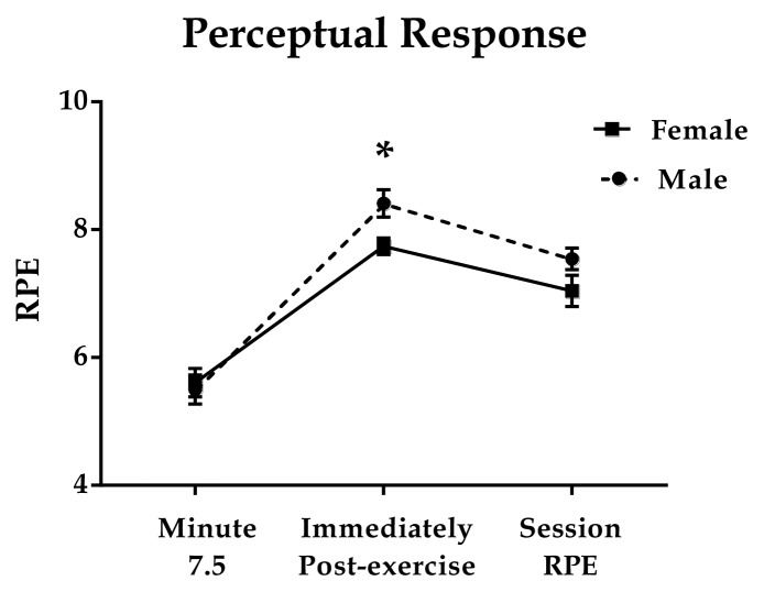
There was a significant time effect for RPE (p<0.01)(Figure 2). The average RPE at minute 7.5 was (5.6±1.1) which was significantly (p<0.01) lower than RPE assessed immediately post-exercise (8.1±0.9). Females reported a lower RPE immediately post-exercise compared to males (females 7.7±0.7 vs. males 8.4±1.0; p=0.01). RPE assessed at minute 7.5 (females 5.6±1.1 vs. males 5.5±1.1; p=0.73) and the overall Session RPE (females 7.0±1.2 vs. males 7.5±0.8; p=0.11) were not significantly different between females and males.
Figure 2. Rate of Perceived Exertion during and after HICT session.
The Omni-RPE (0–10) scale was used to assess perceived exertion at minute 7.5, immediately post-exercise, and 15-minutes post-exercise (Session RPE). To illustrate sex-specific responses, the solid line represents females and the dashed line represents males. Data are presented as means ± SEM.
*Significant difference between males and females (p<0.05).
DISCUSSION
The major findings from this study suggest there are sex-specific responses to a bout of HICT using body weight resistance exercise. There are significant differences between females and males in terms of cardiometabolic demand as measured by %VO2max and RER during the exercise bout. In terms of perceptual responses, RPE assessed immediately post-exercise was significantly higher among males, compared to females. Taken together, these data suggest that there are distinct, sex-specific physiological and perceptual responses to HICT using body weight resistance exercise.
We report sex-specific differences in %VO2max at all three time points during the 15-minute HICT bout (See Figure 1). In a previous study by Laurent et al., (2014), sex-specific physiological and performance responses in response to self-paced high-intensity interval training using a treadmill protocol (14). The training protocol consisted of six 4-minute intervals interspersed with recovery intervals. Females were found to exercise at a significantly higher %HRmax and %VO2max, compared to males during the exercise protocol, despite reporting no differences in RPE (OMNI scale)(14). The authors suggest that a greater proportion of a female’s aerobic capacity may be necessary to maintain moderate-to-vigorous work rate during a high-intensity interval workout (14). Interestingly, our results indicated no sex-specific differences in %HRmax at any of the three time points.
An ancillary finding of the present study was that the overall average %HRmax was approximately 30% higher than the values for %VO2max, and previous studies found a similar response pattern of heart rate and oxygen consumption to HICT (8, 25). The overall average %HRmax and %VO2max values achieved in this study are similar to those reported by Williams et al., in which the participants achieved an average of 87.5%HRmax and 55.7%VO2max in response to a 12-minute, high-intensity circuit using kettlebells (25). Their study findings were similar to our study findings in that %HRmax and %VO2max values differed by approximately 32%. Another study investigating the relationship between %HRmax and %VO2max in response to resistance training also reported similar findings (i.e. %HRmax ~30% higher than %VO2max)(2). Collins et al. suggested a variety of mechanisms that could explain the higher HR. One mechanism could be the performance of the Valsalva maneuver during resistance exercises that increasing sympathetic nerve activity. Another possibility is that upper-body exercises may recruit a greater number of fast-twitch fibers, causing a greater exercise pressor reflex. While the exact mechanisms contributing to the higher HR are unknown, these data and our findings provide additional evidence that the relationship between VO2 and HR in response to HICT and/or resistance training varies from the traditional linear relationship commonly observed in aerobic exercise.
There is a growing body of evidence on mechanisms that may be responsible for the discrepancies observed between males’ and females’ cardiorespiratory and metabolic response to HICT. Plausible explanations for the sex-specific differences in response to HICT include differences in muscle mass, substrate utilization, and muscle morphology (13). While we did not measure blood lactate, other studies have demonstrated sex-specific differences in blood lactate accumulation in response to high-intensity exercise. Laurent et al. (2010) showed that compared to males, females exhibited significantly lower blood lactate concentrations and lower performance decrements during four trials of high-intensity intermittent sprint exercise consisting of three bouts of eight 30 meter sprints (13). Their results suggest females may have a greater resistance to fatigue at high intensities and that there are sex-specific differences in the energy system utilized (aerobic vs. anaerobic) during high-intensity exercise bouts. A previous study investigating physiological responses to a 30-second sprint uncovered both muscle fiber-type- specific and sex-specific metabolic responses (7). Esbjornsson et al. reported significant sex-specific differences in type I muscle fiber (but not type II muscle fiber) glycogen utilization in response to an acute bout of high-intensity exercise (7). These data suggest that females may have a higher relative contribution of the aerobic energy system and rely less on glycolytic processes during high-intensity exercise. This may explain why females in our study maintained higher %VO2max values during the HICT compared to their male counterparts.
Interestingly, when evaluating sex-specific differences in RER, it was found that females had a significantly higher RER than males during the final third of the exercise protocol. These findings are in contrast to other literature demonstrating that females tend to have a greater reliance on fat oxidation than their male counterparts (1, 20). Carter et al. demonstrate that after 7 weeks of endurance training at 60% of VO2peak for 5 days per week, females displayed a significantly lower RER than males during a VO2peak test both before and after endurance training. Although it seems unlikely that any individuals would be capable of exercising at a higher intensity, while accumulating greater amounts of lactate due to a higher anaerobic contribution, it has been suggested females experience greater hypoalgesia (i.e. decreased sensitivity to painful stimuli) during exercise than males (3). Research by Drury et al. suggests that during a graded, exhaustive VO2peak cycling test, females experience a greater pain threshold and pain tolerance at maximum intensity (VO2peak) when compared to baseline, 120 watts, and during recovery (5). This indicates that females experience a significantly elevated hypoalgesic response during maximal exercise. Furthermore, in a study by Dannecker et al., it was determined that after four consecutive days of eccentric arm exercises designed to induce muscular damage and pain, that females reported significantly lower perceived pain ratings at rest and during movement than males (3). This research on hypoalgesic responsiveness during exercise supports the idea that females are capable of exercising at a higher intensity than males, despite having a greater anaerobic contribution during extremely high intensity exercise.
This study has some limitations. First, the use of the Bruce treadmill protocol to determine VO2max may not have yielded the participant’s true VO2max. For example, participants may have prematurely terminated the test due to lower body fatigue. To increase the likelihood of assessing true maximal effort, we included a variety of secondary criteria for achieving VO2max including: an elevated respiratory exchange ratio (RER) ≥ 1.10, a rating of perceived exertion ≥ 17 on the 6–20 Borg scale, and a HR within ± 12 beats per minute of their age-predicted maximum. HRmax was estimated by the formula “220-age” and the validity of this measure has been previously questioned (22). Previous work has suggested no sex-specific bias using this formula to estimate HRmax (16), which provides additional confidence in our overall findings.
In summary, this study measured the physiological and perceptual responses of females and males in response to a 15-minute HICT bout using body weight resistance exercise. Previous research indicates sex-specific exercise prescription considerations are warranted when HICT is incorporated into training regimens of females and males (14, 18); results from this study support this evidence. %VO2max was significantly higher among females compared to males throughout the HICT exercise bout, however there were no differences in %HRmax. These results indicate that heart rate alone may not be the best indicator of relative intensity and/or cardiometabolic demand during this type of exercise. This is important to consider when prescribing this mode of exercise to females and males. Future investigations of sex-specific cardiometabolic responses to HICT using body weight exercise are needed to fully understand the sex-specific cardiovascular and metabolic adjustments made during HICT.
ACKNOWLEDGEMENTS
The authors thank Togy Suren, Nuha Shaker, Brooke Grimes, Alyssa Olenick, Jared Coffell, Caitlin Hesse, Erin McNeil, and Paige Wessel for their assistance with data collection and coaching participants. The authors would like to thank Dr. Grace Lartey for statistical support. Partial funding for this project provided by a grant from the National Institutes of Health (2P20GM103436 RAT and JMM). The authors have no conflicts of interest.
REFERENCES
- 1.Carter SL, Rennie C, Tarnopolsky MA. Substrate utilization during endurance exercise in men and women after endurance training. Am J Physiol Endocrinol Metab. 2001;280(6):E898–907. doi: 10.1152/ajpendo.2001.280.6.E898. [DOI] [PubMed] [Google Scholar]
- 2.Collins MA, Cureton KJ, Hill DW, Ray CA. Relationship of heart rate to oxygen uptake during weight lifting exercise. Med Sci Sports Exerc. 1991;23(5):636–640. [PubMed] [Google Scholar]
- 3.Dannecker EA, Liu Y, Rector RS, Thomas TR, Fillingim RB, Robinson ME. Sex differences in exercise-induced muscle pain and muscle damage. J Pain. 2012;13(12):1242–1249. doi: 10.1016/j.jpain.2012.09.014. [DOI] [PMC free article] [PubMed] [Google Scholar]
- 4.Day ML, McGuigan MR, Brice G, Foster C. Monitoring exercise intensity during resistance training using the session rpe scale. J Strength Cond Res. 2004;18(2):353–358. doi: 10.1519/R-13113.1. [DOI] [PubMed] [Google Scholar]
- 5.Drury DG, Greenwood K, Stuempfle KJ, Koltyn KF. Changes in pain perception in women during and following an exhaustive incremental cycling exercise. J Sports Sci Med. 2005;4(3):215–222. [PMC free article] [PubMed] [Google Scholar]
- 6.Dudley GA, Abraham WM, Terjung RL. Influence of exercise intensity and duration on biochemical adaptations in skeletal muscle. J Appl Physiol Respir Environ Exerc Physiol. 1982;53(4):844–850. doi: 10.1152/jappl.1982.53.4.844. [DOI] [PubMed] [Google Scholar]
- 7.Esbjornsson-Liljedahl M, Sundberg CJ, Norman B, Jansson E. Metabolic response in type I and type II muscle fibers during a 30-s cycle sprint in men and women. J Appl Physiol. 1985;87(4):1326–1332. doi: 10.1152/jappl.1999.87.4.1326. 1999. [DOI] [PubMed] [Google Scholar]
- 8.Farrar RE, Mayhew JL, Koch AJ. Oxygen cost of kettlebell swings. J Strength Cond Res. 2010;24(4):1034–1036. doi: 10.1519/JSC.0b013e3181d15516. [DOI] [PubMed] [Google Scholar]
- 9.Garber CE, Blissmer B, Deschenes MR, Franklin BA, Lamonte MJ, Lee IM, Nieman DC, Swain DP. American college of sports medicine position stand. Quantity and quality of exercise for developing and maintaining cardiorespiratory, musculoskeletal, and neuromotor fitness in apparently healthy adults: Guidance for prescribing exercise. Med Sci Sports Exerc. 2011;43(7):1334–1359. doi: 10.1249/MSS.0b013e318213fefb. [DOI] [PubMed] [Google Scholar]
- 10.Green JM, Crews TR, Bosak AM, Peveler WW. Overall and differentiated ratings of perceived exertion at the respiratory compensation threshold: Effects of gender and mode. Eur J Appl Physiol. 2003;89(5):445–450. doi: 10.1007/s00421-003-0869-4. [DOI] [PubMed] [Google Scholar]
- 11.Klika B, Jordan C. High-intensity circuit training using body weight: Maximum results with minimal investment. Acsms Health & Fitness Journal. 2013;17(3):8–13. [Google Scholar]
- 12.Kravitz L, Robergs RA, Heyward VH, Wagner DR, Powers K. Exercise mode and gender comparisons of energy expenditure at self-selected intensities. Med Sci Sports Exerc. 1997;29(8):1028–1035. doi: 10.1097/00005768-199708000-00007. [DOI] [PubMed] [Google Scholar]
- 13.Laurent CM, Green JM, Bishop PA, Sjokvist J, Schumacker RE, Richardson MT, Curtner-Smith M. Effect of gender on fatigue and recovery following maximal intensity repeated sprint performance. J Sports Med Phys Fitness. 2010;50(3):243–253. [PubMed] [Google Scholar]
- 14.Laurent CM, Vervaecke LS, Kutz MR, Green JM. Sex-specific responses to self-paced, high-intensity interval training with variable recovery periods. J Strength Cond Res. 2014;28(4):920–927. doi: 10.1519/JSC.0b013e3182a1f574. [DOI] [PubMed] [Google Scholar]
- 15.Lind E, Joens-Matre RR, Ekkekakis P. What intensity of physical activity do previously sedentary middle-aged women select? Evidence of a coherent pattern from physiological, perceptual, and affective markers. Prev Med. 2005;40(4):407–419. doi: 10.1016/j.ypmed.2004.07.006. [DOI] [PubMed] [Google Scholar]
- 16.Nes BM, Janszky I, Wisloff U, Stoylen A, Karlsen T. Age-predicted maximal heart rate in healthy subjects: The hunt fitness study. Scand J Med Sci Sports. 2013;23(6):697–704. doi: 10.1111/j.1600-0838.2012.01445.x. [DOI] [PubMed] [Google Scholar]
- 17.O'Connor PJ, Poudevigne MS, Pasley JD. Perceived exertion responses to novel elbow flexor eccentric action in women and men. Med Sci Sports Exerc. 2002;34(5):862–868. doi: 10.1097/00005768-200205000-00021. [DOI] [PubMed] [Google Scholar]
- 18.Panissa VL, Julio UF, Franca V, Lira FS, Hofmann P, Takito MY, Franchini E. Sex-related differences in self-paced all out high-intensity intermittent cycling: Mechanical and physiological responses. J Sports Sci Med. 2016;15(2):372–378. [PMC free article] [PubMed] [Google Scholar]
- 19.Parfitt G, Rose EA, Burgess WM. The psychological and physiological responses of sedentary individuals to prescribed and preferred intensity exercise. Br J Health Psychol. 2006;11(Pt 1):39–53. doi: 10.1348/135910705X43606. [DOI] [PubMed] [Google Scholar]
- 20.Pillard F, Moro C, Harant I, Garrigue E, Lafontan M, Berlan M, Crampes F, de Glisezinski I, Riviere D. Lipid oxidation according to intensity and exercise duration in overweight men and women. Obesity (Silver Spring) 2007;15(9):2256–2262. doi: 10.1038/oby.2007.268. [DOI] [PubMed] [Google Scholar]
- 21.Riebe D, Franklin BA, Thompson PD, Garber CE, Whitfield GP, Magal M, Pescatello LS. Updating acsm's recommendations for exercise preparticipation health screening. Med Sci Sports Exerc. 2015;47(11):2473–2479. doi: 10.1249/MSS.0000000000000664. [DOI] [PubMed] [Google Scholar]
- 22.Tanaka H, Monahan KD, Seals DR. Age-predicted maximal heart rate revisited. J Am Coll Cardiol. 2001;37(1):153–156. doi: 10.1016/s0735-1097(00)01054-8. [DOI] [PubMed] [Google Scholar]
- 23.Thompson WR. Worldwide survey of fitness trends for 2018: The crep edition. ACSM's Health & Fitness Journal. 2017;21(6):10–19. [Google Scholar]
- 24.Utter AC, Robertson RJ, Green JM, Suminski RR, McAnulty SR, Nieman DC. Validation of the adult omni scale of perceived exertion for walking/running exercise. Med Sci Sports Exerc. 2004;36(10):1776–1780. doi: 10.1249/01.mss.0000142310.97274.94. [DOI] [PubMed] [Google Scholar]
- 25.Williams BM, Kraemer RR. Comparison of cardiorespiratory and metabolic responses in kettlebell high-intensity interval training versus sprint interval cycling. J Strength Cond Res. 2015;29(12):3317–3325. doi: 10.1519/JSC.0000000000001193. [DOI] [PubMed] [Google Scholar]
- 26.Williams DM, Lewis BA, Dunsiger S, Whiteley JA, Papandonatos GD, Napolitano MA, Bock BC, Ciccolo JT, Marcus BH. Comparing psychosocial predictors of physical activity adoption and maintenance. Ann Behav Med. 2008;36(2):186–194. doi: 10.1007/s12160-008-9054-7. [DOI] [PMC free article] [PubMed] [Google Scholar]


