Abstract
Mutations in the HNF4A gene cause MODY1 and are associated with an increased risk of Type 2 diabetes mellitus. On the other hand, incretins are hormones that potentiate reductions in blood glucose levels. Given the established role of incretin-based therapy to treat diabetes and metabolic disorders, we investigated a possible regulatory link between intestinal epithelial HNF4α and glucose-dependent insulinotropic polypeptide (GIP), an incretin that is specifically produced by gut enteroendocrine cells. Conditional deletion of HNF4α in the whole intestinal epithelium was achieved by crossing Villin-Cre and Hnf4αloxP/loxP C57BL/6 mouse models. GIP expression was measured by qPCR, immunofluorescence and ELISA. Gene transcription was assessed by luciferase and electrophoretic mobility shift assays. Metabolic parameters were analyzed by indirect calorimetry and dual-energy X-ray absorptiometry. HNF4α specific deletion in the intestine led to a reduction in GIP. HNF4α was able to positively control Gip transcriptional activity in collaboration with GATA-4 transcription factor. Glucose homeostasis and glucose-stimulated insulin secretion remained unchanged in HNF4α deficient mice. Changes in GIP production in these mice did not impact nutrition or energy metabolism under normal physiology but led to a reduction of bone area and mineral content, a well described physiological consequence of GIP deficiency. Our findings point to a novel regulatory role between intestinal HNF4α and GIP with possible functional impact on bone density.
Introduction
Hepatocyte nuclear factor - 4 alpha (HNF4α) is a transcription factor that belongs to the steroid/thyroid hormone receptor superfamily originally identified as a liver-enriched transcription factor but also expressed in gastrointestinal epithelia, the pancreas and kidneys1–3. HNF4α supports the morphogenetic development of the visceral ectoderm as evidenced by deletion of Hnf4a leading to early mouse embryonic death4. This observation has led to the design of conditional deletion strategies for Hnf4a in order to timely define its precise role in various organs during development. For instance, early embryonic hepatic loss of HNF4α has been shown to severely impair hepatocyte differentiation and function leading to premature death4,5 whereas deletion in adulthood resulted in viability with important hepatic dysfunctions6. Similarly, conditional deletion of Hnf4a in the colonic epithelium at a period of time preceding the gut embryonic cytodifferentiation program disrupted colon morphogenesis7 whereas deletion occurring after this developmental transition only partially affected intestinal epithelial homeostasis8. It is therefore assumed that HNF4α acts as a morphogen during early embryonic development rather than being systematically involved in epithelia maintenance.
The pathological consequence of HNF4A mutations was first associated with a hereditary form of diabetes referred to as mature-onset diabetes of the young 1 (MODY1)9. Among the 13 monogenic forms of diabetes, MODY1 particularly evolved from neonatal hypoglycemia to impaired glucose-stimulated insulin secretion (GSIS) and hypoinsulinemia-induced hyperglycemia without being associated with β-cell autoimmunity or obesity phenotypes in humans10. In mouse models, the targeted loss of Hnf4α in pancreatic β-cells was not sufficient to cause hypoinsulinemia and there are conflicting reports as to whether HNF4α is crucial in sustaining GSIS in these murine models11,12. These observations suggest that other HNF4α-defective tissues could be involved to fully recapitulate MODY1 pathogenesis11,12.
HNF4α controls metabolism in the liver where it regulates glycolytic enzymes and glucose transporters13 as well as lipid homeostasis6. As a nuclear receptor, HNF4α harbors ligand-binding affinities for medium to long chain fatty acids and their metabolites that are capable of modulating its transcriptional activity14–16. Given that MODY1 patients display lower circulating triglycerides as part of their pathological phenotype, the hepatic contribution of HNF4α to this metabolic disorder is suspected, although remains to be clarified17,18. The intestinal epithelium represents another crucial tissue regulating whole organism metabolism. To this end, the enterocytes forming the intestinal barrier act as one of the first regulated steps of nutrient absorption and subsequent delivery. Previous observations support a functional role for HNF4α in interfering with these processes. Cultured intestinal epithelial cells made deficient in HNF4α were found less efficient in cellular lipid transport19. Additionally, conditional deletion of Hnf4a in the murine intestine impacted enterocytic fatty acid uptake after lipid gavage20. On the other hand, enteroendocrine cells from the intestinal epithelium produce a wide range of peptides mediating digestive rate, bone remodeling, appetite, GSIS, adipogenesis and global energy homeostasis control. Among these various peptides, incretins that comprise intestinal specific glucose-dependent insulinotropic polypeptide (GIP) actively contribute to GSIS by enhancing insulin secretion upon glucose sensing by pancreatic β-cells, and are also involved in a number of other biological activities including bone metabolism21. The present study aimed to investigate whether intestinal epithelial HNF4α influences GIP regulation, glucose homeostasis and metabolism in mice. We report herein that conditional deletion of Hnf4a in the intestinal murine epithelium significantly reduces GIP production, impacts bone density but does not influence whole body energy metabolism under normal physiological conditions.
Materials and Methods
Animals and analytical procedures
C57BL/6J-Hnf4αloxP/+ mice8 were first crossed with C57BL/6-12.4KbVilCre mice22 to generate 12.4KbVilCre/Hnf4αloxP/+ mice which were subsequently bred with Hnf4αloxP/loxP mice to produce 12.4KbVilCre/Hnf4αloxP/loxP (HNF4αΔIEC) mutant mice and their controls. Mice were genotyped as previously described8,23,24 and treated in accordance with a protocol reviewed and approved by the Institutional Animal Research Review Committee of the Université de Sherbrooke (approval ID number 102–18) and in accordance with relevant guidelines and regulations. All experiments were carried out using male mice maintained on chow diet. Blood glucose values were determined from whole venous blood from fasted mice or during glucose tolerance tests as previously described25. Glucose tolerance tests were performed per os (OGTT) or intraperitoneally (IPGTT) both with a 2 mg/g by weight glucose dose, while insulin tolerance test (ITT) was assessed with a 0.75 mIU/g by weight dose. The following mouse serum hormone levels were measured using rat/mouse ELISA kits according to the manufacturer’s instructions: Total GIP (EMD Millipore, EZRMGIP-55K); GLP-1 (Crystal Chem, 81508) and Insulin (Crystal Chem, 90080). For metabolic analyses, mice were placed in metabolic cages as described previously25. All groups were fed ad libitum throughout the duration of the study. Following a 5-day adaptation period, body weight (g), food intake (g), water intake (ml) as well urine (ml) and feces (g) output were measured daily at the same hour. Fecal pellets were collected freshly and residual gross energy density was determined on dried samples using adiabatic bomb calorimetry (Parr Instruments, Moline, IL, USA). Body composition on post-mortem mice was measured by dual-energy X-ray absorptiometry (DEXA) using the PIXIMUS mouse densitometry apparatus (Lunar Corporation, Madison, WI, USA).
Indirect calorimetry
The Promethion High-Definition Room Calorimetry System was used for indirect calorimetry studies (GA3, Sable Systems. Las Vegas, NV). Data acquisition and instrument control were coordinated by MetaScreen v. 1.6.2 and the obtained raw data was processed using ExpeData v. 1.4.3 (Sable Systems, Las Vegas, NV) using an analysis script detailing all aspects of data transformation. A standard 12 h light/dark cycle (6:00–18:00) was maintained throughout the calorimetry studies. Prior to data collection, all animals were acclimated to cages for 3 days followed by 4 days of data acquisition26. The derived Weir’s equations27 revised by the non-protein assumption28 were used to estimate mouse oxidative rates for carbohydrates (4.585 CO2–3.226 VO2 (mg/min/kg body weight)) and fat (1.695 VO2–1.701 CO2 (mg/min/kg body weight)).
RNA isolation and qRT-PCR
Total RNA from the jejunum and colon was isolated and qRT-PCR was performed as previously described29,30. Target expression was quantitated relatively to TATA box binding protein (Tbp) gene expression. Primer sequences used for qPCR were as follows: Hnf4a sense: 5′-GTGCTGCTCCTAGGCAATGA-3′; Hnf4a antisense: 5′-ACTCAGCCCCTTGGCATCT-3′; Gip sense: 5′-GGCTAGGGGACACAATCTAGG-3′; Gip antisense: 5′-GGATCGGAACTCAACCTCTTC-3′; Gcg sense: 5′-TGATGAACACCAAGAGGAACC-3′; Gcg antisense: 5′CCTGGCCCTCCAAGTAAGA-3′; Tbp sense: 5′GGGGAGCTGTGATGTGAAGT-3′; Tbp antisense: 5′-GGAGAACAATTCTGGGTTTGA-3′.
Immunofluorescence
Mouse pancreas, duodenum, jejunum, ileum and colon biopsies were fixed in 4% paraformaldehyde overnight at 4°C, dehydrated, embedded in paraffin and cut in 5-µm sections. Immunofluorescence studies were performed as previously described25. The following primary affinity-purified antibodies were used: goat anti-GIP (Santa Cruz, sc-23554; diluted 1/200), mouse anti-GLP-1 (Santa Cruz, sc-57166, diluted 1/200), mouse anti-insulin (Santa Cruz, sc-8033, diluted 1/50) and mouse anti-glucagon (Santa Cruz, sc-514592, diluted 1/300). Alexa 488 (Cell Signaling Technology; #4408; diluted 1/400) and Alexa 594 (Cell Signaling Technology; #8890; diluted 1/400) were used as secondary antibodies. Stained sections were analyzed using the NanoZoomer 2.0-RS (Hamamatsu Photonics, Japan) digital slide scanner, the LX2000 fluorescence module (Hamamatsu Photonics, Japan) and NDP.View software.
Plasmid construction, cell culture and luciferase assays
The −192 to +39 region of the mouse Gip promoter was amplified by PCR from purified genomic DNA isolated from C57BL/6 mouse tail with the following primers: 5′-GCCCCAGATAACGCTAGAGA-3′ and 5′-TCTTCTCCTCCTACCTGTTGG-3′. The amplicon was subcloned into the pGL3basic luciferase reporter vector (Promega, Madison, WI). Mutagenesis of the Gip promoter constructs for the HNF4α site (H1) and the GATA site 1 (G1) was performed with the QuickChange Lightning site-directed mutagenesis kit (Agilent Technologies, Santa Clara, CA) while mutagenesis of the GATA site 2 (G2) was performed by GenScript custom services (Piscataway, NJ). The respective integrities of subcloned PCR and mutagenesis products were all confirmed by sequence analysis. 293 T cells were plated in 24-well plates and transfected with 200 ng of wild-type or mutated Gip promoter luciferase constructs, 5 ng of the phRL-CMV Renilla luciferase vector (Promega, Madison, WI), a combination of 100 ng of pBabepuro/HNF4α18 and 100 ng of pBabepuro/GATA-431 expression vectors with a constant total DNA amount of 800 ng per transfected well, and 1.6 µl of Lipofectamine 2000 for a total of 100 µl of OptiMEM (Life Technologies Inc, Burlington, ON). The medium was replaced after 4 h by fresh DMEM supplemented with 10% FBS. Luciferase and Renilla activities were determined 48 h after the transfection with the dual luciferase assay kit (Promega, Madison, WI). Each experiment was repeated three times in triplicate.
EMSA
Electrophoretic mobility shift assays (EMSA) were performed as described previously32. The reactions were performed with 5 µg of nuclear protein extracts from 293 T cells transfected or not with pBabepuro/GATA4 or pBabepuro/HNF4α1 expression vectors. For the supershift analysis, 200 ng of HNF4α C-19 (sc-6556 X) antibody, 200 ng of GATA4 C-20 (sc-1237 X) or 200 ng of irrelevant HNF1α C-19 (sc-6547 X) (Santa Cruz Biotechnology, Santa Cruz, CA, USA) were added and the binding reactions were continued for 10 minutes at room temperature. Retarded complexes were then separated on a 5% polyacrylamide gel at 4 °C during 4 hours, dried for 1 hour at 80 °C and exposed on autography film. The DNA probes consisted of double-stranded oligonucleotides of individual binding sites for both HNF4α (H1) and GATA4 (G1 and G2) within the promoter region of the Gip gene (MatInspector software tool, http://www.genomatix.de)33. Positive controls for GATA4 and HNF4α binding were used from the Sis gene promoter34 and APOC3 gene promoter23.
Statistical analysis
Statistical analyses were performed using GraphPad Prism 7 software. Figures 1B–D, 3B and 4E were analyzed using the Mann-Whitney test while 2-way ANOVA tests were used to analyze Figs 1A,E,F, 2B,E, 3C, 4A–D,F,G, 5 and 6. 2-way ANOVA tests were corrected for multiple comparisons using the Holm-Sidak method. Differences were considered significant with a P value of < 0.05. The data are presented as mean ± standard error of the mean; *P < 0.05, **P < 0.01, ***P < 0.001, ****P < 0.0001.
Figure 1.
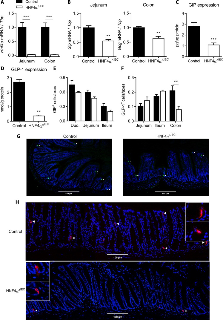
HNF4α regulates GIP and GLP-1 expression in the mouse intestine. (A) Hnf4a gene expression relative to Tbp measured in the jejunum and colon of 1 week-old control (black columns) and HNF4αΔIEC (white columns) mice (n = 4–6). (B) Gip and Gcg gene expression relative to Tbp measured in the jejunum and colon of control (black columns) and HNF4αΔIEC (white columns) mice (n = 4–8). (C) GIP protein level quantified by ELISA in the jejunum of control (black columns) and HNF4αΔIEC (white columns) mice (n = 5–10). (D) GLP-1 protein level quantified by ELISA in the colon of control (black columns) and HNF4αΔIEC (white columns) mice (n = 5–10). Enteroendocrine cells immunopositive for GIP (E) or GLP-1 (F) were counted per crypt-villus axes along the intestinal tract from both control (black columns) and HNF4αΔIEC (white columns) mice (n = 3). Representative immunostaining of enteroendocrine cells expressing GIP in the duodenum (G) and GLP-1 in the colon (H) of control and HNF4αΔIEC mice.
Figure 3.
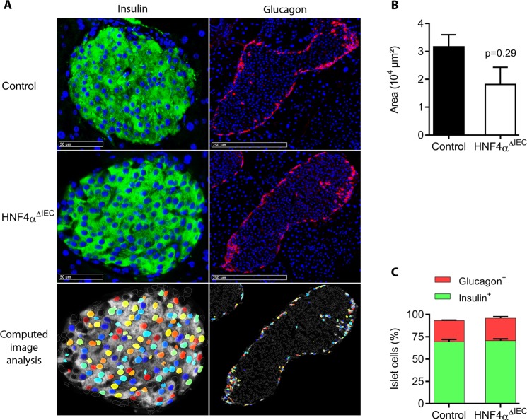
Pancreatic islet structures of HNF4αΔIEC mice. (A) Representative immunofluorescence of insulin and glucagon on two distinct islets of Langerhans sections obtained from adult control and HNF4αΔIEC mice. Computed image analysis using CellProfiler (3.1.5) was designed to quantify labeled cells. Representative image analysis obtained from HNF4αΔIEC mice islet sections (bottom panel) highlights positive detected cells (colored nuclei), overlapping immunofluorescence (white diffuse staining) and non-labeled islet cells (empty and white circled nuclei). (B) Pancreatic islets area measured in adult control (black column) and HNF4αΔIEC (white column) mice (n = 4–5). (C) Relative population of cells positive for insulin (green columns) or glucagon (red columns) signals in pancreatic islets of adult control and HNF4αΔIEC mice (n = 3–5).
Figure 4.
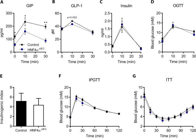
Hormonal and glycemic response in HNF4αΔIEC mice. Circulating GIP (A), GLP-1 (B) and insulin (C) levels measured by ELISA in control (black squares) and HNF4αΔIEC (blue squares) mice following an oral glucose tolerance test (OGTT) (n = 4–10). (D) Blood glucose measured in control (black squares) and HNF4αΔIEC (blue squares) mice during OGTT (n = 4 = 10). (E) Insulinogenic index was calculated in control (black columns) and HNF4αΔIEC (white columns) mice as follows: Δinsulin peak/Δblood glucose peak, where Δ is the time point concentration minus baseline level (n = 6–7). (F) Blood glucose measured in control (black squares) and HNF4αΔIEC (blue squares) mice during an intraperitoneal glucose tolerance test (IPGTT) (n = 7–9). (G) Insulin tolerance test (ITT) performed on control (black squares) and HNF4αΔIEC (blue squares) mice (n = 6–11).
Figure 2.
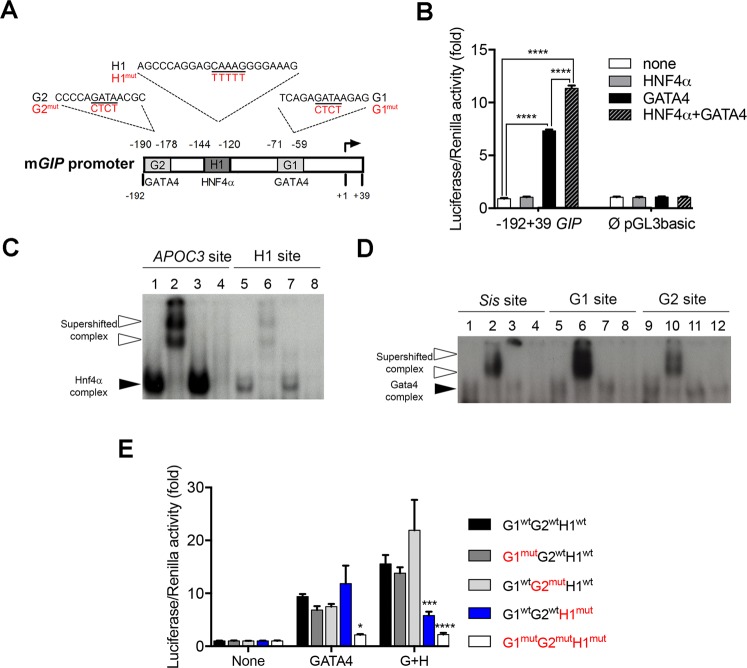
The mouse Gip promoter is regulated by HNF4α. (A) Schematic representation of the mouse Gip promoter with predicted binding sites for HNF4α and GATA4. Mutations introduced in these sites are indicated in red. (B) Luciferase assay using the −192 +39 Gip-PGL3basic and Empty-pGL3basic constructs with a combination of HNF4α and/or GATA4 expression vectors. Luciferase assays are representative of three independent.experiments and are presented as mean ± SEM; ****P < 0.0001. (C) EMSA analysis with HNF4α or (D) GATA4 nuclear extracts of each potential binding site. Lines 1, 5 and 9 show 32P-labelled probes with nuclear extracts; lines 2, 6 and 10 show 32P-labelled probes with nuclear extracts and antibodies; lines 3, 7 and 11 show 32P-labelled probes with nuclear extracts and irrelevant antibodies; lines 4, 8 and 12 show 32P-labelled probe with nuclear extracts from non-transfected empty cells. White arrowheads point to supershifted complexes while black arrowheads depict retarded complexes. Full length gels of these analyses are provided in Supplementary Fig. 2. (E) Luciferase assay using the −192 +39 Gip-PGL3 wild-type or mutated promoter constructs in combination of HNF4α (H) and/or GATA4 (G) expression vectors. Mutation effects on luciferase activity are evaluated against the wild-type promoter for each condition. Luciferase assays are representative of three independent experiments and are presented as mean ± SEM; *P < 0.05, ***P < 0.001, ****P < 0.0001.
Figure 5.
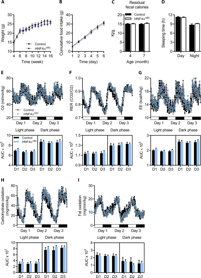
Nutrition behaviors and energy metabolism in HNF4αΔIEC mice. (A) Weight curves for adult control (black squares) and HNF4αΔIEC (blue squares) mice (n = 4–13). (B) Cumulative food intake was monitored in metabolic cages during 6 consecutive days for both adult control (black squares) and HNF4αΔIEC (blue squares) mice (n = 5–6). (C) Calorie density determined from fecal samples collected from 4-month-old and 7-month-old control (black columns) and HNF4αΔIEC (white columns) mice (n = 3–5). (D) Sleeping time monitored in metabolic cages and calculated as the mean of 3 consecutive days for both adult control (black columns) and HNF4αΔIEC (white columns) mice (n = 7). Oxygen consumption (E), respiratory exchange ratio (RER) (F), and energy expenditure (EE) (G) measurements acquired for 3 consecutive days for both adult control (black circles) and HNF4αΔIEC (blue circles) mice (n = 7) with corresponding area under the curve (AUC). Estimated oxidative rates for carbohydrates (H) and fat (I) with AUC calculated for 3 consecutive days for both adult control (black circles) and HNF4αΔIEC (blue circles) mice (n = 7).
Figure 6.
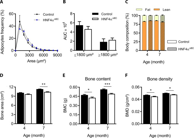
White adipocyte tissue density and body composition in HNF4αΔIEC mice. (A) Ranging of adipocytes according to their size as quantified by CellProfiler (3.1.5) from white adipose tissue sections from adult control (black squares) and HNF4αΔIEC (blue squares) mice (n = 7–8). The AUC calculated from control (black columns) and HNF4αΔIEC (white columns) mice (n = 7–8) is represented in (B). Dual-energy X-ray absorptiometry (DXA) measurements for body composition were acquired post-mortem from 4-month-old and 7-month-old mice. (C) Fat tissues (light yellow) and lean tissues (dark orange) are shown relative to total body weight of control (dark bordered squares) and HNF4αΔIEC (hatched squares) mice (n = 8–13). Osteodensitometry measurements for bone area (D), bone mineral content (BMC) (E) and bone mineral density (BMD) (F) of control (black columns) and HNF4αΔIEC (white columns) mice were calculated from DXA analysis (n = 8–13).
Results
Loss of intestinal HNF4α negatively impacts incretins production from the gut
Mice were conditionally deleted for Hnf4α in the intestinal epithelium as previously described8,23,24. Total RNA isolated from the jejunum and colon of 1 week-old HNF4αΔIEC and control mice were used to confirm that Hnf4α gene transcript expression was decreased in the intestinal epithelium of HNF4αΔIEC mice as determined by qRT-PCR (Fig. 1A). In order to evaluate a possible regulatory link between HNF4α and gut specific incretins, GIP and GLP-1 gene transcript expression was measured from the jejunum and the colon, respectively. A significant reduction was observed for jenunal Gip gene transcript (46%, P < 0.01) and colonic Gcg gene transcript (37%, P < 0.01) of 1 week-old HNF4αΔIEC mice (Fig. 1B). This reduction in Gip and Gcg transcripts was also maintained in adult HNF4αΔIEC mice (data not shown). To assess whether modulation at the gene transcript level impacted mature incretins production, ELISA essays were performed on intestinal tissues. GIP peptide concentration was reduced by more than 61% (P < 0.001) in the jejunum of fasted HNF4αΔIEC mice (Fig. 1C) while GLP-1 peptide concentration dropped by 87% (P < 0.01) in the colon of these mice (Fig. 1D). Immunofluorescence experiments were next performed to assess whether changes observed in incretin production was associated with variations in K (GIP) and L (GLP-1) enteroendocrine cell populations. As expected, the relative number of GIP+ cells was most abundant in the proximal intestine and gradually decreased toward the distal part while GLP-1+cells were more abundant in the distal intestine as previously reported elsewhere35. HNF4αΔIEC mice displayed non-significant changes in the relative number of GIP+ cells along the intestinal segments (Fig. 1E). Intriguingly, loss of intestinal HNF4α led to a reduction in the number of GLP-1+ cells in the colon (62%, P < 0.01) without affecting their number in both the jejunum and the ileum (Fig. 1F). Representative immunostaining results are shown for GIP+ cells within the duodenum (Fig. 1G) and GLP-1+ cells within the colon (Fig. 1H). Collectively, these observations suggest that HNF4α positively impacts Gip gene transcript and peptide expression, while mediating GLP-1 expression in a complex regional manner along the anterior-posterior axis of the gut epithelium.
HNF4α regulates Gip promoter activity
Since the loss of HNF4α was found to correlate well with Gip gene transcript expression without majorly affecting GIP + cell production throughout the small intestine, Gip promoter was further screened for potential HNF4α DNA binding sites. A 193 bp region upstream of the transcription start site (TSS) of the murine Gip gene was analyzed since it was previously reported to represent the minimal promoter region required to direct specific expression in endocrine cells36. This analysis predicted one HNF4α binding site (H1) flanked by 2 GATA DNA binding sites (G1 and G2) (Fig. 2A). Interestingly, comparison of the GIP promoter among different species including human displayed a relatively well conserved distribution pattern of these GATA/HNF4α predicted binding sites (Supplementary Fig. 1). Co-transfection experiments with increasing concentrations of HNF4α did not impact −192 to +39 Gip promoter activity (data not shown). Since GATA-4 has been shown to regulate GIP expression37 and to physically interact with HNF4α38, cooperative interaction of both HNF4α and GATA-4 was tested on Gip promoter activity. GATA-4 alone was able to induce a greater than 7.3-fold increase (P < 0.0001) in Gip promoter activity while HNF4α alone had no effect (Fig. 2B). However, the combination of GATA4 and HNF4α led to a 11.3-fold (P < 0.0001) synergistic induction of the −192 to +39 Gip promoter construct (Fig. 2B). Both GATA-4 and HNF4α expression vector did not influence pGL3basic empty reporter activity (Fig. 2B). To further determine whether the predicted HNF4α and GATA-4 DNA binding sites of the Gip promoter were functional, EMSA was performed with double stranded 32P-labelled probes corresponding to these specific sites. Nuclear extracts isolated from 293 T cells transfected with an HNF4α expression vector were able to generate a complex with both the Apoc3 HNF4α binding site used as a positive control (lane 1, Fig. 2C) and the Gip H1 site (lane 5, Fig. 2C) as opposed to empty nuclear extracts isolated from non-transfected 293 T cells (lanes 4 and 8, Fig. 2C). This complex was specific for HNF4α binding since inclusion of HNF4α antibodies led to the formation of supershifted complexes (lanes 2 and 6, Fig. 2C) which did not form upon substitution with irrelevant antibodies (lanes 3 and 7, Fig. 2C). A similar approach was also used with nuclear extracts isolated from 293 T cells transfected or not with a GATA-4 expression vector. A GATA-4 complex was observed for the Sis GATA binding site used as a positive control (lane 1, Fig. 2D), as well as for the Gip G1 (lane 5, Fig. 2D) and Gip G2 (lane 9, Fig. 2D) sites when GATA-4 positive nuclear extracts were used as opposed to empty 293 T nuclear extracts (lanes 4, 8 and 12, Fig. 2D). Again, this complex was specific for GATA-4 since the use of GATA-4 antibodies produced supershifted complexes (lanes 2, 6 and 10, Fig. 2D) as opposed to the use of irrelevant antibodies (lanes 3, 7 and 11, Fig. 2D). Mutagenesis of H1 (HNF4α), G1 (GATA) and G2 (GATA) sites was next achieved to monitor the importance of these sites for synergistic activation of the Gip promoter from both HNF4α and GATA-4. While mutagenesis of either G2 alone or G1 alone did not significantly impact the combined GATA-4 and HNF4α induction of Gip transcription, mutagenesis of H1 in the presence of both GATA-4 and HNF4α led to a 63% reduction in synergistic activation of the Gip promoter (P < 0.001) (Fig. 2E). Mutagenesis of all G1, G2 and H1 sites reduced the GATA-4 and HNF4α synergistic effect on Gip promoter transcription by more than 86% (P < 0.0001) (Fig. 2E). Collectively, these observations support that the interaction of HNF4α with its binding site is of functional importance to positively regulate Gip promoter activity.
Loss of intestinal HNF4α does not influence glucose homeostasis or glucose-stimulated insulin secretion after oral or intraperitoneal glucose challenges
Given that incretins support pancreatic functions, we next monitored whether loss of intestinal HNF4α and associated reduction of intestinal GIP and GLP-1 production influenced pancreatic islets features. Both control and HNF4αΔIEC pancreatic islets displayed typical structures with inner insulin-expressing cells (β-cells) surrounded by glucagon-expressing cells (α-cells) (Fig. 3A). When compared to control mice, HNF4αΔIEC mice did not show significant change in islets size (Fig. 3B) as well as in β-cell and α-cell distribution (Fig. 3C). We then investigated whether the observed changes in gut incretins expression could impact circulating levels of both GIP and GLP-1 as well as glycemic parameters in HNF4αΔIEC mice following an oral glucose tolerance test (OGTT). Fasting circulating levels of GIP in HNF4αΔIEC mice were reduced by 52% when compared to control mice (P < 0.05) (Fig. 4A). OGTT increased GIP levels reaching a peak at 10 min and then decreasing at 30 min in both control and HNF4αΔIEC mice (Fig. 4A). However, the magnitude of the GIP response remained weaker in HNF4αΔIEC mice with a 32% reduction (P < 0.05) at 10 min and 55% reduction (P < 0.01) at 30 min in circulating GIP levels compared to controls. In contrast to GIP, fasting circulating levels of GLP-1 were not affected in HNF4αΔIEC mice (Fig. 4B). As expected, OGTT transiently increased GLP-1 levels at 10 min in both genotypes with a modest but not significant decrease of peak values for HNF4αΔIEC mice (Fig. 4B). Insulin release (Fig. 4C) and blood glucose concentration (Fig. 4D) remained unchanged between HNF4αΔIEC and control mice during OGTT, consistent with a similar insulinogenic index in these mice (Fig. 4E). In order to gain further insights into glycemic control in these mice, glucose and insulin tolerance tests were achieved intraperitoneally. Both glucose tolerance (Fig. 4F) and insulin tolerance (Fig. 4G) remained similar between HNF4αΔIEC and control mice. These data indicate a defective glucose-stimulated GIP response in HNF4αΔIEC mice that was not counteracted by GLP-1 and without influencing GSIS.
Loss of intestinal HNF4α and coincidental GIP down-regulation do not impact nutrition and energy metabolism but negatively influence bone density
To gain further insight into the physiological impact of reduced GIP production in HNF4αΔIEC mice, metabolic parameters were next assessed. HNF4αΔIEC mice showed similar weight growth curves when compared to control mice (Fig. 5A), consistent with similar food intakes from both groups (Fig. 5B). Gross intestinal absorption ability remained similar in 4-month-old and 7-month-old HNF4αΔIEC and control mice as determined by total residual fecal caloric concentrations (Fig. 5C). Sleep and daily rhythms were also similar between HNF4αΔIEC and control mice (Fig. 5D) with no significant change in energy homeostasis as determined by oxygen consumption (Fig. 5E), respiratory exchange ratio (RER) (Fig. 5F) and energy expenditure (EE) (Fig. 5G). Oxidative rates fluctuations during light and dark phases for both carbohydrate (Fig. 5H) and fat (Fig. 5I) remained also similar between control and HNF4αΔIEC mice. Because incretins can regulate adipogenesis, we also assessed whether adipose density was affected in HNF4αΔIEC mice. Analysis of the white adipocyte tissue showed a similar frequency distribution of adipocyte sizes when compared between HNF4αΔIEC and control mice (Fig. 6A,B). DXA body composition analysis of young adults (4-month-old) and aging (7-month-old) HNF4αΔIEC and control mice also showed similar lean and fat mass among the groups (Fig. 6C). However, DXA analysis revealed a significant reduction in bone area values (8.3% at 7 months, P < 0.01; Fig. 6D), in bone mineral content (9.8% at 4 months, P < 0.05; 12.9% at 7 months, P < 0.001; Fig. 6E) and in bone mineral density values (5.8% at 4 months, P < 0.05; 6% at 7 months, P < 0.05; Fig. 6F) in HNF4αΔIEC mice when compared to controls. Altogether, these data support that HNF4αΔIEC mice retain normal physiology under standard conditions except for altered bone density, a well-described osteotropic effects of GIP deficiency in vivo.
Discussion
Incretins have long been recognized to play a central role in controlling the nutrition metabolism axis. Numerous reports have linked incretins to a number of metabolic features including fat metabolism and obesity39,40, bone metabolism41 and diabetes42. To date, only a small number of transcription factors have been reported to regulate GIP synthesis. GATA-4 regulate GIP expression in cell lines37 and Forkhead box protein O1 (FoxO1) as well as LEF1/β-catenin transcriptional complex mediate glucose and insulin-positive GIP regulation43. The regulatory factor X6 (Rfx6) acts as a positive regulator of GIP44 and coincidently, RFX6 haploinsufficiency was found to be associated with a reduction of GIP in MODY45. The present findings further identify an additional disease-relevant regulatory connection between the transcription factor HNF4α, for which mutations cause MODY1, and the intestinal specific regulation of GIP. Since MODY1 (HNF4A) mutations are somatic, concomitant alterations occurring in other tissues expressing HNF4α such as the intestinal epithelium are thus likely to be functionally involved during the onset of this disease. In support of this, patients with MODY1 showed a correlation between the total of insulin secretion during a test meal and GIP secretion46. In addition, a recent case report described an HNF4A mutation associated with an impaired incretin response during the progression of the MODY phenotype47. Our results open up for a more thorough investigation of this possible regulatory link in the context of MODY1 and other metabolic diseases.
Our results demonstrate that the loss of intestinal epithelial HNF4α impacts glucose-stimulated GIP production. Although a reduction of colonic GLP-1 producing cells was observed exclusively in the colon of HNF4αΔIEC mice, this observation did not significantly impact GLP-1 circulating levels under these conditions. It is possible that other GLP-1 producing sources may contribute to maintain homeostatic circulating GLP-1 levels in HNF4αΔIEC mice. HNF4α possible implication in the regulation of GLP-1 appears complex and will require further investigations. However, no significant difference in glucose tolerance was observed in HNF4αΔIEC mice, an observation reminiscent of the acute loss of K (GIP+) enteroendocrine cells in transgenic mice48. In addition, single deletion of the GIP receptor was found to have only a modest impact on glucose homeostasis in mice49,50. Glucose homeostasis is complex and involves several hormones and factors produced by various tissues. The impact of our findings on glucose homeostasis will have to be measured in multi-tissues knockout models. However, our model based on the loss of HNF4α in the intestinal epithelium recapitulates the findings made in various mouse models impaired for GIP signaling where bone metabolism was the most consistent physiological impairments observed in these mice51–53.
In conclusion, the present study allowed identifying a novel positive regulatory link between HNF4α (MODY1) and GIP incretin in the intestine and paves the way for further studies as to whether this molecular relationship may be therapeutically exploited in metabolism disorders including diabetes.
Supplementary information
Acknowledgements
The authors thank Pierre Pothier for editing the manuscript, the Electron Microscopy & Histology Research Core of the FMSS at the Université de Sherbrooke for histology and phenotyping services and the FRQS-funded « Centre de Recherche du CHUS» (CR-CHUS) for providing access to the indirect calorimetry platform. This research was supported by grants from the Canadian Institutes of Health Research (CIHR) (PJT-156180) and the Natural Sciences and Engineering Research council of Canada (NSERC) (RGPIN-2017-06096) both to FB. MD was a recipient of an NSERC fellowship. ACC is the holder of the CIHR-glaxosmithkline chair in diabetes.
Author Contributions
R.G., M.D., C.J. and F.M.R. designed and performed the experiments, and analysed the results. N.P. and F.B. analysed the results. A.C.C. supervised the indirect calorimetry platform and analysed the data. Y.G. and M.L. supervised DEXA analyses. R.G. and F.B. wrote the manuscript.
Competing Interests
The authors have no competing interests as defined by Nature Publishing Group, or other interests that might be perceived to influence the results and/or discussion reported in this paper.
Footnotes
Publisher’s note: Springer Nature remains neutral with regard to jurisdictional claims in published maps and institutional affiliations.
Supplementary information
Supplementary information accompanies this paper at 10.1038/s41598-019-41061-z.
References
- 1.Duncan SA, et al. Expression of transcription factor HNF-4 in the extraembryonic endoderm, gut, and nephrogenic tissue of the developing mouse embryo: HNF-4 is a marker for primary endoderm in the implanting blastocyst. Proc Natl Acad Sci USA. 1994;91:7598–7602. doi: 10.1073/pnas.91.16.7598. [DOI] [PMC free article] [PubMed] [Google Scholar]
- 2.Sladek FM, Zhong WM, Lai E, Darnell JE., Jr. Liver-enriched transcription factor HNF-4 is a novel member of the steroid hormone receptor superfamily. Genes Dev. 1990;4:2353–2365. doi: 10.1101/gad.4.12b.2353. [DOI] [PubMed] [Google Scholar]
- 3.Taraviras S, Monaghan AP, Schutz G, Kelsey G. Characterization of the mouse HNF-4 gene and its expression during mouse embryogenesis. Mech Dev. 1994;48:67–79. doi: 10.1016/0925-4773(94)90017-5. [DOI] [PubMed] [Google Scholar]
- 4.Chen WS, et al. Disruption of the HNF-4 gene, expressed in visceral endoderm, leads to cell death in embryonic ectoderm and impaired gastrulation of mouse embryos. Genes Dev. 1994;8:2466–2477. doi: 10.1101/gad.8.20.2466. [DOI] [PubMed] [Google Scholar]
- 5.Parviz F, et al. Hepatocyte nuclear factor 4alpha controls the development of a hepatic epithelium and liver morphogenesis. Nat Genet. 2003;34:292–296. doi: 10.1038/ng1175. [DOI] [PubMed] [Google Scholar]
- 6.Hayhurst GP, Lee YH, Lambert G, Ward JM, Gonzalez FJ. Hepatocyte nuclear factor 4alpha (nuclear receptor 2A1) is essential for maintenance of hepatic gene expression and lipid homeostasis. Mol Cell Biol. 2001;21:1393–1403. doi: 10.1128/MCB.21.4.1393-1403.2001. [DOI] [PMC free article] [PubMed] [Google Scholar]
- 7.Garrison WD, et al. Hepatocyte nuclear factor 4alpha is essential for embryonic development of the mouse colon. Gastroenterology. 2006;130:1207–1220. doi: 10.1053/j.gastro.2006.01.003. [DOI] [PMC free article] [PubMed] [Google Scholar]
- 8.Babeu JP, Darsigny M, Lussier CR, Boudreau F. Hepatocyte nuclear factor 4alpha contributes to an intestinal epithelial phenotype in vitro and plays a partial role in mouse intestinal epithelium differentiation. Am J Physiol Gastrointest Liver Physiol. 2009;297:G124–134. doi: 10.1152/ajpgi.90690.2008. [DOI] [PubMed] [Google Scholar]
- 9.Yamagata K, et al. Mutations in the hepatocyte nuclear factor-4alpha gene in maturity-onset diabetes of the young (MODY1) Nature. 1996;384:458–460. doi: 10.1038/384458a0. [DOI] [PubMed] [Google Scholar]
- 10.Fajans SS, Bell GI. MODY: history, genetics, pathophysiology, and clinical decision making. Diabetes Care. 2011;34:1878–1884. doi: 10.2337/dc11-0035. [DOI] [PMC free article] [PubMed] [Google Scholar]
- 11.Gupta RK, et al. The MODY1 gene HNF-4alpha regulates selected genes involved in insulin secretion. J Clin Invest. 2005;115:1006–1015. doi: 10.1172/JCI22365. [DOI] [PMC free article] [PubMed] [Google Scholar]
- 12.Miura A, et al. Hepatocyte nuclear factor-4alpha is essential for glucose-stimulated insulin secretion by pancreatic beta-cells. J Biol Chem. 2006;281:5246–5257. doi: 10.1074/jbc.M507496200. [DOI] [PubMed] [Google Scholar]
- 13.Stoffel M, Duncan SA. The maturity-onset diabetes of the young (MODY1) transcription factor HNF4alpha regulates expression of genes required for glucose transport and metabolism. Proc Natl Acad Sci USA. 1997;94:13209–13214. doi: 10.1073/pnas.94.24.13209. [DOI] [PMC free article] [PubMed] [Google Scholar]
- 14.Dhe-Paganon S, Duda K, Iwamoto M, Chi YI, Shoelson SE. Crystal structure of the HNF4 alpha ligand binding domain in complex with endogenous fatty acid ligand. J Biol Chem. 2002;277:37973–37976. doi: 10.1074/jbc.C200420200. [DOI] [PubMed] [Google Scholar]
- 15.Hertz R, Magenheim J, Berman I, Bar-Tana J. Fatty acyl-CoA thioesters are ligands of hepatic nuclear factor-4alpha. Nature. 1998;392:512–516. doi: 10.1038/33185. [DOI] [PubMed] [Google Scholar]
- 16.Yuan X, et al. Identification of an endogenous ligand bound to a native orphan nuclear receptor. PLoS One. 2009;4:e5609. doi: 10.1371/journal.pone.0005609. [DOI] [PMC free article] [PubMed] [Google Scholar]
- 17.Shih DQ, et al. Genotype/phenotype relationships in HNF-4alpha/MODY1: haploinsufficiency is associated with reduced apolipoprotein (AII), apolipoprotein (CIII), lipoprotein(a), and triglyceride levels. Diabetes. 2000;49:832–837. doi: 10.2337/diabetes.49.5.832. [DOI] [PubMed] [Google Scholar]
- 18.Yin L, Ma H, Ge X, Edwards PA, Zhang Y. Hepatic hepatocyte nuclear factor 4alpha is essential for maintaining triglyceride and cholesterol homeostasis. Arterioscler Thromb Vasc Biol. 2011;31:328–336. doi: 10.1161/ATVBAHA.110.217828. [DOI] [PMC free article] [PubMed] [Google Scholar]
- 19.Marcil V, et al. Modification in oxidative stress, inflammation, and lipoprotein assembly in response to hepatocyte nuclear factor 4alpha knockdown in intestinal epithelial cells. J Biol Chem. 2010;285:40448–40460. doi: 10.1074/jbc.M110.155358. [DOI] [PMC free article] [PubMed] [Google Scholar]
- 20.Frochot V, et al. The transcription factor HNF-4alpha: a key factor of the intestinal uptake of fatty acids in mouse. Am J Physiol Gastrointest Liver Physiol. 2012;302:G1253–1263. doi: 10.1152/ajpgi.00329.2011. [DOI] [PubMed] [Google Scholar]
- 21.Yabe D, Seino Y. Incretin actions beyond the pancreas: lessons from knockout mice. Curr Opin Pharmacol. 2013;13:946–953. doi: 10.1016/j.coph.2013.09.013. [DOI] [PubMed] [Google Scholar]
- 22.Madison BB, et al. Cis elements of the villin gene control expression in restricted domains of the vertical (crypt) and horizontal (duodenum, cecum) axes of the intestine. J Biol Chem. 2002;277:33275–33283. doi: 10.1074/jbc.M204935200. [DOI] [PubMed] [Google Scholar]
- 23.Darsigny M, et al. Loss of hepatocyte-nuclear-factor-4alpha affects colonic ion transport and causes chronic inflammation resembling inflammatory bowel disease in mice. PLoS One. 2009;4:e7609. doi: 10.1371/journal.pone.0007609. [DOI] [PMC free article] [PubMed] [Google Scholar]
- 24.Darsigny M, et al. Hepatocyte nuclear factor-4alpha promotes gut neoplasia in mice and protects against the production of reactive oxygen species. Cancer Res. 2010;70:9423–9433. doi: 10.1158/0008-5472.CAN-10-1697. [DOI] [PubMed] [Google Scholar]
- 25.Brial F, Lussier CR, Belleville K, Sarret P, Boudreau F. Ghrelin Inhibition Restores Glucose Homeostasis in Hepatocyte Nuclear Factor-1alpha (MODY3)-Deficient Mice. Diabetes. 2015;64:3314–3320. doi: 10.2337/db15-0124. [DOI] [PubMed] [Google Scholar]
- 26.Lacraz G, et al. Deficiency of Interleukin-15 Confers Resistance to Obesity by Diminishing Inflammation and Enhancing the Thermogenic Function of Adipose Tissues. PLoS One. 2016;11:e0162995. doi: 10.1371/journal.pone.0162995. [DOI] [PMC free article] [PubMed] [Google Scholar]
- 27.Ferrannini E. The theoretical bases of indirect calorimetry: a review. Metabolism. 1988;37:287–301. doi: 10.1016/0026-0495(88)90110-2. [DOI] [PubMed] [Google Scholar]
- 28.Peronnet F, Massicotte D. Table of nonprotein respiratory quotient: an update. Can J Sport Sci. 1991;16:23–29. [PubMed] [Google Scholar]
- 29.Boudreau F, et al. Loss of cathepsin L activity promotes claudin-1 overexpression and intestinal neoplasia. FASEB J. 2007;21:3853–3865. doi: 10.1096/fj.07-8113com. [DOI] [PubMed] [Google Scholar]
- 30.Langlois MJ, et al. Epithelial phosphatase and tensin homolog regulates intestinal architecture and secretory cell commitment and acts as a modifier gene in neoplasia. FASEB J. 2009;23:1835–1844. doi: 10.1096/fj.08-123125. [DOI] [PubMed] [Google Scholar]
- 31.Lepage D, et al. Identification of GATA-4 as a novel transcriptional regulatory component of regenerating islet-derived family members. Biochim Biophys Acta. 2015;1849:1411–1422. doi: 10.1016/j.bbagrm.2015.10.011. [DOI] [PubMed] [Google Scholar]
- 32.Frechette I, Darsigny M, Brochu-Gaudreau K, Jones C, Boudreau F. The Promyelocytic Leukemia Zinc Finger (PLZF) gene is a novel transcriptional target of the CCAAT-Displacement-protein (CUX1) repressor. FEBS J. 2010;277:4241–4253. doi: 10.1111/j.1742-4658.2010.07813.x. [DOI] [PubMed] [Google Scholar]
- 33.Cartharius K, et al. MatInspector and beyond: promoter analysis based on transcription factor binding sites. Bioinformatics. 2005;21:2933–2942. doi: 10.1093/bioinformatics/bti473. [DOI] [PubMed] [Google Scholar]
- 34.Boudreau F, et al. Hepatocyte nuclear factor-1 alpha, GATA-4, and caudal related homeodomain protein Cdx2 interact functionally to modulate intestinal gene transcription. Implication for the developmental regulation of the sucrase-isomaltase gene. J Biol Chem. 2002;277:31909–31917. doi: 10.1074/jbc.M204622200. [DOI] [PubMed] [Google Scholar]
- 35.Suzuki K, et al. Distribution and hormonal characterization of primary murine L cells throughout the gastrointestinal tract. J Diabetes Investig. 2018;9:25–32. doi: 10.1111/jdi.12681. [DOI] [PMC free article] [PubMed] [Google Scholar]
- 36.Boylan MO, Jepeal LI, Jarboe LA, Wolfe MM. Cell-specific expression of the glucose-dependent insulinotropic polypeptide gene in a mouse neuroendocrine tumor cell line. J Biol Chem. 1997;272:17438–17443. doi: 10.1074/jbc.272.28.17438. [DOI] [PubMed] [Google Scholar]
- 37.Jepeal LI, Boylan MO, Michael Wolfe M. GATA-4 upregulates glucose-dependent insulinotropic polypeptide expression in cells of pancreatic and intestinal lineage. Mol Cell Endocrinol. 2008;287:20–29. doi: 10.1016/j.mce.2008.01.024. [DOI] [PMC free article] [PubMed] [Google Scholar]
- 38.Sumi K, et al. Cooperative interaction between hepatocyte nuclear factor 4 alpha and GATA transcription factors regulates ATP-binding cassette sterol transporters ABCG5 and ABCG8. Mol Cell Biol. 2007;27:4248–4260. doi: 10.1128/MCB.01894-06. [DOI] [PMC free article] [PubMed] [Google Scholar]
- 39.Miyawaki K, et al. Inhibition of gastric inhibitory polypeptide signaling prevents obesity. Nat Med. 2002;8:738–742. doi: 10.1038/nm727. [DOI] [PubMed] [Google Scholar]
- 40.Ding X, Saxena NK, Lin S, Gupta NA, Anania FA. Exendin-4, a glucagon-like protein-1 (GLP-1) receptor agonist, reverses hepatic steatosis in ob/ob mice. Hepatology. 2006;43:173–181. doi: 10.1002/hep.21006. [DOI] [PMC free article] [PubMed] [Google Scholar]
- 41.Ramsey W, Isales CM. Intestinal Incretins and the Regulation of Bone Physiology. Adv Exp Med Biol. 2017;1033:13–33. doi: 10.1007/978-3-319-66653-2_2. [DOI] [PubMed] [Google Scholar]
- 42.Gamble JM, et al. Incretin-based medications for type 2 diabetes: an overview of reviews. Diabetes Obes Metab. 2015;17:649–658. doi: 10.1111/dom.12465. [DOI] [PubMed] [Google Scholar]
- 43.Garcia-Martinez JM, Chocarro-Calvo A, De la Vieja A, Garcia-Jimenez C. Insulin drives glucose-dependent insulinotropic peptide expression via glucose-dependent regulation of FoxO1 and LEF1/beta-catenin. Biochim Biophys Acta. 2014;1839:1141–1150. doi: 10.1016/j.bbagrm.2014.07.020. [DOI] [PubMed] [Google Scholar]
- 44.Suzuki K, et al. Transcriptional regulatory factor X6 (Rfx6) increases gastric inhibitory polypeptide (GIP) expression in enteroendocrine K-cells and is involved in GIP hypersecretion in high fat diet-induced obesity. J Biol Chem. 2013;288:1929–1938. doi: 10.1074/jbc.M112.423137. [DOI] [PMC free article] [PubMed] [Google Scholar]
- 45.Patel KA, et al. Heterozygous RFX6 protein truncating variants are associated with MODY with reduced penetrance. Nat Commun. 2017;8:888. doi: 10.1038/s41467-017-00895-9. [DOI] [PMC free article] [PubMed] [Google Scholar]
- 46.Ekholm E, Shaat N, Holst JJ. Characterization of beta cell and incretin function in patients with MODY1 (HNF4A MODY) and MODY3 (HNF1A MODY) in a Swedish patient collection. Acta Diabetol. 2012;49:349–354. doi: 10.1007/s00592-011-0312-y. [DOI] [PubMed] [Google Scholar]
- 47.Arya VB, et al. HNF4A mutation: switch from hyperinsulinaemic hypoglycaemia to maturity-onset diabetes of the young, and incretin response. Diabet Med. 2014;31:e11–15. doi: 10.1111/dme.12369. [DOI] [PMC free article] [PubMed] [Google Scholar]
- 48.Pedersen J, et al. Glucose metabolism is altered after loss of L cells and alpha-cells but not influenced by loss of K cells. Am J Physiol Endocrinol Metab. 2013;304:E60–73. doi: 10.1152/ajpendo.00547.2011. [DOI] [PubMed] [Google Scholar]
- 49.Miyawaki K, et al. Glucose intolerance caused by a defect in the entero-insular axis: a study in gastric inhibitory polypeptide receptor knockout mice. Proc Natl Acad Sci USA. 1999;96:14843–14847. doi: 10.1073/pnas.96.26.14843. [DOI] [PMC free article] [PubMed] [Google Scholar]
- 50.Pamir N, et al. Glucose-dependent insulinotropic polypeptide receptor null mice exhibit compensatory changes in the enteroinsular axis. Am J Physiol Endocrinol Metab. 2003;284:E931–939. doi: 10.1152/ajpendo.00270.2002. [DOI] [PubMed] [Google Scholar]
- 51.Xie D, et al. Glucose-dependent insulinotropic polypeptide receptor knockout mice have altered bone turnover. Bone. 2005;37:759–769. doi: 10.1016/j.bone.2005.06.021. [DOI] [PubMed] [Google Scholar]
- 52.Mieczkowska A, Irwin N, Flatt PR, Chappard D, Mabilleau G. Glucose-dependent insulinotropic polypeptide (GIP) receptor deletion leads to reduced bone strength and quality. Bone. 2013;56:337–342. doi: 10.1016/j.bone.2013.07.003. [DOI] [PubMed] [Google Scholar]
- 53.Mansur SA, et al. Stable Incretin Mimetics Counter Rapid Deterioration of Bone Quality in Type 1 Diabetes Mellitus. J Cell Physiol. 2015;230:3009–3018. doi: 10.1002/jcp.25033. [DOI] [PubMed] [Google Scholar]
Associated Data
This section collects any data citations, data availability statements, or supplementary materials included in this article.


