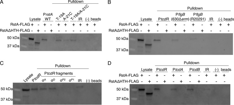
FIG 4.
RstA binds to the rstA, tcdR, flgB, tcdA, and tcdB promoters. Western blot analysis using FLAG M2 antibody to detect recombinant RstA-3XFLAG or RstAΔHTH-3XFLAG in cell lysates or following biotin-labeled DNA pulldown assays. As a control, cell lysate expressing the RstA-3XFLAG construct (MC1004) or the RstAΔHTH-3XFLAG construct (MC1028) is included in the first lane or two of each Western blot shown. Additional negative controls in each panel include unbiotinylated full-length rstA promoter (−) and beads-only controls to ensure that RstA does not interact with the beads nonspecifically. The biotin-labeled fragments used as bait are of the 115-bp wild-type, T-19A, A-21C, or T-19A/A-21C rstA promoters or of the 380-bp intergenic region upstream of the rstA promoter (IR; see Fig. 2; present in all panels) (A), the full-length tcdR (446-bp) or the 630Δerm or R20291 flgB (229-bp) promoters (B), the full-length tcdR (446-bp), σA-dependent (92-bp), σD-dependent (116-bp), σTcdRP2-dependent (188-bp), or σTcdRP1-dependent (112-bp) promoters (C), or the full-length tcdR (446-bp), tcdA (511-bp), or tcdB (501-bp) promoters (D). All promoter fragments were bound to streptavidin-coated magnetic beads and incubated with C. difficile cell lysates grown in TY medium (pH 7.4) supplemented with 2 µg/ml thiamphenicol and 1 µg/ml nisin to mid-log phase (OD600 of 0.5 to 0.7), expressing either the RstA-3XFLAG construct (MC1004) or the RstAΔHTH-3XFLAG construct (MC1028).

