

To the Editor: Mucous gland cyst in nasal sinus is common, but large mucous gland cyst in uncinate process has not been reported home and abroad. We report a rare case of mucous gland cyst in uncinate process, to improve the awareness of mucous gland cyst in uncinate process.
A patient (46 years old, male) with a 5-month history of progressive right nasal obstruction was admitted in June 12, 2015. He appeared rhinobyon on right side because of a cold 5 months ago. His nasal congestion gradually increased, until the right nasal cavity was completely blocked. He rarely had a runny nose, but sometimes had a headache on the right side. Sinoscopy revealed a light reddish, rough and tough mass which totally occupied his right limen nasi and could not be contracted by decongestant. Nasal endoscopy could not enter the right nasal cavity after using decongestant. Sinus computed tomography (CT) scan showed a homogeneous mass in the right nasal cavity without bony erosion [Figure 1A]. The patient had no history of hypertension, diabetes and coronary heart disease. Diagnosis: benign tumor of the nasal cavity (right). Making sure no taboo, we arranged endoscopic exploratory operation for this patient in June 15, 2015. During the operation, the tumor was broken when pulled by the cutting forceps. The tumor soon shrunk after about 5 to 6 mL milky fluid flowed out from it. With the assistance of elevator, the pedicle of the tumor was found in the right uncinate process [Figure 1B] and then was removed [Figure 1C]. The uncinate process stump was properly handled with electrocautery [Figure 1D]. Gelatin sponge was used to pack the operation cavity. The pathology report proved the diagnosis of mucous gland cyst [Figure 1E]. The patient was discharged 5 days later. Twelve months after operation, the nasal mucosa was recovered well.
Figure 1.
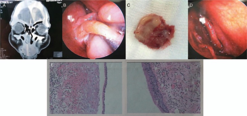
(A) CT showed a soft tissue shadow on the anterior part of the right nasal cavity. (B) The pedicle of the tumor was found locating in the uncinate process after part of fluid flowed out from the cyst. (C) Macroscopic observation: a grey-white mass, 3.0 × 2.0 × 1.8 cm in volume, with a cystic section, milky white liquid inside, rough inner wall and 0.1 to 0.2 cm in thickness. (D) The wound in the uncinate process was visible after excision of the cyst. (E) Microscopic observation: The tumor is mainly composed of glands and connective tissue, covered with pseudostratified ciliary columnar epithelium. The tumor is infiltrated by lymphocytes, plasma cells, eosinophils, and neutrophils, and the interstitium is hyperemia and edema. (H&E staining, original magnification × 40).
Nasal mucous gland cyst, which belongs to mucosal cyst, mostly occurs in the sinuses, of which the maxillary sinus is the most common, and the maxillary sinus floor and inner wall are the most common. Mucous gland cysts occurring in nasal mucosa, especially in uncinate process, are rare in clinic and have not been reported home and abroad yet.
The etiology of this disease is not completely clear. The author believes that it is caused by inflammation or allergy of nasal mucosa, which blockages the mouth of mucous glands, causes mucus to accumulate in mucous glands and enlarges the glandular cavity.[1] The cyst wall is the epithelium of mucous glandular ducts. It is also believed that the disease is caused by polyp cystic degeneration. Because the cyst is located in the mucosa, it has the names of mucosal cyst, submucosal cyst or storage cyst.
Surgical excision under nasal endoscope is the main treatment of mucous gland cyst in uncinate process. The lesions should be completely removed during the operation, and the normal nasal mucosa should be preserved as far as possible.
The pedicle of the tumor could not be discovered because the tumor had blocked the anterior naris. In addition, CT showed that the density of the nasal anterior mass was homogeneous, because the anterior part of the mass was ductile. Cyst could not be diagnosed until cyst fluid run from the ruptured mass during operation. Enhanced CT or MRI examination preoperatively is recommended to determine the nature of the tumor.[2] The milky liquid is the sufficient evidence for the diagnosis of mucous gland cyst.
Mucous gland cyst in uncinate process is a rare disease. Mucous gland cyst in uncinate process is easily misdiagnosed, because of the atypical clinical features and imaging findings. Surgical resection is an effective method for treating large mucous gland cyst in uncinate process. Diagnosis of this disease depends on pathology.
Conflicts of interest
None.
Footnotes
How to cite this article: Wang CY, Wang BB, Yao XL, Zhang F, Zhao YY, Li ZY, Zhang QQ. Mucous gland cyst in uncinate process. Chin Med J 2019;00:00–00. doi:10.1097/CM9.0000000000000104
References
- 1.Moriyama H, Nakajima T, Honda Y. Studies on mucocoeles of the ethmoid and sphenoid sinuses:analysis of 47 case. Laryngol Otol 1992; 106:23–27. doi: 10.1017/S002221510011850X. [DOI] [PubMed] [Google Scholar]
- 2.Malhotra R, Wormald PJ, Selva D. Bilateral dynamic proptosis due to frontoethmoidal sinus mucocele. Ophthal Plast Recon str Surg 2003; 19:156–157. doi: 10.1097/01.IOP.0000055829.79494.37. [DOI] [PubMed] [Google Scholar]