Abstract
Anterior communicating artery (A.com. A) aneurysm projection is an important factor in determining the outcome of aneurysm clipping. The objective of this study was to analyze the outcome of A.com.A aneurysm projection and prognostic factors influencing it and comparing them with Glasgow outcome scale. A retrospective analysis of 47 patients from hospital records who have got admitted in the Banbuntanke Hotokokai Hospital, Nagoya, Japan, from 2014 to 2017, with unruptured A.com.A aneurysm and subsequently operated in the hospital. Demographic factors such as age, sex, and associated with other aneurysms and the morphological characteristics such as aneurysm size, projection, and height were analyzed with postoperative complications and Glasgow outcome scale. Totally 47 cases have been operated in which 26 (55.3%) are female and 21 (44.6%) are male, and the median age is 68 years, 7 (14.89%) patients had middle cerebral artery aneurysm along with A.com.A aneurysm and 1 had internal carotid artery-posterior communicating artery junction aneurysm. Four (8.5%) had chronic subdural hematoma and 1 (2.12%) had epilepsy, 1 (2.12%) case got reoperated, and 1 (2.12%) had hydrocephalus. Moreover, the overall complication rate is 14.89%. For six patients, motor-evoked potential monitoring was used. Forty-six patients had Glasgow outcome scale of 5 and 1 patient had Glasgow outcome scale of 4. There was no mortality in this study. Mean size of the aneurysm was 6.68 mm and the range was 2–25 mm. Mean height was 4.14 mm, 26 (56.52%) A.com.A aneurysm were anteriorly projecting, 9 (19.56%) were superiorly projecting, 8 (17.32%) were inferiorly projecting, and 3 (6.38%) were posteriorly projecting. Morphological parameters such as size, height, and projection were not only highly associated with A.com.A aneurysm rupture and also complications due to clipping of aneurysm.
Keywords: Clipping, morphology, outcome, unruptured anterior communicating artery aneurysm
Introduction
Anterior communicating artery (A.com.A) aneurysms are among the most common aneurysms in different case series.[1] They comprise 20.6% in our latest case series of unruptured aneurysms coming 2nd only to middle cerebral artery (MCA) aneurysm. Unruptured intracranial aneurysms are diagnosed with greater frequency as imaging techniques improve. The management of unruptured intracranial aneurysms remains controversial because of incomplete and conflicting data about the natural history of these lesions and the risks associated with their repair the increasing use of noninvasive intracranial imaging, an increasing number of unruptured intracranial aneurysms are being incidentally discovered.[2] The optimal procedural management of these lesions is still being debated, which can carry significant risk, with morbidity and mortality rates up to 10 and 2.5%, respectively.[3] The International Study of Unruptured Intracranial Aneurysm (ISUIA) concluded that aneurysms <7 mm in size in the anterior circulation have an annual rupture risk of 0%–0.1% per year.[3] Anterior projection of an A. com. A aneurysm presents of blebs and size more than 5 mm increase the risk of rupture.[4] The aim of microneurosurgical management of A.com.A aneurysm is total occlusion of the aneurysm sac with preservation of flow in all branching and perforating arteries. This task necessitates perfect surgical strategy based on review of the three-dimensional (3D) angioarchitecture and abnormalities of the patient's A.com.A complex with its A.com.A aneurysm and to orientate accordingly during the microsurgical dissection.[5]
Materials and Methods
Patient population
We performed a retrospective analysis of 47 medical records of patients at Banbuntane Hotokukai Hospital, Nagoya, Japan, who were diagnosed to have unruptured A.com.A aneurysm and subsequently clipping was done from May 2014 to Decmber 2017. All patients were asymptomatic and diagnosed while screening for unruptured A.com.A aneurysm.
Clinical analysis
In clinical analysis, we measured the outcome based on Glasgow outcome scale and complications arising out of surgical clipping were analyzed. When the patient recovery is good given a maximum score of 5, with moderate disability as 4, with severe disability as 3, vegetative state as 2, and dead is 1.
Anatomical analysis
To evaluate any patient with subarachnoid hemorrhage (SAH) or any candidate for aneurysm screening, we request 3D-computerized tomography angiogram (3D-CTA). Aneurysm morphology was analyzed using the 3D-CTA. There are important anatomical points which should be paid attention, on 3D-CTA that may affect our decision-making before or during the operation.
Aneurysm projection, the location of neck and dome of aneurysm, its height and distance from the planum sphenoidale, and most condition of A2 fork are the factors affecting our surgical planning. Here, we describe the most important factors and explain how they are pivotal for decision-making.
Aneurysm size (S) is defined as maximum aneurysm diameter, aneurysm size was classified as <7 mm, 7–12 mm, 13–25 mm, and >25 mm
Maximum height of aneurysm is defined as furthest distance from the center of the aneurysm neck to the aneurysm dome
Shape of the aneurysm, with regular shape defined as simple saccular aneurysms and irregular shape defined as saccular aneurysms with additional daughter domes or blebs
Number of aneurysms, divided into single or multiple aneurysms
Variation of the A1 segment, including dominance of the A1 segment, defined as >33% of the difference between the A1 segment diameters as well as hypoplasia and absence of an A1 segment
A.com.A fenestration is defined as the A.com.A trunk dividing into two branches and then converging into only 1 trunk
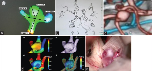
The direction [Figure 1] of the dome around the A.com.A was classified according to 6 directions: superior [Figure 1a–c], anterior [Figure 2a–c], posterior, inferior, right, left, and also as anterosuperior, right-superior when they are projection in multiple directions. First, we drew a line through the A.com.A and parallel to the anterior skull base (“parallel line”) on the sagittal plane of the 3D-CTA, perpendicular to the parallel line (“perpendicular line”). An aneurysm was defined as having anterior or posterior projections according to its positional relation to the perpendicular line. The projection was defined as superior and inferior according to its positional relation to the parallel line. The projection was said to be medial and lateral according to its direction to the aneurysmal side A1–A2 junction on the coronal or axial plane of the images.[6]
Figure 1.
(a) Various projection of anterior communicating artery aneurysm, (b) three dimensional computerized tomography angiogram of superior projection unruptured anterior communicating artery aneurysm, (c) magnified view of superior projection A.com.A Aneurysm, (d) computational fluid dynamics image showing high wall pressure, low wall shear stress, diverging vectors, and streamline velocity spiraling at the dome, (e) intraoperative picture
Figure 2.
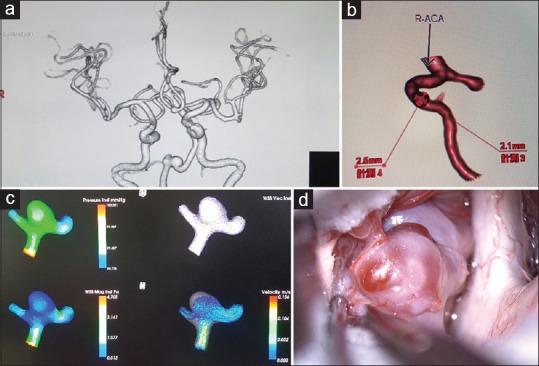
(a) Three-dimensional computerized tomography angiogram of anterior projection of unruptured anterior communicating artery aneurysm, (b) Magnified view of anterior projection aneurysm, (c) computational fluid dynamics showing yellow colored with high wall pressure and thick neck divergent vectors and streamline hitting wall with high speed, (d) intraoperative picture showing Red wall which implies low wall shear stress
The direction of each aneurysm dictated different kind of procedure:
Aneurysms with anterior and inferior projection have more favorable relationship to the infundibula and hypothalamic perforators. Attention must be paid while retracting the frontal lobe because the dome of these aneurysms is attached to the tuberculum sella and the probability of rupture is high
For aneurysms with superior projection, the A2 segment may be detected hardly or it is necessary to use fenestrated clipping. The posterior wall of their neck usually is associated intimately with the infundibula and hypothalamic perforators, which must be cleared and displaced below the path of the clip blade
Aneurysms with posterior projection were the most complicated type of aneurysms. The majority of perforator vessels are placed adjacent to the neck or in the inferior surface of the dome of aneurysms; they have to be cleared and released before permanent clipping. An extended dissection of the perforators and more creative clip configurations were required for these aneurysms. When approached from the side with A2 posterior to the aneurysm, the neck or the posterior stretched perforating cannot be reached and dissected efficiently.
A2 fork
Bilateral A2s and the A.com.A are named A2 fork at the level of A.com.A, one A2 is usually anterior to the other, making A.com.A run obliquely instead of in the coronal plane.
When the complex is approached from the side with anteriorly located A2, the A.com.A is hidden behind it and so, the complex is called closed A2 fork. In contrary, when the same complex is approached from the side with the A2 located posteriorly, the A.com.A is very visible, and the complex is named open A2 fork.
We approach the A.com.A aneurysm from the side with the open A2 fork because the neck of the aneurysm is usually anterior or superior to the A.com.A and visible from this side. Posterior looking aneurysm are exception to this rule as their neck is located posterior to the A.com.A and should be approached from the side with the closed fork.
Computational fluid dynamics
A 3D surface model of the aneurysm and adjacent vasculature was created. Mathematical cross sections in the computational fluid dynamics (CFD) model at the same locations as the scan planes were defined and intra-aneurismal flow patterns based on the blood flow velocity component to these cross-sections were calculated. Wall pressure, wall shear stress (WSS), and WSS vector and velocity in streamline flow images were constructed and studied. Moreover, CFD has potentials to allow prediction about aneurysm wall weakness. Adjunct to 3D-CTA, Haemsocope a Software integrated with 3D-CTA was used.
Treatment analysis
Most of the authors have reported better management results with early than with delayed surgery as early surgery avoids re-bleeding and helps treatment of ischemic events of vasospasm. We selected the pterional approach for these reasons: (1) the distance between skull and anterior communicating region will be shortened, (2) approach to the aneurysm is more vertical, (3) the proximal control is performed easily, and (4) the majority of surgeons are more familiar and experienced with this approach. All the cases surgical clipping was done to the unruptured A.com.A aneurysm, intraoperative motor-evoked potential monitoring was done in 4 cases, and all cases were visualized with dual-image video angiography (VA) and with endoscope before and after clipping to assess the perforator orientation and relationship with the aneurysm.
In their study of 60 fixed human brains, indicated that all perforating branches followed a posterior direction and formed an angle with the pericallosal arteries that ranged between 30 and 180.[7] Perlumtter and Rhoton showed that 90% of perforating arteries arising from the A.com.A were branched on the superior and posterior facing.[8] Yasargil considered this to be a predominant anatomical factor. Yasargil observed that projection of fundus is an important factor-predicting outcome.[7]
Anterior, superior, and posterior projecting aneurysms had good results of 93.7%, 91.8%, and 88.4% respectively, while patients with inferiorly projecting aneurysms had only 79.2% good results.[9] Observed most of the posterosuperior pointing aneurysms presented in poorer grades and outcome was unsatisfactory. In comparison, most of the anteroinferior pointing aneurysms presented with better grades and results were satisfactory.[7]
Statistical methods
Statistical analysis was performed with Python language tool and analyses Continuous variables were presented as means ± standard deviation and categorical variables as frequencies (percentages). Clinical and morphological characteristics were compared between the various aneurysm projection groups. Pearson correlation coefficient analysis was done.
Results
Demography
Out of 47 cases for which analysis was done, 26 (55.31%) were females and 21 (44.68%) were males, aged between 38 and 79 and the mean age was 71.14 with standard deviation + 9.58. All the patients underwent surgical clipping of the unruptured A.com.A aneurysm (100%) [Table 1].
Table 1.
Demography
| Factors | Number |
|---|---|
| Total number of cases | 47 |
| Age | |
| Mean±SD | 71.14±9.58 |
| Range (years) | 38–79 |
| Sex (%) | |
| Female | 26 (55.31) |
| Male | 21 (44.68) |
SD – Standard deviation
Aneurysm morphology
Out of 47 A.com.A aneurysm, 19 (40.42%) were simple saccular shaped and 20 (42.553%) cases were irregular in shape which are complex with either fusiform or bleb aneurysm, 9 (19.14%) were bleb aneurysm, and 1 (2.12%) patient had fenestrated type of aneurysm. Neck of aneurysm was wide in 32 (68%) and narrow neck in 15 (31.9%) [Table 2]. Aneurysm size [Table 3] ranged between 2 mm to 25 mm in which the mean size was 6.68 mm. The aneurysm projection noticed was anterior 10 (21.27%), superior 6 (12.76%), posterior 3 (6.38%), inferior 8 (17.02%), right 10 (21.27%), left 6 (12.76%), anterosuperior 1 (2.12%), and right superior 2 (4.25%). Mean height of the aneurysm was 4.14 mm. Five (10.63%) patients had multiple aneurysm of which 4 (8.51%) patients had MCA aneurysm and 1 (2.12%) patient had internal carotid artery-posterior communicating artery junction aneurysm along with A.com.A aneurysm [Table 4]. Twelve (25.53) patients had contralateral A1 segment were either hypoplastic or absent. Side of the aneurysm and type of aneurysm were not associated with outcome [Table 5]. However, as age advances, the size of the aneurysm has a linear correlation and at the same time, aneurysm with <7 mm was more associated with complications. To investigate the inter-dependency of the morphological parameters, we used 2D-scatterplots to analyze the interactions among the morphological parameters with age and postoperative complications which showed a positive correlation.
Table 2.
Aneurysm morphology
| Morphology | Number |
|---|---|
| Shape (%) | |
| Saccular | 19 (40.42) |
| Irregular | 20 (42.553) |
| Bleb | 9 (19.14) |
| Fenestrated type | 1 (2.12) |
| Neck (%) | |
| Wide | 32 (68) |
| Narrow | 15 (31.9) |
| Contralateral A1 hypoplastic (%) | 12 (25.53) |
Table 3.
Size of A.com.A aneurysm
| Size | n (%) |
|---|---|
| <7 | 31 (65.95) |
| 7-12 | 14 (29.78) |
| 13-24 | 1 (2.12) |
| >25 | 1 (2.12) |
Table 4.
Multiple aneurysm
| Number (%) | MCA (%) | IC-PC (%) |
|---|---|---|
| 5 (10.63) | 4 (8.51) | 1 (2.12) |
MCA – Middle cerebral artery; IC-PC – Internal carotid artery-posterior communicating artery junction
Table 5.
Age correlation with height and size of aneurysm
| Age | Height | Size |
|---|---|---|
| <45 | 3.4 | 3 |
| 45-54 | 3.9 | 5.25 |
| 55-64 | 3.512 | 6.4 |
| 65-74 | 4.46 | 6.97 |
| >75 | 4.22 | 7.2 |
Computational fluid dynamics
Out of 19 A.com.A unruptured aneurysm which were surgically clipped, the CFD study showed that the difference of diameters in the A1 segments of both sides were more than 50 % and the induced high WSS in the A.com.A (25–70 dyne/cm2) [Chart 1] suggesting a possible explanation why A.com.A are more frequent to rupture than aneurysms in other locations.[10] In CFD analysis, the volumetric flow rates measured in the A1 segments of the left and right anterior cerebral artery (30 ml/min and 60 ml/min, respectively) [Chart 2]. The thickness of the aneurysm wall within the aneurysm sac can be predicted as the high WSS (28 dyne/cm2) which occurred at aneurysm neck, slow flow and low WSS (<5 dyne/cm2) [Chart 3] were in the dome. Moreover yellow thick wall were observed in 17 (89.47%) patients which implicates high pressure and red thin wall in 16 (84.21%), showing low WSS [Figures 1d and 2d]. Divergent vectors and streamline flow hitting the wall with high speed were also observed. Interestingly, sustained low WSS levels have also been related to aneurysm rupture in several studies[11] [Chart 4]. Almost 90% of the aneurysms had maximum WSS higher than or similar to the WSS in the parent artery. Hence, locally elevated WSS could contribute to the focalized wall damage that formed blebs.[12]
Chart 1.
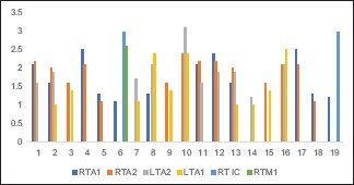
Differences in the diameters of the A1 segments of more than 50%, induced high wall shear stress in the anterior communicating artery (25–70 dyne/cm2)
Chart 2.

Volumetric flow rates measured in the A1 segments of the left and right anterior cerebral artery (30 ml/min and 60 ml/min, respectively)
Chart 3.
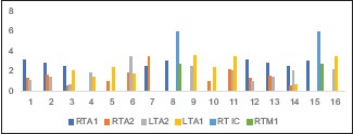
The highest wall shear stress (about 6 Pa) occurred at the aneurysm neck while slow flow and low wall shear stress (below. 5 Pa) were observed within the aneurysm sac
Chart 4.
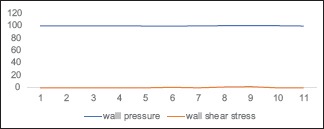
High wall pressure and low wall shear stress denotes impending rupture of the aneurysm
Surgical outcome
All the patients with unruptured A.com.A aneurysm were surgically clipped (100%) with pterional operative approach. There was no mortality in this study and the Glasgow outcome scale was Good outcome in 46 (97.87%) patients and with moderate disability in 1 (2.12%) patient.
Multi-modality monitoring of the patients during aneurysm surgeries has been associated with improved outcome in recent years. Endoscope-assisted microsurgery of aneurysm is a common practice in many centers treating such pathologies. Visualizing the corners concealed behind the superficial structures gives some information about the contra-lateral A2 and perforating arteries and their relations to the hidden side of the aneurysm. Furthermore, after inserting the clip, the endoscopic view shows the relation of the clip blades to the perforators and confirms complete occlusion of the aneurysm neck. Another monitoring modality is indocyanine green VA (ICG-VA): it confirms appropriate occlusion of the aneurysm and the patency of the bilateral A2 and perforating arteries. Although there are emerging data considering a role for FLOW 800 software of ICG-VA to quantify blood flow before and after clipping, we still do not rely on such semi-quantitative data. Instead, whenever we want to measure blood flow and its changes before and after clipping (e.g., in aneurysms with severely atherosclerotic parent arteries), Doppler ultrasound (20 MHz probe) is used. Although recommended by some, we do not advocate routine intraoperative monitoring of somatosensory or motor-evoked potentials for these patients.
Postoperative complications
Symptomatic complications [Table 6] leading to acute neurological impairment were found in 6 (12.76%) patients out of which 3 (6.38%) patients had chronic subdural hematoma for which burr-hole and tapping evacuation was done, 1 (2.12%) patient had epilepsy which was managed conservatively, 1 (2.12%) patient had communicating hydrocephalus for which shunt was done, and 1 (2.12%) patient was reoperated for surgical clipping of the aneurysm. Aneurysm projection and complication [Table 7] were not statistically associated but Pearson correlation coefficient for size and height of patients with aged from 55 to 75 is 0.83710882 [Chart 5], and there exist a correlation between aneurysm height, size, and age of the patients with complications.
Table 6.
Postoperative complication
| Complication | n (%) |
|---|---|
| Chronic subdural hematoma | 3 (6.38) |
| Epilepsy | 1 (2.12) |
| Hydrocephalus | 1 (2.12) |
| Reoperation | 1 (2.12) |
| Total | 6 (12.76) |
Table 7.
Aneurysm morphology and complication
| Projection | n (%) | Mean height | Mean size | Complication |
|---|---|---|---|---|
| Anterior | 10 (21.27) | 4.13 | 6.45 | CSDH |
| Superior | 6 (12.76) | 3.84 | 6.5 | Reoperation |
| Right | 10 (21.27) | 4.37 | 8.1 | Hydrocephalus |
| Left | 6 (12.76) | 3.766 | 5.833 | CSDH |
| Posterior | 3 (6.38) | 3.05 | 4.66 | 0 |
| Anterosuperior | 1 (2.12) | 5.5 | 10 | 0 |
| Inferior | 8 (17.02) | 4.91 | 7.675 | Epilepsy |
| Right superior | 2 (4.25) | 2.7 | 3.5 | 0 |
CSDH – Chronic subdural hematoma
Chart 5.
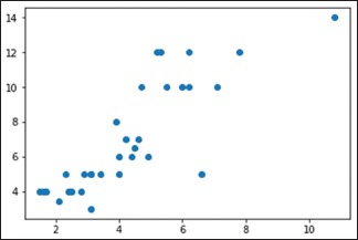
Scatter diagram showing correlation between aneurysm height and size with age of the population and complication with Glasgow outcome scale of the unruptured anterior communicating artery aneurysm
Pearson correlation coefficient for size and height of patients with aged from 55 to 75 is 0.83710882.
Discussion
The available literature evaluating unruptured aneurysms regarding basic patient population, clinical practice, and even aneurysm characteristics studied, a meta-analysis is nearly impossible to perform. This study will instead focus on the various anatomical and morphological characteristics of aneurysms reported in the literature with an attempt to draw broad inferences and serve to highlight pressing questions for the future in our continued effort to improve clinical management of unruptured intracranial aneurysms. Gerlach et al. in a series of prospective study of unruptured intracranial aneurysm finds approximately 48% of treated unruptured intracranial aneurysm were smaller than 7 mm[13] which is similar 31 (65.95%) in this series, although the risk of bleeding based on the ISUIA data seems to be low. Hypoplasia of the A1 segment of the anterior cerebral artery has been found more frequently in patients with A.com.A aneurysm than in those with aneurysms in other locations,[10] similar to our study 12 (25.53%). In addition, in patients with A.com.A aneurysm, larger necks on the dominant side have been observed,[10] similar to our study 32 (68%). In CFD analysis, the volumetric flow rates measured in the A1 segments of the left and right anterior cerebral artery (30 ml/min and 60 ml/min, respectively) are in good agreement with values reported in the literature, the WSS values found in our study (1–11 Pa) agree well with what is commonly found for saccular aneurysms: 5–13 Pa. High wall pressure and low WSS implies impending rupture of the A.com.A aneurysm. Streamline flow in the vessel denies the turbulent flow in the aneurysmal sac. New surgical techniques, such as indocyanine green angiography combined with continuous intraoperative electrophysiological monitoring and new endovascular methods, may contribute to increased safety and efficacy of unruptured intracranial aneurysm repair as well as the concentration of such elective procedures in dedicated cerebrovascular centers. After treatment, no patient suffered a SAH. No patient died as a result of treatment of an unruptured intracranial aneurysm, resulting in a mortality rate of 0%,[13] our study also endorses the finding. Assessment of overall clinical outcome after 6 months proved favorable (mRS 0–2) in 97.7% of all 133 aneurysms (97.9%) after surgery.[13] Our study also has a Glasgow outcome scale of good in 46 (97.87%) patients. Age was the only independent predictor of a poor surgical outcome. Surgery-related morbidity and mortality at 1 year among patients younger than 45 years was 6.5%, as compared with 14.4% for those between 45 and 64 years old and 32% for those over 64 (P < 0.001)[14] which is goes hand in hand with our study findings. Pearson correlation coefficient for size and height of patients aged from 55 to 75 is 0.83710882. The rates of surgery-related morbidity and mortality were substantially lower for younger patients than for older patients and also in fact, a recent prospective, population-based cohort study showed that this tendency is more prominent in women than men and that at least one pathway in the development of SAH requires both smoking and female sex.[15] Which is similar to our study, females are 26 (55.31%). In Shao et al. series no significant differences in patient characteristics between the anterior and posterior projection aneurysm groups but significant differences in aneurysm morphologies,[16] which corroborates our study. Although the hemodynamic flow features between the anterior and posterior projection aneurysms still require additional study, the aneurysm projection and other morphologies may be considered in the use of CFD in the aneurysm formation or rupture.[16] Castro et al. analyzed the image-based CFD in 26 A.com.A aneurysm and they found that smaller impaction zones, higher flow rates, and increased maximum WSS were associated with rupture status.[17]
Study limitations
Ours was conducted retrospectively at a single institution, and the number of patients was too small to draw definite conclusions about morphological and clinical characteristics, possibly leading to selection bias and wide confidence intervals. Second, most patients were diagnosed with the A.com.A aneurysm and were treated with surgical clipping only. Observed differences may reflect racial differences in the patient population given that recent genetic analyses have indicated that Japanese and/or Finnish patients are at higher risk for aneurysm rupture. Hence, the external validity applied only to a Japanese population and may not be generalizable to other ethnic groups. To exclude any such limitations in future research, we intend to expand our series and gather data on a larger number of patients with A.com.A aneurysm to verify the present findings.
Conclusion
Optimal decision for the treatment of A.com.A aneurysm cannot be determined by anyone anatomic characteristic, but all morphological features need to be considered and that microsurgical clipping remains a valid treatment modality with the use of CFD study, motor-evoked potential, 3D-CTA. Aneurysm size and height are important morphological factors with age for determining the postoperative outcome of the patient. CFD can help in predicting the nature of aneurysm wall and probability of rupture; our study is of small number of patient, so further studies with greater patient cohort are required to evaluate our findings.
Financial support and sponsorship
Nil.
Conflicts of interest
There are no conflicts of interest.
References
- 1.Bunevicius A, Cikotas P, Steibliene V, Deltuva VP, Tamsauskas A. Unruptured anterior communicating artery aneurysm presenting as depression: A case report and review of literature. Surg Neurol Int. 2016;7:S495–8. doi: 10.4103/2152-7806.187489. [DOI] [PMC free article] [PubMed] [Google Scholar]
- 2.Ajiboye N, Chalouhi N, Starke RM, Zanaty M, Bell R. Unruptured cerebral aneurysms: Evaluation and management. Sci World J 2015. 2015 doi: 10.1155/2015/954954. 954954. [DOI] [PMC free article] [PubMed] [Google Scholar]
- 3.Lall RR, Eddleman CS, Bendok BR, Batjer HH. Unruptured intracranial aneurysms and the assessment of rupture risk based on anatomical and morphological factors: Sifting through the sands of data. Neurosurg Focus. 2009;26:E2. doi: 10.3171/2009.2.FOCUS0921. [DOI] [PubMed] [Google Scholar]
- 4.Fujimoto K, Yano S, Shinojima N, Hide T, Kuratsu JI. Endoscopic endonasal transsphenoidal surgery for patients aged over 80 years with pituitary adenomas: Surgical and follow-up results. Surg Neurol Int. 2017;8:213. doi: 10.4103/sni.sni_189_17. [DOI] [PMC free article] [PubMed] [Google Scholar]
- 5.Cai W, Hu C, Gong J, Lan Q. Anterior communicating artery aneurysm morphology and the risk of rupture. World Neurosurg. 2018;109:119–26. doi: 10.1016/j.wneu.2017.09.118. [DOI] [PubMed] [Google Scholar]
- 6.Matsukawa H, Uemura A, Fujii M, Kamo M, Takahashi O, Sumiyoshi S, et al. Morphological and clinical risk factors for the rupture of anterior communicating artery aneurysms. J Neurosurg. 2013;118:978–83. doi: 10.3171/2012.11.JNS121210. [DOI] [PubMed] [Google Scholar]
- 7.Agrawal NR. Factors Influencing Outcome Following Intraoperative Rupture During Surgical Clipping of Anterior Communicating Artery Aneurysms; October. 2000 [Google Scholar]
- 8.Rhoton AL., Jr The supratentorial arteries. Neurosurgery. 2002;51:S53–120. [PubMed] [Google Scholar]
- 9.Park JH, Park SK, Kim TH, Shin JJ, Shin HS, Hwang YS, et al. Anterior communicating artery aneurysm related to visual symptoms. J Korean Neurosurg Soc. 2009;46:232–8. doi: 10.3340/jkns.2009.46.3.232. [DOI] [PMC free article] [PubMed] [Google Scholar]
- 10.Cebral JR, Raschi M. Suggested connections between risk factors of intracranial aneurysms: A review. Ann Biomed Eng. 2013;41:1366–83. doi: 10.1007/s10439-012-0723-0. [DOI] [PMC free article] [PubMed] [Google Scholar]
- 11.Wong GK, Poon WS. Current status of computational fluid dynamics for cerebral aneurysms: The clinician's perspective. J Clin Neurosci. 2011;18:1285–8. doi: 10.1016/j.jocn.2011.02.014. [DOI] [PubMed] [Google Scholar]
- 12.Cebral J, Mut F, Sforza D, Löhner R, Scrivano E, Lylyk P, et al. Clinical application of image-based CFD for cerebral aneurysms. Int J Numer Method Biomed Eng. 2011;27:977–92. doi: 10.1002/cnm.1373. [DOI] [PMC free article] [PubMed] [Google Scholar]
- 13.Gerlach R, Beck J, Setzer M, Vatter H, Berkefeld J, Du Mesnil de Rochemont R, et al. Treatment related morbidity of unruptured intracranial aneurysms: Results of a prospective single centre series with an interdisciplinary approach over a 6 year period (1999-2005) J Neurol Neurosurg Psychiatry. 2007;78:864–71. doi: 10.1136/jnnp.2006.106823. [DOI] [PMC free article] [PubMed] [Google Scholar]
- 14.Choi JH, Kang MJ, Huh JT. Influence of clinical and anatomic features on treatment decisions for anterior communicating artery aneurysms. J Korean Neurosurg Soc. 2011;50:81–8. doi: 10.3340/jkns.2011.50.2.81. [DOI] [PMC free article] [PubMed] [Google Scholar]
- 15.Raaymakers TW, Rinkel GJ, Limburg M, Algra A. Mortality and morbidity of surgery for unruptured intracranial aneurysms: A meta-analysis. Stroke. 1998;29:1531–8. doi: 10.1161/01.str.29.8.1531. [DOI] [PubMed] [Google Scholar]
- 16.Shao X, Wang H, Wang Y, Xu T, Huang Y, Wang J, et al. The effect of anterior projection of aneurysm dome on the rupture of anterior communicating artery aneurysms compared with posterior projection. Clin Neurol Neurosurg. 2016;143:99–103. doi: 10.1016/j.clineuro.2016.02.023. [DOI] [PubMed] [Google Scholar]
- 17.Castro MA, Putman CM, Sheridan MJ, Cebral JR. Hemodynamic patterns of anterior communicating artery aneurysms: A possible association with rupture. AJNR Am J Neuroradiol. 2009;30:297–302. doi: 10.3174/ajnr.A1323. [DOI] [PMC free article] [PubMed] [Google Scholar]


