Abstract
Tunicates (Ascidians, sea squirts) are marine protochordates, which live sedentary or sessile in colonial or solitary forms. These invertebrates have to protect themselves against predators and invaders. A most successful strategy, to not being eaten by predators and prevent pathogenic microorganisms to settle, is the usage of chemical molecules for defence. To accomplish this, tunicates take advantage of the specialized metabolites produced by the bacteria associated with them. Therefore, the microbiome of the tunicates can be regarded as a promising bioresource for bacterial strains producing compounds with antibacterial activity. The aim of this study was to test this hypothesis by (i) isolation of tunicate-associated bacteria, (ii) analysis of the antibacterial activities of these strains, and (iii) purification and structure elucidation of an active compound derived from this bioresource. In total, 435 bacterial strains were isolated and thereof 71 (16%) showed antibacterial activity against multidrug resistant (MDR) bacteria. Therefrom, the ethyl acetate crude extracts from liquid fermentations of 25 strains showed activity against MDR Extended-Spectrum Beta-Lactamase (MDR-ESBL) Escherichia coli, MDR Bacillus cereus, Micrococcus luteus, and Bacillus megaterium. Phenotypic analysis based on 16S rDNA sequencing revealed the active strains belonging to different genera and phyla, like Bacillus, Pantoea, Pseudoalteromonas, Salinicola, Streptomyces, Vibrio and Virgibacillus. To obtain first insights into the molecules responsible for the antibacterial activities observed, strain Pseudoalteromonas rubra TKJD 22 was selected for large-scale fermentation and the active compound was isolated. This allowed the purification and structure elucidation of isatin, a compound known for its strong biological effects, thereunder inhibition of Gram-positive and Gram-negative pathogens.
Introduction
The exploration of new bioresources for the discovery of novel antibiotic lead structures is of importance due to the increase of antibiotic resistant pathogenic bacteria [1, 2]. In nowadays, already a multitude of pathogenic bacteria have become resistant to many kinds of antibiotics, summarized under the term Multi-Drug Resistant (MDR) bacteria [3, 4]. Cases of infection are increasing each year, leading to serious problems in clinical as well as economic aspects [5–8]. The antibiotics available for treatment of bacterial infections in hospital and outpatient settings are no more effective to combat MDR strains [9, 10]. Therefore, novel compounds must be developed and exploration of new resources can be regarded as a must.
The marine environment is the largest habitat on earth, representing more than 70% of the planet´s surface. However, it still must be regarded as understudied and unexplored if compared with terrestrial habitats [11]. Indonesia, having the second largest marine environment, is a major house of diverse and unique marine invertebrates; thereby, promoting the potential for diverse chemical substances with putative bio(techno)logical activities and applications [12–14]. The Marine National Park of Karimunjawa is a conservation area with good water conditions and an intact marine biota [15]. The latter should be regarded as a main target for the discovery of novel marine natural products, since their associated microbiomes occupy a unique niche in the ocean´s biota.
One well-investigated subphylum of marine protochordata are tunicates. Already in the early 70ies, even before Marine Natural Products have been named as a scientific discipline [16], Sigel and colleagues reported that the ethanolic extract from the Caribbean tunicate Ecteinascidia turbinata possessed anti-proliferative properties [17]. Later, exactly from this tunicate species, an anti-proliferative compound was isolated, which was the lead to develop the anticancer drug Yondelis (Trabectedin/ ET-743) [18]. The latter molecule shows structural similarities with the natural products saframycin and safracin, which were isolated from Pseudomonas fluorescens. This fact pointed towards the fact that bacteria associated with the macroorganism represent the original producer [19]. This hypothesis was finally verified by the discovery of a biosynthetic gene cluster, encoding the biosynthetic machinery responsible for ET-743 biosynthesis in the bacterial symbiont Candidatus Endoecteinascidia frumentesis, a γ-proteobacterium [20]. The fact that microbes associated with invertebrates are the producers of molecules of interest became well known by further examples and is also advantageous for bioprospecting approaches, since they will be more sustainable and cultivation will be possible, rendering the process faster, easier and less expensive [21].
Materials and methods
Sample collection and preliminary identification of the tunicates
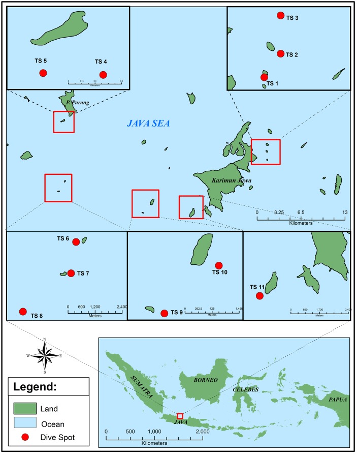
Sampling was performed at the Marine National Park of Karimunjawa, North Java Sea, Indonesia in March, May and July 2017 (Fig 1). All samples were collected by scuba diving at 10–18 m depths. Underwater photo documentation and labelling was conducted before the samples were stored in sterile plastic zip lock bags and cooled. They were either directly processed, or cooled until further processing in the laboratory. Preliminary identification of the specimen was based on the morphological appearance of the specimen, according to the tunicate atlas [22, 23].
Fig 1. Sampling site in Karimunjawa Sea, Indonesia.
TS1-5, and TS11 represent diving sites at sampling site 1 (March 2017). TS 6,7,8 represent diving sites at sampling site 2 (May 2017) and TS 9,10 represent the last sampling site (July 2017).
Isolation of associated bacteria
First, the environmental samples were processed under sterile condition as follows: 1 g of tunicate sample tissue was rinsed 3 times with sterile natural seawater, grinded with a sterile mortar and homogenized with 5 mL sterile natural seawater. The homogenized tunicate tissue was serially diluted and spread (30 μL) onto different media (PYA, ISP2, M1). The composition of the media used were (i) PYA medium: peptone (2.5 g), yeast extract (0.5 g), and agar (15 g), ingredients were mixed with sterile natural seawater (1 L); (ii) ISP2 medium: malt extract (10 g), yeast extract (4 g), glucose (4 g) and agar (15 g), ingredients were mixed with sterile natural sea water (1 L); (iii) M1 medium: starch (10 g), yeast extract (4 g), peptone (2 g), agar (15 g), mixed with sterile natural seawater (1 L). All plates were incubated for 2 days for fast growing bacteria and 7 days for slow growing bacteria (e.g., actinobacteria) at room temperature (24±2 °C). According to the morphological features, colonies were randomly picked to agar plates containing the same medium as before and purified using the streak plate method [24]. Purification on agar plates was continued until axenic cultures were obtained. The resulting bacterial strains were provisionally named with the initial according to the tunicate they were isolated from.
Screening for antibacterial activities
To investigate the potential of the isolated bacterial strains for the production of compounds inhibiting the growth of MDR pathogenic bacteria, the antibacterial activity of the strains was tested against clinical isolates, which were obtained from the Medical Microbiology Laboratory Dr. Kariadi Hospital, Semarang, Central Java, Indonesia. Therefore, the isolates were incubated for 6 days on peptone yeast agar with natural seawater. Agar blocks (diameter: 6 mm) were taken from these grown cultures and were placed on Mueller Hinton Agar (MHA), which was inoculated with 30 μL of a solution containing 108 CFU of the pathogen to be tested. The human pathogens used were ESBL-MDR Escherichia coli, MDR Bacillus cereus, and MDR Escherichia coli. Cultures were incubated at 37 °C for 24 h, after an initial incubation of 1 h at 4 °C. Antibacterial potential was determined by measuring the complete zone of inhibition (ZOI, in mm) [25]. A ZOI of 7–11 mm was categorized as low, a ZOI of 12–16 mm as moderate, a ZOI of 17–21 mm as strong, and a ZOI ≥ 21 mm as very strong antimicrobial activity.
Generation of extracts
Bacteria were inoculated from solid medium in 20 mL Erlenmeyer flasks containing 5 mL of Marine Broth Difco medium, and fermentation was done for 24 h at 30 °C. From this pre-culture 1% (v/v) was transferred to 100 mL of the same medium in 300 mL Erlenmeyer flasks. Incubation was carried out at 30 °C for 2 days, shaking at 110 rpm. Afterwards, 100 mL ethyl acetate were added to the culture broth and the mixture was shaken thoroughly. The organic phase was collected using a separation funnel, transferred to a round bottom flask and evaporated until complete dryness under reduced pressure. The resulting extracts were dissolved to 10 mg/mL in ethyl acetate for antibacterial assays and to 1 mg/mL in methanol for LC-HRMS analysis.
Antibacterial assays
Agar diffusion assay: LB agar plates (10g peptone, 5g yeast, 5g NaCl, 15 g agar, mixed with 1 L distilled water) were prepared and the respective test bacteria were swapped on to it. 15 μl of an ethyl acetate extract (10 mg/mL) were added to a paper disk, which was positioned on the agar plate. Incubation was performed at 37 °C overnight.
Microtiter plate assay: Samples were adjusted to a concentration of 5 mg/mL in HPLC grade MeOH and 100 μL were injected. Separation was achieved using a EC 250/4.6 Nucleodur C18 Gravity-SB 5μ REF 760619.46 column with a H2O:MeOH gradient (0–5 min 5% MeOH, 5–30 min gradient from 5 to 100% MeOH, 30–45 min 100% MeOH; flow rate of 0.8 mL/min). Every 30 s, equivalent to 400 μL, were collected in one well, and the microtiter plates were dried completely. Then, medium inoculated with the respective test bacteria (OD600 adjusted to 0.1, 200 μL per well) was added. Incubation was performed shaking at 220 rpm and 30 °C, and for the read out of the assay, a monochromator CLARIOstar BMG LABTECH’s microplate reader was used. Antimicrobial activity was determined by comparison of the OD600 before and after overnight incubation. The growth rate of the negative control (8 μL of the solvent ethyl acetate) was set to 100%. A final OD600 value <50% was considered as positive result. Carbenicillin was used as positive control.
Identification of the bacteria and phylogenetic analysis
The bacteria were identified by 16S r DNA gene sequencing. The amplificate was either generated directly by colony PCR, or by using isolated genomic DNA as template. For the latter case, DNA extraction was performed using the DNA extraction kit from Analytik Jena following the manufacturer´s protocol. The PCR reaction (total volume 40 μL) contained 1 μL dNTPs, 2 μL of each primer, 4 μL of 10x Dream Taq Buffer (includes 20 mM MgCl2), 0.2 μL of GoTaq G2 Flexi DNA Polymerase (Promega, Madison, USA), 26.8 μL ddH2O, 2 μL DMSO and 2 μL DNA template. Primers used: PA 5’-AGA GTT TGA TCC TGG CTC AG-3’ and PH 5’-AAG GAG GTG ATC CAG CCG CA-3’, or if the first pair did not provide good results: 27F 5’-AGAGTTTGATCMTGGCTCAG-3 and 1492R 5’-GGTTACCTTGTTACGACTT-3’ [26]. PCR was done in a Biometra TRIO Thermal Cycler (Analytik Jena) using the following program: initial denaturation at 95 °C for 10 min; 34 cycles of denaturation at 95 °C for 45 s, annealing at 50 °C for 60 s, extension at 72 °C for 90 s; final extension at 72 °C for 5 min. PCR results were examined on a 1% agarose gel, visualized and documented using an INTAS UVIDoc instrument. The amplificate with the desired size of ~1.5 kbp was excised from the gel and purification was done using the Promega SV Gel and PCR Clean-Up System kit. Purified amplificates were sent to Eurofins Genomics for sequencing. The program Bioedit was used to assemble the two sequences before BLAST search, which resulted in determination of the closest relative strain, based on 16S rDNA comparison. The phylogenetic tree of the bacterial strains was constructed by the Neighbor-joining analysis, using the program MEGA 6.0 and bootstrap values are given.
Medium optimization and large-scale cultivation
To optimize production of the antibacterial compounds, an OSMAC approach using the media MB, ISP2, SNB, NB, TSB, M1, MYE, PYB was performed. The composition of media was as follows. (i) MB (Marine Broth Carl Roth GmbH+Co.KG, Karlsruhe, Germany): 40 g mixed with distilled water 1 L, (ii) ISP2: malt extract (10 g), yeast extract (4 g), glucose (4 g) and artificial sea water (ASW) 1 L; (iii) Medium SNB (Starch Nitrate Broth): starch (20 g), KNO3 (1 g), K2HPO4 (0.5 g), MgSO4·7H2O (0.5 g), NaCl (0.5 g), FeSO4·7H2O (0.01 g) mixed with 1 L ASW; (iv) Medium NB: peptone (5 g), malt extract (3 g), NaCl (5 g) and mixed with distilled water 1 L; (v) Medium TSB: casein peptone (17 g), K2HPO4 (2.5 g), glucose (2.5 g), NaCl (5 g), soya peptone (3 g), and mixed with 1 L ASW; (vi) Medium M1: starch (10 g), yeast extract (4 g), peptone (2 g), and mixed 1 L ASW; (vii) medium MYE: glucose (10 g), yeast extract (3 g), malt (3 g) peptone (5 g), and mixed with 1 L ASW; (viii) medium PYB: peptone (2.5 g), yeast extract (0.5 g), and 1 L ASW.
The best medium for large fermentation was selected based on the result of the antibacterial assay; therefore, MYE was chosen to cultivate the most active strain Pseudoalteromonas rubra TKJD 22. The pre-culture was cultivated in a 300 mL Erlenmeyer flask, containing 100 mL MYE medium. After 2 days, the pre-culture was used to inoculate 5 L Erlenmeyer flasks, each containing 1.5 L MYE medium and fermentation was performed in a shaker with 110 rpm at 30 °C for 8 days.
Crude extracts were obtained through separation of cells and supernatant by centrifugation. The cell-free supernatant was extracted with ethyl acetate 1:1 (v/v) and the cell pellet was extracted using MeOH. Both crude extracts were concentrated using a rotary evaporator and tested for antimicrobial activity. The more active cell-free supernatant extract, was used for further purification.
Isolation and structure elucidation of the active compound
The cell-free supernatant was fractionated using the Interchim Puriflash 4125 Chromatography system applied with a Puriflash C18-HP 30 μm F0080 Flash column. The crude extract was fractionated into 9 fractions using the following gradient: 0–10 min, 20% MeOH; 10–50 min, 20–70% MeOH; 50–60 min, 70–100% MeOH. The activity-guided isolation resulted in 3 fractions active against E. coli and M. luteus, i.e. F1, F2, and F3, which were further fractionated using Sephadex LH-20 chromatography. The final purification was achieved by HPLC using a semi-preparative C18 column (VP 250/10 Nucleodur C18 Gravity-SB, 5 μm). Analysis of the active crude extract, fractions, and pure compounds was done by HPLC (column EC 250/4.6 Nucleodur C18 Gravity-SB, 5 μm) with the following conditions: gradient 0–10 min, 10% MeOH; 10–40 min, 10%-100% MeOH; 40–50 min, 100%; 50–60 min, 10% MeOH at 1mL/min flowrate. Mass spectra were recorded on a micrOTOF-Q mass spectrometer (Bruker, Billerica, MA, USA) with ESI-source coupled with a HPLC Dionex Ultimate 3000 (Thermo Scientific, Darmstadt, Germany) using an EC10/2 Nucleoshell C18 2.7 μm column (Macherey-Nagel, Düren, Germany). The column temperature was 25 °C. MS data were acquired over a range from 100 to 1000 m/z in positive mode. Auto MS/MS fragmentation was achieved with rising collision energy (35–50 keV over a gradient from 500 to 2000 m/z) with a frequency of 4 Hz for all of the ions over a threshold of 100. The injection volume was 2 μl with a concentration of 1 mg/mL. The pure compound was analyzed by 1D and 2D NMR measurements in aceton-d6, as well as by LC-HRMS.
Results
Isolation of tunicate-associated bacteria
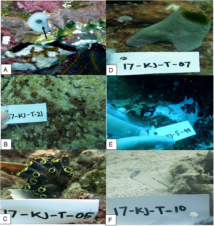
During 11 dives (Fig 1), 37 tunicate specimen were collected (Fig 2). The well-grown and adult zooid was picked from the colony. The preliminary identification of the specimens resulted in the genera Ascidia 5.4%, Atriolum 13.5%, Clavelina 16.2%, Didemnum 10.8%, Lissoclinum 13.5%, and Rhopalaea 18.9%. The rest of the samples (21.6%) remained unidentified (Fig A in S1 File).
Fig 2. Underwater documentation of the samples.
(A) Genus Ascidia, (B) genus Atriolum, (C) genus Clavelina, (D) genus Didemnum, (E) genus Lissoclinum, (F) genus Rhopalaea.
Bacteria were isolated from the holobionts, which resulted in 435 axenic strains. From the specimen preliminary identified as Rhopalaea, the majority (i.e., 22%) of the bacterial strains was obtained, followed by Clavelina, from which 16% were isolated. The lowest percentage of strains was isolated from Ascidia (5%). There was no difference if isolation was performed less than 6 hours from sampling, or if cooled samples were processed after few days in the laboratory, since a share of 52% and 48% of the bacteria was obtained, respectively. In contrast, the media, which have been used for isolation, had an impact on the number of isolated strains. Most bacteria (43%) were isolated on M1 medium (Fig B in S1 File). PYA medium, a medium based on peptone, resulted only in 22% of the strains. Remaining strains were recovered from ISP2 agar plates.
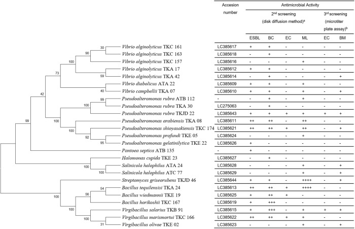
After an initial primary activity screening (for details see below; Table A in S1 File), the 16S rDNA gene of the 25 active strains, which showed at least activity against one of the test strains, was sequenced and a phylogenetic tree was generated (Fig 3). Active strains belonged to the following genera: Bacillus, Halomonas, Pantoea, Pseudoalteromonas, Salinicola, Streptomyces, Vibrio and Virgibacillus (Fig 4). About 2/3 of these strains (n = 16) were Gram-negative bacteria, while the remaining were Gram-positives. In this collection, most of the active strains were classified as Vibrio (28%), followed by Pseudoalteromonas (20%). Halomonas, Pantoea and Streptomyces were solely represented by one strain (equal to 4%), respectively (Fig B in S1 File).
Fig 3. Phylogenetic tree and antimicrobial activities of the 25 active strains.
Left side: Phylogenetic tree. Right side: Activity screen with the following test microorganisms: ESBL (Extended Spectrum Beta-Lactamase Escherichia coli), BC (Bacillus cereus), EC (Escherichia coli), ML (Micrococcus luteus), BM (Bacillus megaterium). a: The signs used indicate:— = no Zone of Inhibition (ZOI), + = ZOI 7–11 mm, ++ = ZOI 12–16 mm, +++ = ZOI 17–21 mm, and ++++ = ZOI ≥ 21mm. b: Signs used indicate:— = no inhibition, + = inhibition (OD600 <50% compared to a negative control).
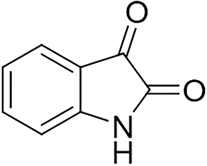
Fig 4. Structure of isatin.
Analysis by HPLC and LC-HRMS revealed that fractions F2 and F3 also contained isatin, and therefore these were purified as well. In total 46.8 mg of isatin were isolated from a 9.8 L fermentation. Isatin showed the same moderate inhibitory effect against B. megaterium DSM32, E. coli K12 derivative, and ESBL-MDR E. coli strain, with an MIC value of 200 μg/mL.
Antimicrobial activity
To get a first insight into the antimicrobial activities, all strains were tested against MDR-ESBL E. coli and MDR B. cereus using the agar plug method. Thereby, 71 strains (16% of the collection) showed activity (Table A in S1 File) and 45 bacterial strains showed activity against the Gram-negative MDR strain.
The 71 strains, which showed activity in the primary screening, were tested in a secondary screen by evaluating the antibacterial activity of the extracts resulting from liquid fermentation. As a result, 25 out of these 71 strains were positive in the secondary screen and the extract of 13 strains showed activity against both, Gram-positives and Gram-negative MDR-ESBL E. coli (Fig 3).
The next goal was to identify a compound responsible for the Gram-negative activity observed. Therefore, first, it was tested whether the activity can be linked to a specific peak in HPLC analysis. Hence, 13 active extracts were fractionated using a microtiter plate assay. In this fractionation experiment, only for 9 strains activity was observed. Pseudoalteromonas rubra TKJD 22 was prioritized, due to its strong activity against both, Gram-positive and Gram-negative test strains.
Isolation and structure elucidation of the active compound
The active compound of fraction F1 of Pseudoalteromonas rubra TKJD 22 was isolated as red-orange crystalline solid compound. The molecular formula was determined as C8H5NO2 (calculated mass 147.1311 Da) based on the prominent ion peak at m/z 148.0410 [M+H]+ and m/z 170.0240 [M+Na]+ in the LC-HRMS (ESI) spectrum. The compound exhibited UV absorption maxima at λmax (MeOH) = 201, 208, 238 and 301 nm (Fig C in S1 File). The structure was elucidated by NMR analysis to be isatin (Fig 4), as well as by comparison with reported literature data [27]. 1H NMR (aceton-d6, 400 MHz) δH 7.62 ppm (1H, td, J = 7.77, 1.26), δH 7.53 ppm (1H, d, J = 7.49), δH 7.12 ppm (1H, td, J = 7.53, 0.85), δH 7.03 ppm (1H, d, J = 7.92).
Discussion
The filter feeding tunicates are exposed to many microorganisms, which enter through the branchial siphon to the atrial siphon [28]. Therefore, it is of utmost importance to the animal that the normal non-pathogenic microbiome is kept intact. It can be speculated that both, the animal and the bacteria, contribute to maintaining and shaping of the microbiome. Bacteria are subject of intra- and interspecies competition. Chemical molecules inhibiting the growth of others in this competition might become lead molecules for drug development based on their antibacterial activities.
Most of the active bacteria isolated from the microbiomes in this project belonged to the phylum proteobacteria, class γ-proteobacteria. The highest proportion of γ-proteobacteria belonged to the genus Vibrio (28%) and Pseudoalteromonas (20%). The Vibrionaceace family consist of 128 species, with many potential pathogenic species [29]. Many antimicrobial compounds have been reported from this genus, such as andrimid, holomycin, and prodigiosin [29, 30]. Unlike Vibrio, which is known for pathogenicity, the genus Pseudoalteromonas is well known for production of antibacterial compounds. This genus consists of 41 species, of which 16 are documented as antimicrobial metabolite producers. Around 70 antimicrobial compounds were reported, ranging from alkaloids over polyketides to peptides [31]. The occurrence of the genus Pantoea (Enterobacteriaceae), which is commonly found in terrestrial habitats, might be explained by cells which were washed into the ocean, e.g., from the harbor. The strain Pantoea agglomerans EH318 was reported as producer of the antibiotic compounds pantocin A and B, which affect mainly other enteric bacteria [32]. Furthermore, broad-spectrum compounds like the antibacterial peptide herbicolin [33], microcin MccEh252 [34], and redox-active phenazines [35] were reported from this genus. In addition, strains of the genera Halomonas and Salinicola were isolated in this project. However, only few antibacterial metabolites have been reported from the family Halomonadaceae to date, e.g., maminophenoxazinones [36], which renders them interesting for further chemical investigation.
Other active bacteria isolated in this project belonged to the Firmicutes phylum (32%), specifically Bacillus and Virgibacillus strains. Both genera belong to the family Bacillaceae, which showed a high percentage of antimicrobial activity against gram-positive and/or gram-negative bacteria in previous studies [12, 37]. Bacilli produce a wide range of natural products, including lipopeptides, polypeptides, macrolactams, fatty acids, polyketides, and isocoumarins, with a series of bioactivities [38, 39]. The genus Streptomyces is well known for proliferative producers of specialized metabolites. To date, around 80% of actinobacteria-derived natural products are reported from Streptomyces species [40]. Therefore, it was not surprising that strain Streptomyces griseorubens TKJD 46 showed activity against MDR bacteria in the screening.
Only 35% (25 of 71) of the active strains cultivated on agar plate retained antimicrobial activity in liquid fermentation, which is in the range generally observed in such bioprospecting projects. However, the fact that only nine strains showed antibacterial activity after fractionation using the 96-well microtiter plate assay, might indicate that some of the activities observed are based on different compounds acting synergistically.
The activity-guided purification of Pseudoalteromonas rubra TKJD22 extract resulted in the isolation of isatin, which was first reported in 1841 by Erdman and Laurent as a product from the oxidation of indigo by nitric and chromic acids [41]. In this project, isatin inhibited the growth of E. coli test strains, a standard lab as well as an ESBL-MDR strain. It was considered to be a synthetic compound for almost 140 years until it was found to be present in plants from the genus Isatis, and in secretions from the parotid gland of Bufo toads. In humans and other mammals, isatin can be detected as an endogenous molecule. Modified analogues showed stronger activity and good potential against various cancer lines [42]. For example, 6-bromoisatin showed anticancer activity against the human lymphoma cell line U937 with an IC50 of 0.02 mg/mL and 5,7-dibromo-N-(P-methylbenzyl)-isatin showed an IC50 of 490 nM against U937 and Jurkat leukemic cell lines [43]. For the compound isatin-3-phenylhydrazone activities against various bacteria and fungi were reported, e.g., Staphylococcus aureus (MIC 140 μg.cm-3), Bacillus subtilis (MIC 45 μg.cm-3), E. coli ATCC95 (MIC 100 μg.cm-3), Pseudomonas aeruginosa ATCC 2853 (MIC 240 μg.cm-3), P. vulgaris (MIC 200 μg.cm-3), and Candida albicans (MIC 160 μg.cm-3) [44]. Furthermore, the hybrid isatin-indole molecules showed strong antifungal activity, i.e. N’-[(3Z)-1-Benzyl-5-methoxy-2-oxo-1,2-dihydro-3H-indol-3-ylidene]-5-methoxy-1H-indole-2-carbohydrazide against C. albicans with MIC 3.9 μg/mL, N’-[(3Z)-1-Benzyl-5-bromo-2-oxo-1,2-dihydro-3H-indol-3-ylidene]-5-methoxy-1H-indole-2-carbohydrazide against Aspergillus niger with MIC 15.6 μg/mL and N’-[(3Z)-1-Benzyl-5-chloro-2-oxo-1,2-dihydro-3H-indol-3-ylidene]-5-methoxy-1H-indole-2-carbohydrazide against Penicillium notatum with MIC 7.8 μg/mL [45]. Majik et al. reported the antifouling activity against Planococcus donghaensis, Erythrobacter litoralis, Alivibrio salmonicida and Vibrio furnisii of the isatin-based compound (E)-3-(2-Oxopropylidene)indolin-2-one [42].
In general, sessile didemnid species have to protect themselves not only against predators like fish or sea urchin, but also against the settlement by micro- and/or macroorganisms. This can be achieved by chemical molecules like the alkaloids didemnimides A-D, which were reported as chemical weapon of Didemnum conchyliatum against predation by carnivorous fish in mangrove habitats [46]. However, the structure of these compounds indicates that they are produced by bacteria. In addition, other molecules produced by cyanobacterial symbionts, i.e., Prochloron and Synechosystis species, are beneficial for Didemnum [47–49]. It was speculated that cytotoxic constituents produced by these symbionts prevent marine animals to feed on the tunicates [50]. The algal-containing larvae of Trididemnum solidum induced vomiting in fish, resulting in a rapid learned aversion to this toxic food source. Furthermore, the patellamides, which are generated by the host-symbiont relationship between Prochloron and didemnid ascidian (Lissoclinum patella), fulfill this ecological function as feeding deterrents [48]. Therefore, it can be speculated that isatin plays a role in protecting the ascidian against predation due to the cytotoxicity. Hence, the bacteria producing isatin might be beneficial for the tunicate microbiome as defense mechanism.
In this regard, tunicate-associated bacteria can be regarded as promising bioresource for the discovery of biological active compounds, which might have the potential to serve as drug leads for antibiotic development. Further research is required to identify the specialized metabolites used as chemical weapon by the holobiont and to evaluate their potential for development.
Supporting information
Table A. Primary screening for antimicrobial activity from tunicate-associated bacteria against Multidrug resistant (MDR) bacteria. Fig A. Number of collected tunicates sample from each sampling site. Sampling site 1 resulted 26 samples, sampling site 2 resulted in 7 samples and sampling site 3 resulted in 4 samples. Genus Rhopalaea is the highest, followed by Claveliana and Atriolum. Fig B. Percentage of tunicate-associated bacteria isolated from different media. Fig C. Percentage of genera with antimicrobial activity. The genus Vibrio is the most active (28%) among others, followed by genus Pseudoalteromonas (20%), genera Bacillus and Virgibacillu sharing the same percentage (16%), and the least number from genera Halomonas, Streptomyces and Pantoea (4%). Fig D. UV absorption of isatin.
(DOCX)
Acknowledgments
The samples were collected legally by the permission from the Balai Taman Nasional Karimunjawa, Indonesia. Those samples are not listed in IUCN or CITES. The authors thank the Republic of Indonesia for making this research possible, the PEER team for their help to collect the samples, the Endang Sri Lestari, Medical Microbiology Laboratory of Rumah Sakit Nasional Diponegoro (RSND), Diponegoro University for the assistance in handling the human pathogens, and the Dr. Kariadi Hospital, Semarang, Central Java for provision of the MDR strains.
Data Availability
All relevant data are within the manuscript and its Supporting Information files.
Funding Statement
DA was supported by No. 315-10/UN7.5.1/PP/2017 Ministry of Research, Technology and Higher Education, Indonesia, under the scheme Master Program of Education Leading to Doctoral Degree for Excellent Graduates (PMDSU), and No. 2064.23/D3/PG/2017 Enhancing International Publication Program (PKPI). TFS was supported by Federal Ministry of Education and Research (BMBF, grant 16GW0117K). The funders had no role in study design, data collection and analysis, decision to publish, or preparation of the manuscript.
References
- 1.CDC. Antibiotic resistance threats in the United States. 2013. https://www.cdc.gov/drugresistance/biggest_threats.html Cited 2 October 2018.
- 2.WHO. Antimicrobial Resistance Global Report on Surveillance. 2014. https://apps.who.int/iris/bitstream/handle/10665/112642/9789241564748_eng.pdf?sequence=1 Cited 2 October 2018
- 3.Magiorakos AP, Srinivasan A, Carey RB, Carmeli Y, Falagas ME, Giske CG, et al. Multidrug-resistant, extensively drug-resistant and pandrug-resistant bacteria: an international expert proposal for interim standard definitions for acquired resistance. Clin Microbiol Infect. 2012;18(3):268–81. 10.1111/j.1469-0691.2011.03570.x [DOI] [PubMed] [Google Scholar]
- 4.Nikaido H. Multidrug resistance in bacteria. Annu Rev Biochem. 2009;78:119–46. 10.1146/annurev.biochem.78.082907.145923 [DOI] [PMC free article] [PubMed] [Google Scholar]
- 5.Lestari ES, Severin JA. Antimicrobial resistance in Indoensia prevalence, determinants and genetic basis. Rotterdam: Erasmus University; 2009. [Google Scholar]
- 6.Mendes RE, Mendoza M, Banga Singh KK, Castanheira M, Bell JM, Turnidge JD, et al. Regional resistance surveillance program results for 12 Asia-Pacific nations (2011). Antimicrob Agents Chemother. 2013;57(11):5721–6. 10.1128/AAC.01121-13 [DOI] [PMC free article] [PubMed] [Google Scholar]
- 7.Nangino GO, Oliveira CD, Correia PC, Machado NM, ATB D. Financial impact of nosocomial infections in the intensive care units of a charitable hospital in Minas Gerais, Brazil. Rev Bras Ter Intensiva. 2012;24(4): 357–361. 10.1590/S0103-507X2012000400011 [DOI] [PMC free article] [PubMed] [Google Scholar]
- 8.Peleg AY, Hooper DC. Hospital-acquired infections due to gram-negative bacteria. N Engl J Med. 2010;362(19):1804–13. 10.1056/NEJMra0904124 [DOI] [PMC free article] [PubMed] [Google Scholar]
- 9.Cornejo-Juarez P, Vilar-Compte D, Perez-Jimenez C, Namendys-Silva SA, Sandoval-Hernandez S, Volkow-Fernandez P. The impact of hospital-acquired infections with multidrug-resistant bacteria in an oncology intensive care unit. Int J Infect Dis. 2015;31:31–4. 10.1016/j.ijid.2014.12.022 [DOI] [PubMed] [Google Scholar]
- 10.Shaikh S, Fatima J, Shakil S, Rizvi SM, Kamal MA. Antibiotic resistance and extended spectrum beta-lactamases: Types, epidemiology and treatment. Saudi J Biol Sci. 2015;22(1):90–101. 10.1016/j.sjbs.2014.08.002 [DOI] [PMC free article] [PubMed] [Google Scholar]
- 11.Joint I, Muhling M, Querellou J. Culturing marine bacteria—an essential prerequisite for biodiscovery. Microb Biotechnol. 2010;3(5):564–75. 10.1111/j.1751-7915.2010.00188.x [DOI] [PMC free article] [PubMed] [Google Scholar]
- 12.Ayuningrum D, Kristiana R, Asagabaldan MA, Sabdono A, Radjasa OK, Nuryadi H, et al. Isolation, Characterisation and Antagonistic Activity of Bacteria Symbionts Hardcoral Pavona sp. Isolated from Panjang Island, Jepara Against Infectious Multi-drug Resistant (MDR) Bacteria. IOP Conference Series: Earth and Environmental Science. 2017;55:012029. [Google Scholar]
- 13.Kristiana R, Ayuningrum D, Asagabaldan MA, Nuryadi H, Sabdono A, Radjasa OK, et al. Isolation And Partial Characterization Of Bacteria Activity Associated With Gorgonian Euplexaura sp. Against Methicillin-Resistant Staphylococcus aureus (MRSA). IOP Conference Series: Earth and Environmental Science. 2017;55:012056. [Google Scholar]
- 14.Asagabaldan MA, Ayuningrum D, Kristiana R, Sabdono A, Radjasa OK, Trianto A. Identification and Antibacterial Activity of Bacteria Isolated from Marine SpongeHaliclona (Reniera)sp. against Multi-Drug Resistant Human Pathogen. IOP Conference Series: Earth and Environmental Science. 2017;55:012019. [Google Scholar]
- 15.Sulardiono B, Purnomo PW, Haeruddin. Environmental Suitability for Holothuroidea Habitat in Karimunjawa. Indonesian Journal of Fisheries and Technology. 2017;12(2):5. [Google Scholar]
- 16.Carter GT, Bernan VS. Marine natural products in Pharma In: B BJ, editor. Marine biomedicine from beach to bedside. Florida: CRC Press Tayor & Francis Group; 2016. p. 612. [Google Scholar]
- 17.Sigel MM, McCumber LJ, Hightower JA, Hayasara SS, Huggins EM Jr, Davis JF. Ecteinascidia turbinata extract activates components of inflammatory responses throughout the phylogenetic spectrum. Amer Zool. 1983;24:8. [Google Scholar]
- 18.Sakai R, Rinehart KL, Guan Y, Wang AHJ. Additional antitumor ecteinascidins fron a Caribbean tunicate: crystal structures and activities in vivo. Proc Nati Acad Sci. 1992;89:5. [DOI] [PMC free article] [PubMed] [Google Scholar]
- 19.Piel J. Bacterial symbionts: prospects for the sustainable production of invertebrate-derived pharmaceuticals. Current Medicinal Chemistry. 2006;13(1):12 10.2174/092986706775197944 [DOI] [PubMed] [Google Scholar]
- 20.Rath CM, Janto B, Earl J, Ahmed A, Hu FZ, Hiller L, et al. Meta-omic characterization of the marine invertebrate microbial consortium that produces the chemotherapeutic natural product ET-743. ACS Chem Biol. 2011;6(11):1244–56. 10.1021/cb200244t [DOI] [PMC free article] [PubMed] [Google Scholar]
- 21.Fuhrman JA. Microbial community structure and its functional implications. Nature. 2009;459(7244):193–9. 10.1038/nature08058 [DOI] [PubMed] [Google Scholar]
- 22.Kott P. A new species od Didemnum (Ascidiacea, Tunicata) from the Atlantic coast of North America. Zootaxa. 2004;732:10. [Google Scholar]
- 23.Monniot F, Monniot C. Ascidians Collected in Tanzania. Journal of East African Natural History. 1997;86(1):1–35. 10.2982/0012-8317(1997)86[1:acit]2.0.co;2 [DOI] [Google Scholar]
- 24.Benson HJ. Microbiological Applications A Laboratory Manual in General Microbiology 8th Edition United States: The McGraw-Hill Company; 2002. [Google Scholar]
- 25.Burgess JG, Boyd KG, Armstrong E, Jiang Z, Yan L, Berggren M, et al. The development of a marine natural product-based antifouling paint. Biofouling. 2003;19 Suppl:197–205. Epub 2003/11/19. 10.1080/0892701031000061778 [DOI] [PubMed] [Google Scholar]
- 26.Weisburg WG, Barns SM, Pelletier DA, Lane DJ. 16S Ribosomal DNA amplification for phylogenetic study. Journal of bacteriology. 1991;173(2):7. [DOI] [PMC free article] [PubMed] [Google Scholar]
- 27.Kalinovskaya NI, Ivanova EP, Alexeeva YV, Gorshkova NM, Kuznetsova TA, Dmitrenok AS, et al. Low-molecular-weight, biologically active compounds from marine Pseudoalteromonas species. Curr Microbiol. 2004;48(6):441–6. 10.1007/s00284-003-4240-0 [DOI] [PubMed] [Google Scholar]
- 28.Ali HAJ, Tamilselvi M. Ascidians in Coastal Water A Comprehensive Inventory of Ascidian Fauna from the Indian Coast. Switzerland: Springer; 2016. 165 p. [Google Scholar]
- 29.Mansson M, Gram L, Larsen TO. Production of bioactive secondary metabolites by marine vibrionaceae. Mar Drugs. 2011;9(9):1440–68. 10.3390/md9091440 [DOI] [PMC free article] [PubMed] [Google Scholar]
- 30.Wietz M, Mansson M, Gotfredsen CH, Larsen TO, Gram L. Antibacterial compounds from marine Vibrionaceae isolated on a global expedition. Mar Drugs. 2010;8(12):2946–60. 10.3390/md8122946 [DOI] [PMC free article] [PubMed] [Google Scholar]
- 31.Offret C, Desriac F, Le Chevalier P, Mounier J, Jegou C, Fleury Y. Spotlight on Antimicrobial Metabolites from the Marine Bacteria Pseudoalteromonas: Chemodiversity and Ecological Significance. Mar Drugs. 2016;14(7). 10.3390/md14070129 [DOI] [PMC free article] [PubMed] [Google Scholar]
- 32.Wright SA, Zumoff CH, Schneider L, Beer SV. Pantoea agglomerans strain EH318 produces two antibiotics that inhibit Erwinia amylovora in vitro. Appl Environ Microbiol. 2001;67(1):284–92. 10.1128/AEM.67.1.284-292.2001 [DOI] [PMC free article] [PubMed] [Google Scholar]
- 33.Kamber T, Lansdell TA, Stockwell VO, Ishimaru CA, Smits TH, Duffy B. Characterization of the biosynthetic operon for the antibacterial peptide herbicolin in Pantoea vagans biocontrol strain C9-1 and incidence in Pantoea species. Appl Environ Microbiol. 2012;78(12):4412–9. 10.1128/AEM.07351-11 [DOI] [PMC free article] [PubMed] [Google Scholar]
- 34.Vanneste JL, Cornish DA, Yu J, Voyle MD, editors. The peptide antibiotic produced by Pantoea agglomerans Eh252 is a microcin. 9th Int Workshop on Fire Blight Eds; 2002: Acta Hort.
- 35.Giddens SR, Feng Y, Mahanty HK. Characterization of a novel phenazine antibiotic gene cluster in Erwinia herbicola Eh 1087. Molecular Microbiology. 2002;45(3):15. [DOI] [PubMed] [Google Scholar]
- 36.Bitzer J, Grosse T, Wang L, Lang S, Beil W, A Z. New aminophenoxazinones from a marine Halomonas sp.: fermentation, structure elucidation, and biological activity. J Antibiot. 2006;59(2):7. [DOI] [PubMed] [Google Scholar]
- 37.Arunachalam K, Amirtham Jacob Appadorai R. Antioxidant potential and biochemical evaluation of metabolites from the marine bacteria Virgibacillus sp. associated with the sponge Callyspongia diffusa. Free Radicals and Antioxidants. 2013;3(1):47–51. 10.1016/j.fra.2013.04.004 [DOI] [Google Scholar]
- 38.Mondol MA, Shin HJ, Islam MT. Diversity of secondary metabolites from marine Bacillus species: chemistry and biological activity. Mar Drugs. 2013;11(8):2846–72. 10.3390/md11082846 [DOI] [PMC free article] [PubMed] [Google Scholar]
- 39.Mohan G, Thipparamalai Thangappanpillai AK, Ramasamy B. Antimicrobial activities of secondary metabolites and phylogenetic study of sponge endosymbiotic bacteria, Bacillus sp. at Agatti Island, Lakshadweep Archipelago. Biotechnol Rep (Amst). 2016;11:44–52. 10.1016/j.btre.2016.06.001 [DOI] [PMC free article] [PubMed] [Google Scholar]
- 40.Manivasagan P, Venkatesan J, Sivakumar K, Kim SK. Pharmaceutically active secondary metabolites of marine actinobacteria. Microbiol Res. 2014;169(4):262–78. 10.1016/j.micres.2013.07.014 [DOI] [PubMed] [Google Scholar]
- 41.da Silva JFM, Garden SJ, Pinto AC. The chemistry of isatins: a review from 1975 to 1999. J Braz Chem Soc. 2001;12(3):52. [Google Scholar]
- 42.Majik MS, Rodrigues C, Mascarenhas S, D’Souza L. Design and synthesis of marine natural product-based 1H-indole-2,3-dione scaffold as a new antifouling/antibacterial agent against fouling bacteria. Bioorg Chem. 2014;54:89–95. 10.1016/j.bioorg.2014.05.001 [DOI] [PubMed] [Google Scholar]
- 43.Vine KL. An investigation into the cytotoxic properties of isatin-derived compounds: potential for use in targeted cancer theraphy: University of Wollongong; 2007. [Google Scholar]
- 44.Konstantinovic S, Radovanovic B, Sovilj S, Stanojevic S. Antimicrobial activity of some isatin-3-thiosemicarbazone complexes. Journal of the Serbian Chemical Society. 2008;73(1):7–13. [Google Scholar]
- 45.Al-Wabli RI, Zakaria AS, Attia MI. Synthesis, Spectroscopic Characterization and Antimicrobial Potential of Certain New Isatin-Indole Molecular Hybrids. Molecules. 2017;22(11). 10.3390/molecules22111958 [DOI] [PMC free article] [PubMed] [Google Scholar]
- 46.Vervoort HC, Richards-Gross SE, Fenical W. Didemnimides A-D: novel, predator-deterrent alkaloids from the Carribean mangrove ascidian Didemnum conchyliatum. J Org Chem. 1997;62:5. [Google Scholar]
- 47.Sings HL, Rinehart KL. Compounds produced from potential tunicate-blue-green algal symbiosis: a review. Journal of industrial microbiology. 1996;17:12. [Google Scholar]
- 48.Schmidt EW, Nelson JT, Rasko DA, Sudek S, Eisen JA, Haygood MG, et al. Patellamide A and C biosynthesis by a microcin-like pathway in Prochloron didemni, the cyanobacterial symbiont of Lissoclinum patella. Proc Natl Acad Sci U S A. 2005;102(20):7315–20. 10.1073/pnas.0501424102 . [DOI] [PMC free article] [PubMed] [Google Scholar]
- 49.Schmidt EW, Donia MS, McIntosh JA, Fricke WF, Ravel J. Origin and variation of tunicate secondary metabolites. J Nat Prod. 2012;75(2):295–304. 10.1021/np200665k [DOI] [PMC free article] [PubMed] [Google Scholar]
- 50.Hirose E, Maruyama T. What are the benefits in the ascidian-Prochloron symbiosis. Endocytobiosis Cell Res. 2004;15:12. [Google Scholar]
Associated Data
This section collects any data citations, data availability statements, or supplementary materials included in this article.
Supplementary Materials
Table A. Primary screening for antimicrobial activity from tunicate-associated bacteria against Multidrug resistant (MDR) bacteria. Fig A. Number of collected tunicates sample from each sampling site. Sampling site 1 resulted 26 samples, sampling site 2 resulted in 7 samples and sampling site 3 resulted in 4 samples. Genus Rhopalaea is the highest, followed by Claveliana and Atriolum. Fig B. Percentage of tunicate-associated bacteria isolated from different media. Fig C. Percentage of genera with antimicrobial activity. The genus Vibrio is the most active (28%) among others, followed by genus Pseudoalteromonas (20%), genera Bacillus and Virgibacillu sharing the same percentage (16%), and the least number from genera Halomonas, Streptomyces and Pantoea (4%). Fig D. UV absorption of isatin.
(DOCX)
Data Availability Statement
All relevant data are within the manuscript and its Supporting Information files.


