Black esophagus (BE) is an entity characterized by a striking circumferential black coloration of the esophageal mucosa, which is generally depicted on endoscopy due to acute necrotizing esophagitis. This entity was first described in 1990 by Goldenberg.1-3 Despite several articles describing the BE as a rare entity, the actual incidence is uncertain due to the great variability of the studied population; therefore, many cases seem to run underdiagnosed. It tends to be more frequent in critically ill patients, particularly those with refractory shock or systemic inflammatory disease. The estimated incidence ranges between 0.001% and 0.028% based on autopsy and endoscopic series.4-8
The etiology of the BE is not well understood. Chemical injury by gastric contents associated with esophageal ischemia, severe infectious diseases, emergency surgery, and hypovolemic shock are present in most cases.2,3,9,10 However, drug abuse (e.g. cocaine11 and alcohol12), and diabetic ketoacidosis were reported, and are considered to be triggering events in the young.13,14 Hypothermia,15,16 anticardiolipin antibody syndrome,17 herpes simplex virus,18 cytomegalovirus,19,20 and candida21 infections were also reported, although they are less common. Anecdotal cases of BE associated with drugs such as haloperidol and erythromycin have also been reported.22,23
The typical BE is most frequently seen in the distal esophagus until the gastroesophageal junction, but the lesion may occasionally extend to the proximal part of the esophagus. Rare cases have been described involving only the proximal esophagus.3,24 Concomitant duodenal lesions can be found in a large number of patients,25 which has been attributed to a vascular watershed area and a lesser degree of vascularization of the distal esophagus. The esophageal vascularization is represented by three different vascular zones: (i) the proximal, which depends on the inferior thyroid arteries with support by subclavian and carotid branches; (ii) the median zone supplied by bronchial and intercostal arteries, which are aortic branches; (iii) the distal zone supported by branches of gastric and phrenic arteries, with possible contribution from hepatic and splenic vessels. This distal zone seems particularly vulnerable to the impairment of vascular support, thereby justifying the almost invariable implication of distal esophagus in the cases of BE.26 Histologically, the mucosa in the BE is necrotic with disrupted muscle fibers without identifiable causative organisms or agents.
Generally, BE involves the elderly, with a small predominance of male, and more than two comorbidities are usually present. Gastrointestinal bleeding, with hematemesis or melena occurs in about 70% to 90% of patients.3,5 A series with 310 consecutive autopsy cases found an incidence of esophageal necrosis of 10.3%27 and showed that the most important differential diagnosis of the BE is hematin coloration; however, black pigmentation is described as a longitudinally striated appearance limited to the lower parts of the organ, and does not usually affect the entire esophagus in a circumferential pattern. Melanoma of the esophagus is rare and is a possible cause of black lesions on the esophagus, but it does not follow a vascular or circumferential pattern.28 There is a report of coal dust deposition simulating a BE.29
No specific treatment for BE is available. However, in the patient with this condition, the esophagus should never be used as a feeding route, and if an endoscopic examination is unavoidable, it should be performed with exceptional caution.
Treatment should be directed to any underlying clinical entity, and any complications should be controlled as the treatment of shock, infections, and surgery may be indicated.2 The standard treatment for gastrointestinal bleeding should be considered.2,25 BE has a poor outcome with a mortality of 30%.2
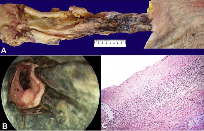
The images above (Figure 1) refer to an autopsy specimen of a 75-year-old woman who died because of multiple organ failure due to septicemia. Her past medical history included hypertension, insulin-dependent diabetes mellitus, heart failure, and chronic renal failure. She had a 2-year history of anemia and progressive weight loss, and was recently complaining of diffuse abdominal colic pain and intermittent diarrhea. She attended the emergency care unit after presenting hematemesis. On admission, she was cachectic, obtunded, pale, hypotensive, and hypoxemic. The physical examination depicted a flat abdomen with an easy palpable hardened mass in the right upper quadrant, along with lower limbs edema. The diagnostic work-up evidenced the presence of BE and duodenal ulcers on the upper digestive endoscopy, and unilateral deep venous thrombosis (popliteal vein). The abdominal ultrasound showed a mass in the topography of the right colon, and multiple hepatic images consistent with metastatic disease.
Figure 1. A – A picture composition of the gross aspect of esophagus and adjacent organs. Note the dark appearance of esophageal mucosa and the sharp limit at the gastroesophageal junction. B – Endoscopic view of the distal esophagus. Note the darkened mucosa in every circumference of the organ. C – Microscopic aspect of the esophagus at autopsy showing superficial necrosis of the mucosa and neutrophilic inflammation (H&E 100x). Image courtesy Anatomic Pathology Service – Hospital Universitário - USP.
The autopsy revealed a 5.5cm fungating adenocarcinoma at the cecum, invasion of subserosa (stage pT3), with metastases to the regional lymph nodes (pN2) and the liver, with extensive portal invasion. There was a microscopic peritumoral abscess, bronchopneumonia, and systemic signs of ischemia and shock. Diabetic nodular glomerulosclerosis and systemic arteriosclerosis were other remarkable autopsy findings. The BE was confirmed in the context of a severely ill patient with advanced cancer, septic shock, diabetes, and blockade of the portal system.
Footnotes
How to cite: Monteiro JMC, Castelo LF, Fischer WGG, Felipe-Silva A. Black esophagus. Autops Case Rep [Internet]. 2019;9(1):e20180077. https://doi.org/10.4322/acr.2018.077
Financial Support: None
References
- 1.Goldenberg SP, Wain SL, Marignani P. Acute necrotizing esophagitis. Gastroenterology. 1990;98(2):493-6. 10.1016/0016-5085(90)90844-Q. [DOI] [PubMed] [Google Scholar]
- 2.Gurvits GE. Black esophagus: acute esophageal necrosis syndrome. World J Gastroenterol. 2010;16(26):3219-25. 10.3748/wjg.v16.i26.3219. [DOI] [PMC free article] [PubMed] [Google Scholar]
- 3.Gurvits GE, Shapsis A, Lau N, Gualtieri N, Robilotti JG. Acute esophageal necrosis: a rare syndrome. J Gastroenterol. 2007;42(1):29-38. 10.1007/s00535-006-1974-z. [DOI] [PubMed] [Google Scholar]
- 4.Ramos R, Mascarenhas J, Duarte P, Vicente C, Casteleiro C. Acute esophageal necrosis: a retrospective case series. Rev Esp Enferm Dig. 2008;100(9):583-5. [DOI] [PubMed] [Google Scholar]
- 5.Augusto F, Fernandes V, Cremers MI, et al. Acute necrotizing esophagitis: a large retrospective case series. Endoscopy. 2004;36(5):411-5. 10.1055/s-2004-814318. [DOI] [PubMed] [Google Scholar]
- 6.Moretó M, Ojembarrena E, Zaballa M, Tánago JG, Ibánez S. Idiopathic acute esophageal necrosis: not necessarily a terminal event. Endoscopy. 1993;25(8):534-8. 10.1055/s-2007-1009121. [DOI] [PubMed] [Google Scholar]
- 7.Lacy BE, Toor A, Bensen SP, Rothstein RI, Maheshwari Y. Acute esophageal necrosis: report of two cases and a review of the literature. Gastrointest Endosc. 1999;49(4 Pt 1):527-32. 10.1016/S0016-5107(99)70058-1. [DOI] [PubMed] [Google Scholar]
- 8.Etienne JP, Roge J, Delavierre P, Veyssier P. Esophageal necrosis of vascular origin. Sem Hop. 1969;45(23):1599-606. [PubMed] [Google Scholar]
- 9.Haviv YS, Reinus C, Zimmerman J. “Black esophagus”: a rare complication of shock. Am J Gastroenterol. 1996;91(11):2432-4. [PubMed] [Google Scholar]
- 10.Grudell AB, Mueller PS, Viggiano TR. Black esophagus: report of six cases and review of the literature, 1963-2003. Dis Esophagus. 2006;19(2):105-10. 10.1111/j.1442-2050.2006.00549.x. [DOI] [PubMed] [Google Scholar]
- 11.Ullah W, Abdullah HMA, Rauf A, Saleem K. Acute oesophageal necrosis: a rare but potentially fatal association of cocaine use. BMJ Case Rep. 2018;2018:bcr-2018-225197 10.1136/bcr-2018-225197. [DOI] [PMC free article] [PubMed] [Google Scholar]
- 12.Katsinelos P, Pilpilidis I, Dimiropoulos S, et al. Black esophagus induced by severe vomiting in a healthy young man. Surg Endosc. 2003;17(3):521. [DOI] [PubMed] [Google Scholar]
- 13.Usmani A, Samarany S, Nardino R, Shaib W. Black esophagus in a patient with diabetic ketoacidosis. Conn Med. 2011;75(8):467-8. [PubMed] [Google Scholar]
- 14.Talebi-Bakhshayesh M, Samiee-Rad F, Zohrenia H, Zargar A. Acute Esophageal Necrosis: A Case of Black Esophagus with DKA. Arch Iran Med. 2015;18(6):384-5. [PubMed] [Google Scholar]
- 15.Brennan JL. Case of extensive necrosis of the oesophageal mucosa following hypothermia. J Clin Pathol. 1967;20(4):581-4. 10.1136/jcp.20.4.581. [DOI] [PMC free article] [PubMed] [Google Scholar]
- 16.Živković V, Nikolić S. The unusual appearance of black esophagus in a case of fatal hypothermia: a possible underlying mechanism. Forensic Sci Med Pathol. 2013;9(4):613-4. 10.1007/s12024-013-9445-3. [DOI] [PubMed] [Google Scholar]
- 17.Cappell MS. Esophageal necrosis and perforation associated with the anticardiolipin antibody syndrome. Am J Gastroenterol. 1994;89(8):1241-5. [PubMed] [Google Scholar]
- 18.Nagri S, Hwang R, Anand S, Kurz J. Herpes simplex esophagitis presenting as acute necrotizing esophagitis (“black esophagus”) in an immunocompetent patient. Endoscopy. 2007;39(Suppl 1):E169. 10.1055/s-2007-966619. [DOI] [PubMed] [Google Scholar]
- 19.Trappe R, Pohl H, Forberger A, Schindler R, Reinke P. Acute esophageal necrosis (black esophagus) in the renal transplant recipient: manifestation of primary cytomegalovirus infection. Transpl Infect Dis. 2007;9(1):42-5. 10.1111/j.1399-3062.2006.00158.x. [DOI] [PubMed] [Google Scholar]
- 20.Barjas E, Pires S, Lopes J, et al. Cytomegalovirus acute necrotizing esophagitis. Endoscopy. 2001;33(8):735. 10.1055/s-2001-16215. [DOI] [PubMed] [Google Scholar]
- 21.Gaissert HA, Breuer CK, Weissburg A, Mermel L. Surgical management of necrotizing Candida esophagitis. Ann Thorac Surg. 1999;67(1):231-3. 10.1016/S0003-4975(98)01144-8. [DOI] [PubMed] [Google Scholar]
- 22.Mangan TF, Colley AT, Wytock DH. Antibiotic-associated acute necrotizing esophagitis. Gastroenterology. 1990;99(3):900. 10.1016/0016-5085(90)90997-F. [DOI] [PubMed] [Google Scholar]
- 23.Hejna P, Ublová M, Voříšek V. Black esophagus: acute esophageal necrosis in fatal haloperidol intoxication. J Forensic Sci. 2013;58(5):1367-9. 10.1111/1556-4029.12151. [DOI] [PubMed] [Google Scholar]
- 24.Neumann DA 2nd, Francis DL, Baron TH. Proximal black esophagus: a case report and review of the literature. Gastrointest Endosc. 2009;70(1):180-1. 10.1016/j.gie.2008.09.055. [DOI] [PubMed] [Google Scholar]
- 25.Gurvits GE, Cherian K, Shami MN, et al. Black esophagus: new insights and multicenter international experience in 2014. Dig Dis Sci. 2015;60(2):444-53. 10.1007/s10620-014-3382-1. [DOI] [PubMed] [Google Scholar]
- 26.Manno V, Lentini N, Chirico A, Perticone M, Anastasio L. Acute esophageal necrosis (black esophagus): a case report and literature review. Acta Diabetol. 2017;54(11):1061-3. 10.1007/s00592-017-1028-4. [DOI] [PubMed] [Google Scholar]
- 27.Jacobsen NO, Christiansen J, Kruse A. Incidence of oesophageal necrosis in an autopsy material. APMIS. 2003;111(5):591-4. 10.1034/j.1600-0463.2003.1110509.x. [DOI] [PubMed] [Google Scholar]
- 28.Yamamoto S, Makuuchi H, Kumaki N, et al. A Long Surviving Case of Multiple Early Stage Primary Malignant Melanoma of the Esophagus and a Review of the Literature. Tokai J Exp Clin Med. 2015;40(3):90-5. [PubMed] [Google Scholar]
- 29.Khan HA. Coal dust deposition--rare cause of “black esophagus”. Am J Gastroenterol. 1996;91(10):2256. [PubMed] [Google Scholar]


