Abstract
Phelan-McDermid syndrome (PMS) is a neurodevelopmental disorder characterized by varying degrees of intellectual disability, severely delayed language development and specific facial features, and is caused by a deletion within chromosome 22q13.3. SHANK3, which is located at the terminal end of this region, has been repeatedly implicated in other neurodevelopmental disorders and deletion of this gene specifically is thought to cause much of the neurologic symptoms characteristic of PMS. However, it is still unclear to what extent SHANK3 deletions contribute to the PMS phenotype, and what other genes nearby are causal to the neurologic disease. In an effort to better understand the functional landscape of the PMS region during normal neurodevelopment, we assessed RNA-sequencing (RNA-seq) expression data collected from post-mortem brain tissue from developmentally normal subjects over the course of prenatal to adolescent age and analyzed expression changes of 65 genes on 22q13. We found that the majority of genes within this region were expressed in the brain, with ATNX10, MLC1, MAPK8IP2, and SULT4A1 having the highest overall expression. Analysis of the temporal profiles of the highest expressed genes revealed a trend towards peak expression during the early post-natal period, followed by a drop in expression later in development. Spatial analysis revealed significant region specific differences in the expression of SHANK3, MAPK8IP2, and SULT4A1. Region specific expression over time revealed a consistently unique gene expression profile within the cerebellum, providing evidence for a distinct developmental program within this region. Exon-specific expression of SHANK3 showed higher expression within exons contributing to known brain specific functional isoforms. Overall, we provide an updated roadmap of the PMS region, implicating several genes and time periods as important during neurodevelopment, with the hope that this information can help us better understand the phenotypic heterogeneity of PMS.
Introduction
Phelan-McDermid Syndrome (PMS) is a rare genetic disorder, characterized predominately by a neurodevelopmental phenotype. The disorder is typically considered in children with varying degrees of intellectual disability, severely delayed language development, and a typical facies that may include dolichocephaly, full brows, and a flat midface among other features [1–4]. However, the phenotypic spectrum is wide and thus, the diagnosis is made with laboratory testing to establish a deletion at chromosome 22q13.3. Relatively little is known about the relationship between genotype and corresponding phenotype in PMS, especially as it relates to the neurologic manifestations of the disease. There is evidence to suggest that larger deletions on 22q13 tend to be associated with a more severe phenotype [5–10], however the relationship is not consistent, as patients with similar size deletions can have varied disease presentations [1, 10–12]. SHANK3, located at the terminal end of the Phelan-McDermid region, has been repeatedly implicated in the pathogenesis of neurodevelopmental disorders such as autism and intellectual disability [13], as well as psychiatric diseases such as schizophrenia [14, 15] and bipolar disorder [16]; its protein product known to play an important role in synaptic plasticity and maintenance of long term potentiation [17, 18]. While several groups have implicated SHANK3 as the causative gene for the neurologic deficits in Phelan-McDermid patients [7–9, 12, 19–22], a deletion or variant in this gene is not considered necessary for diagnosis [11, 23].
It is likely that other genes also influence the neurologic sequelae of PMS as patients with similar neurologic phenotypes and 22q13 deletions proximal to SHANK3 have been reported [11, 24–28]. MAPK8IP2, located about 70 kb proximal to SHANK3, and frequently co-deleted in PMS, is one such gene that has been implicated due to the high expressivity of the protein product in the brain at the post synaptic density, and studies showing mice with absence of MAPK8IP2 have abnormal dendritic morphology, as well as motor and cognitive deficits [29]. Despite this work, it is still unclear how deletions in SHANK3 and MAPK8IP2 specifically, contribute to the neurologic phenotype of PMS and what other genes or combination of genes contribute to the complex and varied symptomology. In an attempt to better characterize the functional landscape of the 22q13 region, we assessed the expression patterns of genes mapped within the Phelan-McDermid region in neurologically normal human brain samples over developmental time. Our results provide insight into the importance of 22q13 during normal human neurodevelopment and suggest other genes, with similar expression patterns to MAPK8IP2 and SHANK3, that may contribute the neurologic phenotype in PMS.
Materials and methods
Developing human brain transcriptome data
The BrainSpan transcriptional atlas was downloaded from http://www.brainspan.org, where specific details regarding tissue acquisition and processing can be found [30, 31]. The online dataset contains next-generation RNA-sequencing data obtained from 41 donors ranging from pre-natal development (8 postconception weeks) to adulthood (40 years of age). All donor brains came from patients who until time of death were considered to be neurologically and developmentally normal. For each donor, sequencing data is available from several distinct brain regions (maximum 16 regions), however several donors have missing data from certain brain regions, and donors with more than 6 regions missing were excluded. For those donors with six or less missing brain regions, the missing data was imputed using a nearest neighbor approach, as we previously described [32]. After excluding the donors with missing data, a total of 30 brains with sequencing data for 16 brain regions were analyzed: cerebellar cortex, medial dorsal nucleus of thalamus, striatum, amygdala, hippocampus, and 11 regions of the neocortex. For comparative purposes, we binned the 30 brain samples into 7 discrete developmental time periods (16 post-conception weeks– 17 post-conception weeks, 19 post-conception weeks– 24 post-conception weeks, 4 months– 1 year, 2 years– 4 years, 8 years– 13 years, 15 years– 21 years, 23 years– 40 years) as has been previously used to analyze this dataset [32–35]. The resulting dataset consisted of RNA-sequencing expression values for 524 tissue samples given in units of reads per kilobase of exon model per million mapped reads (RPKM) [36].
22q13 gene set
The UCSC Genome Browser, genome assembly GRCh38/hg38 released December 2013 (https://genome.ucsc.edu/)), was used to identify genes within the PMS 22q13 region (coordinates: hg 19 chr22: 37,600,001–51,304,566) [37]. After exclusion of genes and other RNA species not present in the BrainSpan dataset, 65 protein-coding genes remained for inclusion in the final dataset (S3 Table). Average RPKM per gene was calculated for all developmental time periods and all brain regions. An extensive literature search was performed on all genes with at least one read greater than five RPKM (S1 and S2 Tables), which we considered to be biologically significant brain expression [32]. The four genes with the highest expression were further analyzed. The expression profile of SHANK3 was also analyzed given the importance of this gene in the neurologic manifestations of disease.
Analysis of highly expressed genes
Total average expression per gene per developmental time period was calculated and one-way analysis of variance (ANOVA) were run to assess significance. Pre-natal and post-natal expression were compared using student t-test analysis, with p<0.05 set as significant. Region specific expression (total and over developmental time) was also calculated and ANOVA analyses were performed to test difference between region. Expression was assessed specifically in the amygdala, cerebellar cortex, hippocampus, dorsolateral prefrontal cortex, ventrolateral prefrontal cortex, and striatum as these regions have been consistently implicated in neurodevelopmental disorders [38–42]. Additionally, for the SHANK3 gene, average RPKM was calculated for each exon and compared.
Results
Expression of genes on 22q13 reveals subset with high brain tissue specific expression
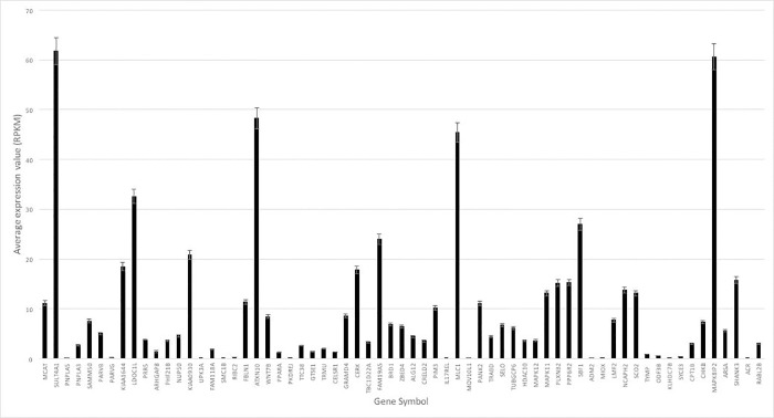
The average expression value (in RPKM) for each of the 65 genes used in the analysis was calculated over all developmental time periods and all brain regions assessed (Fig 1). Six (9%) of the genes did not have at least one read greater than five RPKM, and gene expression of those genes was interpreted as noise, as reported previously [32]. The mean total expression for SHANK3 was 15.82 RPKM (standard deviation (SD) = 12.10) vs 9.59 (SD = 13.60) for the entire set of 65 genes over all time periods and brain regions. Four genes, ATXN10 (mean (M) = 48.24, SD = 17.30), MAPK8IP2 (M = 60.61, SD = 26.40), MLC1 (M = 45.40, SD = 56.38), and SULT4A1 (M = 61.81, SD = 50.15), had average expression at least two standard deviations above the average expression for the entire gene set (Figs 1 and 2).
Fig 1. Average expression of 22q13 genes.
The average expression value for each of the 65 genes on 22q13 is shown, arranged from most proximal (left) to most distal (right) on chromosome. Error bars represent the standard error of the mean.
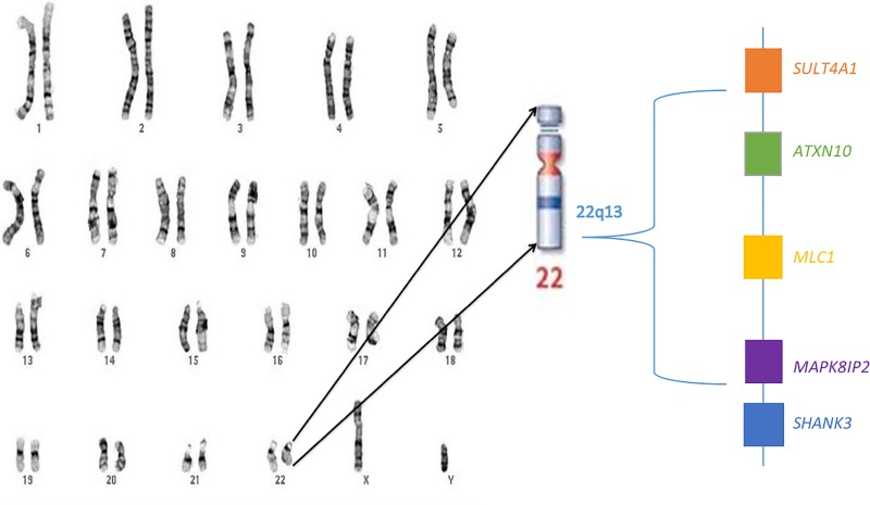
Fig 2. 22q13 region.
The location of the highest expressed genes in relation to SHANK3 is shown.
Expression of highly expressed genes reveals shared temporal profile with SHANK3
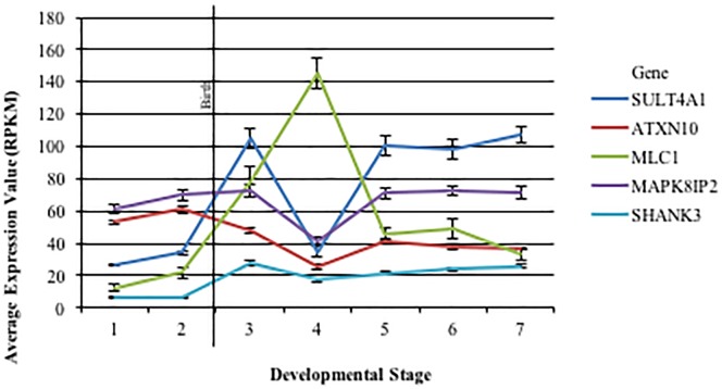
Average expression of ATXN10, MAPK8IP2, MLC1, SULT4A1 and SHANK3, incorporating all brain regions, over developmental time was calculated (Fig 3). Overall the tendency was for a bimodal pattern, with highest expression during infancy and a decrease in expression over early childhood, followed by gradual increase and eventual plateau later in childhood to adulthood. Notably, MLC1 and ATXN10 expression did not follow a bimodal pattern, expression did not increase later in childhood. Additionally, MLC1 showed a delay in peak expression, with highest expression being in early childhood, while ATXN10 showed early peak expression, with highest expression during gestation.
Fig 3. Average expression across developmental time.
The average expression of each gene assessed across seven developmental stages is shown (1: 16 pcw– 17 pcw, 2: 19 pcw– 24 pcw, 3: 4 mos– 1 yr, 4: 2 yrs– 4 yrs, 5:8 yrs– 13 yrs, 6: 15 yrs– 21 yrs, 7: 23 yrs– 40 yrs). Error bars represent the standard error of the mean. pcw = post-conception years; mos = months; yrs = years.
One-way ANOVA test showed a significant main effect of developmental time on gene expression for all five genes assessed, p-value <0.05 (S4 Table). When pre-natal vs post-natal expression was compared, post-natal expression was found to be significantly higher than pre-natal expression for SHANK3, MAPK8IP2, and SULT4A1. Pre-natal expression was significantly higher than post-natal expression for ATXN10, while no significant difference between pre- and post-natal expression was found for MLC1 (S4 Table).
Expression of highly expressed genes reveals unique spatial profiles
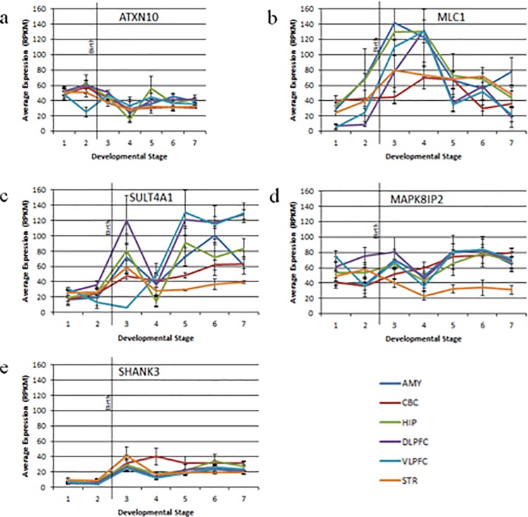
Average brain region specific expression of the four highest expressed genes as well as SHANK3 was analyzed (S5 Table). Expression of SHANK3, MAPK8IP2, and SULT4A1 was significantly different between brain regions when all developmental time periods were included. SHANK3 showed highest expression in the cerebellum, while MAPK8IP2 and SULT4A1 showed highest expression in the dorsolateral prefrontal cortex. We also assessed region specific expression over developmental time. Overall, spatial patterns of expression over time were largely consistent with average expression over time, although gene specific expression within the cerebellum did not seem to follow this trend (Fig 4).
Fig 4. Average expression across time and space.
The expression profile for each candidate gene across developmental time in six different brain regions (AMY, CBC, HIP, DLPFC, VLPFC, STR) is shown in a-e (a: ATXN10, b: MLC1, c: SULT4A1, d: MAPK8IP2, e: SHANK3). The black vertical line represents birth. Error bars represent the standard error of the mean. AMY = amygdala, CBC = cerebellum, HIP = hippocampus, DLPFC = dorsolateral prefrontal cortex, VLPFC = ventrolateral prefrontal cortex, STR = striatum.
Expression of SHANK3 exons
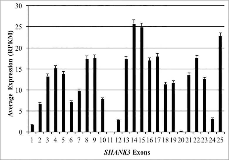
SHANK3 exon-specific average expression was calculated for all regions and all time periods (Fig 5). Notably, 5/25 exons (Exons 1, 11, 12, 20, and 24) did not reach five RPKM of average expression. The exons with the highest brain specific expression were 14, 15 and 25.
Fig 5. Average expression of SHANK3 exons.
The average expression of each individual SHANK3 exon across all brain regions and developmental time periods is shown. Error bars represent the standard error of the mean.
Discussion
Patients with Phelan-McDermid Syndrome can present with a wide range of neurologic symptoms of varying severity, making clinical diagnosis difficult. Appropriate diagnosis relies upon molecular testing to confirm a deletion on 22q13. While evidence supports the role of SHANK3 in the neuropathogenesis of the disease, it is unclear how other genes on 22q13 also contribute to the neurologic phenotype [7, 8, 10, 11, 21, 26, 28]. Traditional approaches have relied on rodent knock-out models to study the correlation between genotype and phenotype, however these studies are cumbersome and the subtle neurologic symptoms typical of PMS are not ideally suited for animal behavioral models [17, 18, 29]. More recently, candidate genes have been identified with detailed phenotype mapping, using array comparative genome hybridization data to correlate deletions of a specific location and size with clinical symptoms [11, 43]. While, this approach results in the identification of many candidate genes, in the absence of tissue specific data, the developmental context and significance of these genes in the pathology of human diseases remains unknown. In this manuscript we describe a complementary approach; we use gene expression data to examine the Phelan-McDermid region during normal development, which we show provides insight into the functional landscape of this region.
Our analysis of gene expression trends over developmental time revealed specific expression patterns of known functional significance. The gene ATXN10 showed a pattern of increased gene expression during gestation and decreased expression after birth, while SULT4A1, SHANK3, and MAPK8IP2 expression remained high throughout fetal life and infancy. Interestingly, large genome-wide transcriptome analyses have shown these distinct patterns of gene expression have functional significance, and specifically, enrich for genes related to axonal growth and synaptic function, respectively, highlighting the importance of the PMS region during normal neurodevelopment [44]. Notably, in a detailed review of 22q13.3 genes likely to be haploinsufficient, Mitz et al. also identified SULT4A, MAPK8IP2, and ATXN10 as likely candidates for the PMS phenotype. [45].
Our analysis of gene expression trends over developmental time also provide insight into potential mechanisms of disease pathogenesis. For instance, the gene MLC1 showed a delayed pattern of peak gene expression, with highest expression profiles during early childhood, a period typically characterized by decreased overall expression in the brain [44]. We can speculate that this pattern of expression may explain, in part, the childhood developmental regression typical of megalencephalic leukoencephalopathy (MLC), a disorder caused by variants in MLC1 [46]. Moreover, as childhood developmental regression is described in up to 50% of PMS patients [47], future work may focus on further characterizing the relationship between MLC1 deletions and regression symptoms in PMS patients.
Spatial analysis revealed that in most of the genes assessed, there was significantly different region-specific expression. This is likely a product of differences in function between brain regions, however it highlights the importance of spatial context for understanding normal neurodevelopment. Overall, region specific gene expression showed largely conserved trends in gene expression over developmental time, with the exception of the cerebellum. This finding however, is not unique to our study, as several groups have shown whole genome cerebellar expression to be distinct as compared to other brain regions [48, 49]. Additionally, while the cerebellum is consistently implicated in neurodevelopmental disorders and as having a role in higher cognitive function, how the unique developmental timeline of this region plays a role in the pathology of such diseases is still unknown [50, 51].
Given the importance of SHANK3 in the pathogenesis of PMS and known complex cell type and region specific transcriptional regulation, we analyzed exon specific expression, which confirmed high brain-specific expression of known functional isoforms [52]. Exons encoding known functional domains that act at the post-synaptic density (PSD), notably ankryrin repeat domain (exons 4–7), the PSD protein/Drosophila disc large tumor suppressor/zonula occludens-1 protein (PDZ; exons 13–16), and the Homer binding domain (exon 21), were all highly expressed [17, 53, 54].
For this study we specifically chose to analyze the expression of genes within the PMS region with the highest overall gene expression however it is likely that genes with lower baseline expression also contribute to the pathophysiology of PMS, and we have reviewed these genes in the supplemental information (S1 and S2 Tables). For instance, the gene ARSA, which had a total average gene expression of 5.6 RPKM, encodes the enzyme arylsulfatase A and is situated near SHANK3 at the terminal end of the chromosome 22q13.33, a region commonly in deleted in PMS [21]. However as variants in this gene lead to metachromatic leukodystrophy, a devastating disease characterized by severe neurologic symptoms including progressive mental deterioration, hypotonia, weakness and seizures, how exactly deletions in this gene may contribute to the neurological sequelae of PMS should be further explored [55].
Our manuscript is limited by our hypothesis generating approach. However, it is our intention that our data be used to draw insight into the developmental and spatial relationships of genes within the PMS region, which may then be used to test hypotheses using traditional functional studies. Additionally, while we focus on protein-coding genes specifically, non-coding RNAs (ncRNAs) are known to play an active role in transcriptional regulation during neurodevelopment and so understanding the dynamic expression of these genes in the PMS region and how they modulate the local transcriptional landscape will be important and deserves further study [34].
Conclusion
The broad Phelan-McDermid phenotype encompasses a range of neurologic symptoms of varying severity and consequence; the etiology of which remains poorly understood. In this brief report we use gene expression data from normal controls to glean insight into the functional landscape of the 22q13.3 region. This work begins to show how the PMS region is involved in normal neurodevelopment, and specifically how dynamic temporal and spatial expression profiles may hint at gene function and mechanisms of disease, and moreover may be used to guide future functional studies.
Supporting information
(DOCX)
Developmental time period when RPKM >5 also shown. (1: 16 pcw– 17 pcw, 2: 19 pcw– 24 pcw, 3: 4 mos– 1 yr, 4: 2 yrs– 4 yrs, 5:8 yrs– 13 yrs, 6: 15 yrs– 21 yrs, 7: 23 yrs– 40 yrs). (pcw = post-conception weeks, mos = months, yrs = years).
(DOCX)
The average expression in RPKM and SD of each of the 65 protein coding genes within the PMS region is shown. Genes are ordered by most proximal to most distal on chromosome.
(DOCX)
Average expression of each gene assessed by developmental stage shown. Results of ANOVA testing and t-test also displayed. (1: 16 pcw– 17 pcw, 2: 19 pcw– 24 pcw, 3: 4 mos– 1 yr, 4: 2 yrs– 4 yrs, 5:8 yrs– 13 yrs, 6: 15 yrs– 21 yrs, 7: 23 yrs– 40 yrs). (pcw = post-conception weeks, mos = months, yrs = years).
(DOCX)
Average brain region specific expression of each gene assessed is shown. Results of ANOVA testing is also displayed. Significant p-values are bolded. (AMY = amygdala, CBC = cerebellum, HIP = hippocampus, DLPFC = dorsolateral prefrontal cortex, VLPFC = ventrolateral prefrontal cortex, STR = striatum).
(DOCX)
Acknowledgments
This work was supported by the Intramural Research Programs of the National Institute of Child Health and Human Development, NIH and the National Institute of Mental Health, NIH. MNZ was also supported by the Baylor College of Medicine MSTP grant. The funders had no role in this work or the decision to publish. The authors declare no conflicts of interest.
Data Availability
All gene expression files are available from the Allen Brain Institute BrainSpan Dataset at http://www.brainspan.org/.
Funding Statement
This work was supported by the Intramural Research Programs of the National Institute of Child Health and Human Development, NIH and the National Institute of Mental Health, NIH. MNZ was supported by the Baylor College of Medicine MSTP grant. The funders had no role in study design, data collection and analysis, decision to publish, or preparation of the manuscript.
References
- 1.Dhar SU, del Gaudio D, German JR, Peters SU, Ou Z, Bader PI, et al. 22q13.3 deletion syndrome: clinical and molecular analysis using array CGH. American journal of medical genetics Part A. 2010;152a(3):573–81. Epub 2010/02/27. 10.1002/ajmg.a.33253 [DOI] [PMC free article] [PubMed] [Google Scholar]
- 2.Cusmano-Ozog K, Manning MA, Hoyme HE. 22q13.3 deletion syndrome: a recognizable malformation syndrome associated with marked speech and language delay. American journal of medical genetics Part C, Seminars in medical genetics. 2007;145c(4):393–8. Epub 2007/10/11. 10.1002/ajmg.c.30155 . [DOI] [PubMed] [Google Scholar]
- 3.Phelan MC Rogers SG. Deletion 22q13 syndrome: Phelan-McDermid syndrome In: Cassidy SB AJ, editor. The Management of Genetic Syndromes. 3rd ed. Hoboken, NJ: John Wiley and Sons; 2010. p. 285–97. [Google Scholar]
- 4.Phelan K, McDermid HE. The 22q13.3 Deletion Syndrome (Phelan-McDermid Syndrome). Molecular syndromology. 2012;2(3–5):186–201. Epub 2012/06/07. 000334260. 10.1159/000334260 [DOI] [PMC free article] [PubMed] [Google Scholar]
- 5.Sarasua SM, Boccuto L, Sharp JL, Dwivedi A, Chen CF, Rollins JD, et al. Clinical and genomic evaluation of 201 patients with Phelan-McDermid syndrome. Human genetics. 2014;133(7):847–59. Epub 2014/02/01. 10.1007/s00439-014-1423-7 . [DOI] [PubMed] [Google Scholar]
- 6.Sarasua SM, Dwivedi A, Boccuto L, Rollins JD, Chen CF, Rogers RC, et al. Association between deletion size and important phenotypes expands the genomic region of interest in Phelan-McDermid syndrome (22q13 deletion syndrome). Journal of medical genetics. 2011;48(11):761–6. Epub 2011/10/11. 10.1136/jmedgenet-2011-100225 . [DOI] [PubMed] [Google Scholar]
- 7.Luciani JJ, de Mas P, Depetris D, Mignon-Ravix C, Bottani A, Prieur M, et al. Telomeric 22q13 deletions resulting from rings, simple deletions, and translocations: cytogenetic, molecular, and clinical analyses of 32 new observations. Journal of medical genetics. 2003;40(9):690–6. Epub 2003/09/10. 10.1136/jmg.40.9.690 [DOI] [PMC free article] [PubMed] [Google Scholar]
- 8.Jeffries AR, Curran S, Elmslie F, Sharma A, Wenger S, Hummel M, et al. Molecular and phenotypic characterization of ring chromosome 22. American journal of medical genetics Part A. 2005;137(2):139–47. Epub 2005/08/02. 10.1002/ajmg.a.30780 . [DOI] [PubMed] [Google Scholar]
- 9.Bonaglia MC, Giorda R, Borgatti R, Felisari G, Gagliardi C, Selicorni A, et al. Disruption of the ProSAP2 gene in a t(12;22)(q24.1;q13.3) is associated with the 22q13.3 deletion syndrome. American journal of human genetics. 2001;69(2):261–8. Epub 2001/06/30. 10.1086/321293 [DOI] [PMC free article] [PubMed] [Google Scholar]
- 10.Zwanenburg RJ, Ruiter SA, van den Heuvel ER, Flapper BC, Van Ravenswaaij-Arts CM. Developmental phenotype in Phelan-McDermid (22q13.3 deletion) syndrome: a systematic and prospective study in 34 children. Journal of neurodevelopmental disorders. 2016;8:16 Epub 2016/04/28. 10.1186/s11689-016-9150-0 [DOI] [PMC free article] [PubMed] [Google Scholar]
- 11.Tabet AC, Rolland T, Ducloy M, Levy J, Buratti J, Mathieu A, et al. A framework to identify contributing genes in patients with Phelan-McDermid syndrome. NPJ genomic medicine. 2017;2:32 Epub 2017/12/22. 10.1038/s41525-017-0035-2 [DOI] [PMC free article] [PubMed] [Google Scholar]
- 12.Macedoni-Luksic M, Krgovic D, Zagradisnik B, Kokalj-Vokac N. Deletion of the last exon of SHANK3 gene produces the full Phelan-McDermid phenotype: a case report. Gene. 2013;524(2):386–9. Epub 2013/04/25. 10.1016/j.gene.2013.03.141 . [DOI] [PubMed] [Google Scholar]
- 13.Leblond CS, Nava C, Polge A, Gauthier J, Huguet G, Lumbroso S, et al. Meta-analysis of SHANK Mutations in Autism Spectrum Disorders: a gradient of severity in cognitive impairments. PLoS genetics. 2014;10(9):e1004580 Epub 2014/09/05. 10.1371/journal.pgen.1004580 [DOI] [PMC free article] [PubMed] [Google Scholar]
- 14.de Sena Cortabitarte A, Degenhardt F, Strohmaier J, Lang M, Weiss B, Roeth R, et al. Investigation of SHANK3 in schizophrenia. American journal of medical genetics Part B, Neuropsychiatric genetics: the official publication of the International Society of Psychiatric Genetics. 2017;174(4):390–8. Epub 2017/04/04. 10.1002/ajmg.b.32528 . [DOI] [PubMed] [Google Scholar]
- 15.Gauthier J, Champagne N, Lafreniere RG, Xiong L, Spiegelman D, Brustein E, et al. De novo mutations in the gene encoding the synaptic scaffolding protein SHANK3 in patients ascertained for schizophrenia. Proceedings of the National Academy of Sciences of the United States of America. 2010;107(17):7863–8. Epub 2010/04/14. 10.1073/pnas.0906232107 [DOI] [PMC free article] [PubMed] [Google Scholar]
- 16.Alexandrov PN, Zhao Y, Jaber V, Cong L, Lukiw WJ. Deficits in the Proline-Rich Synapse-Associated Shank3 Protein in Multiple Neuropsychiatric Disorders. Frontiers in neurology. 2017;8:670 Epub 2018/01/13. 10.3389/fneur.2017.00670 [DOI] [PMC free article] [PubMed] [Google Scholar]
- 17.Bozdagi O, Sakurai T, Papapetrou D, Wang X, Dickstein DL, Takahashi N, et al. Haploinsufficiency of the autism-associated Shank3 gene leads to deficits in synaptic function, social interaction, and social communication. Molecular autism. 2010;1(1):15 Epub 2010/12/21. 10.1186/2040-2392-1-15 [DOI] [PMC free article] [PubMed] [Google Scholar]
- 18.Sheng M, Kim E. The postsynaptic organization of synapses. Cold Spring Harbor perspectives in biology. 2011;3(12). Epub 2011/11/03. 10.1101/cshperspect.a005678 [DOI] [PMC free article] [PubMed] [Google Scholar]
- 19.Hamdan FF, Gauthier J, Araki Y, Lin DT, Yoshizawa Y, Higashi K, et al. Excess of de novo deleterious mutations in genes associated with glutamatergic systems in nonsyndromic intellectual disability. American journal of human genetics. 2011;88(3):306–16. Epub 2011/03/08. 10.1016/j.ajhg.2011.02.001 [DOI] [PMC free article] [PubMed] [Google Scholar]
- 20.Moessner R, Marshall CR, Sutcliffe JS, Skaug J, Pinto D, Vincent J, et al. Contribution of SHANK3 mutations to autism spectrum disorder. American journal of human genetics. 2007;81(6):1289–97. Epub 2007/11/14. 10.1086/522590 [DOI] [PMC free article] [PubMed] [Google Scholar]
- 21.Wilson HL, Wong AC, Shaw SR, Tse WY, Stapleton GA, Phelan MC, et al. Molecular characterisation of the 22q13 deletion syndrome supports the role of haploinsufficiency of SHANK3/PROSAP2 in the major neurological symptoms. Journal of medical genetics. 2003;40(8):575–84. Epub 2003/08/16. 10.1136/jmg.40.8.575 [DOI] [PMC free article] [PubMed] [Google Scholar]
- 22.De Rubeis S, Siper PM, Durkin A, Weissman J, Muratet F, Halpern D, et al. Delineation of the genetic and clinical spectrum of Phelan-McDermid syndrome caused by SHANK3 point mutations. Molecular autism. 2018;9:31 Epub 2018/05/03. 10.1186/s13229-018-0205-9 [DOI] [PMC free article] [PubMed] [Google Scholar]
- 23.Phelan K, Boccuto L, Rogers RC, Sarasua SM, McDermid HE. Letter to the editor regarding Disciglio et al.: interstitial 22q13 deletions not involving SHANK3 gene: a new contiguous gene syndrome. American journal of medical genetics Part A. 2015;167(7):1679–80. Epub 2015/08/22. 10.1002/ajmg.a.36788 . [DOI] [PubMed] [Google Scholar]
- 24.Wilson HL, Crolla JA, Walker D, Artifoni L, Dallapiccola B, Takano T, et al. Interstitial 22q13 deletions: genes other than SHANK3 have major effects on cognitive and language development. European journal of human genetics: EJHG. 2008;16(11):1301–10. Epub 2008/06/05. 10.1038/ejhg.2008.107 . [DOI] [PubMed] [Google Scholar]
- 25.Fujita Y, Mochizuki D, Mori Y, Nakamoto N, Kobayashi M, Omi K, et al. Girl with accelerated growth, hearing loss, inner ear anomalies, delayed myelination of the brain, and del(22)(q13.1q13.2). American journal of medical genetics. 2000;92(3):195–9. Epub 2000/05/19. . [PubMed] [Google Scholar]
- 26.Palumbo P, Accadia M, Leone MP, Palladino T, Stallone R, Carella M, et al. Clinical and molecular characterization of an emerging chromosome 22q13.31 microdeletion syndrome. American journal of medical genetics Part A. 2018;176(2):391–8. Epub 2017/12/02. 10.1002/ajmg.a.38559 . [DOI] [PubMed] [Google Scholar]
- 27.Ha JF, Ahmad A, Lesperance MM. Clinical characterization of novel chromosome 22q13 microdeletions. International journal of pediatric otorhinolaryngology. 2017;95:121–6. Epub 2017/06/04. 10.1016/j.ijporl.2016.12.008 . [DOI] [PubMed] [Google Scholar]
- 28.Disciglio V, Lo Rizzo C, Mencarelli MA, Mucciolo M, Marozza A, Di Marco C, et al. Interstitial 22q13 deletions not involving SHANK3 gene: a new contiguous gene syndrome. American journal of medical genetics Part A. 2014;164a(7):1666–76. Epub 2014/04/05. 10.1002/ajmg.a.36513 . [DOI] [PubMed] [Google Scholar]
- 29.Giza J, Urbanski MJ, Prestori F, Bandyopadhyay B, Yam A, Friedrich V, et al. Behavioral and cerebellar transmission deficits in mice lacking the autism-linked gene islet brain-2. The Journal of neuroscience: the official journal of the Society for Neuroscience. 2010;30(44):14805–16. Epub 2010/11/05. 10.1523/jneurosci.1161-10.2010 [DOI] [PMC free article] [PubMed] [Google Scholar]
- 30.Hawrylycz MJ, Lein ES, Guillozet-Bongaarts AL, Shen EH, Ng L, Miller JA, et al. An anatomically comprehensive atlas of the adult human brain transcriptome. Nature. 2012;489(7416):391–9. Epub 2012/09/22. 10.1038/nature11405 [DOI] [PMC free article] [PubMed] [Google Scholar]
- 31.BrainSpan Atlas of the Developing Human Brain.: Allen Institute for Brain Science; 2010. [cited 2018]. Available from: http://www.brainspan.org/. [Google Scholar]
- 32.Mahfouz A, Ziats MN, Rennert OM, Lelieveldt BP, Reinders MJ. Shared Pathways Among Autism Candidate Genes Determined by Co-expression Network Analysis of the Developing Human Brain Transcriptome. Journal of molecular neuroscience: MN. 2015;57(4):580–94. Epub 2015/09/25. 10.1007/s12031-015-0641-3 [DOI] [PMC free article] [PubMed] [Google Scholar]
- 33.Kang HJ, Kawasawa YI, Cheng F, Zhu Y, Xu X, Li M, et al. Spatio-temporal transcriptome of the human brain. Nature. 2011;478(7370):483–9. Epub 2011/10/28. 10.1038/nature10523 [DOI] [PMC free article] [PubMed] [Google Scholar]
- 34.Ziats MN, Rennert OM. Identification of differentially expressed microRNAs across the developing human brain. Molecular psychiatry. 2014;19(7):848–52. Epub 2013/08/07. 10.1038/mp.2013.93 [DOI] [PMC free article] [PubMed] [Google Scholar]
- 35.Cogill SB, Srivastava AK, Yang MQ, Wang L. Co-expression of long non-coding RNAs and autism risk genes in the developing human brain. BMC systems biology. 2018;12(Suppl 7):91 Epub 2018/12/15. 10.1186/s12918-018-0639-x [DOI] [PMC free article] [PubMed] [Google Scholar]
- 36.Mortazavi A, Williams BA, McCue K, Schaeffer L, Wold B. Mapping and quantifying mammalian transcriptomes by RNA-Seq. Nature methods. 2008;5(7):621–8. Epub 2008/06/03. 10.1038/nmeth.1226 . [DOI] [PubMed] [Google Scholar]
- 37.Kent WJ, Sugnet CW, Furey TS, Roskin KM, Pringle TH, Zahler AM, et al. The human genome browser at UCSC. Genome research. 2002;12(6):996–1006. Epub 2002/06/05. 10.1101/gr.229102 [DOI] [PMC free article] [PubMed] [Google Scholar]
- 38.Courchesne E, Redcay E, Morgan JT, Kennedy DP. Autism at the beginning: microstructural and growth abnormalities underlying the cognitive and behavioral phenotype of autism. Development and psychopathology. 2005;17(3):577–97. Epub 2005/11/03. 10.1017/S0954579405050285 . [DOI] [PubMed] [Google Scholar]
- 39.Haznedar MM, Buchsbaum MS, Hazlett EA, LiCalzi EM, Cartwright C, Hollander E. Volumetric analysis and three-dimensional glucose metabolic mapping of the striatum and thalamus in patients with autism spectrum disorders. The American journal of psychiatry. 2006;163(7):1252–63. Epub 2006/07/04. 10.1176/appi.ajp.163.7.1252 . [DOI] [PubMed] [Google Scholar]
- 40.Munson J, Dawson G, Abbott R, Faja S, Webb SJ, Friedman SD, et al. Amygdalar volume and behavioral development in autism. Archives of general psychiatry. 2006;63(6):686–93. Epub 2006/06/07. 10.1001/archpsyc.63.6.686 . [DOI] [PubMed] [Google Scholar]
- 41.Schumann CM, Hamstra J, Goodlin-Jones BL, Lotspeich LJ, Kwon H, Buonocore MH, et al. The amygdala is enlarged in children but not adolescents with autism; the hippocampus is enlarged at all ages. The Journal of neuroscience: the official journal of the Society for Neuroscience. 2004;24(28):6392–401. Epub 2004/07/16. 10.1523/jneurosci.1297-04.2004 . [DOI] [PMC free article] [PubMed] [Google Scholar]
- 42.Palmen SJ, van Engeland H, Hof PR, Schmitz C. Neuropathological findings in autism. Brain: a journal of neurology. 2004;127(Pt 12):2572–83. Epub 2004/08/27. 10.1093/brain/awh287 . [DOI] [PubMed] [Google Scholar]
- 43.Sarasua SM, Dwivedi A, Boccuto L, Chen CF, Sharp JL, Rollins JD, et al. 22q13.2q13.32 genomic regions associated with severity of speech delay, developmental delay, and physical features in Phelan-McDermid syndrome. Genetics in medicine: official journal of the American College of Medical Genetics. 2014;16(4):318–28. Epub 2013/10/19. 10.1038/gim.2013.144 . [DOI] [PubMed] [Google Scholar]
- 44.Colantuoni C, Lipska BK, Ye T, Hyde TM, Tao R, Leek JT, et al. Temporal dynamics and genetic control of transcription in the human prefrontal cortex. Nature. 2011;478(7370):519–23. Epub 2011/10/28. 10.1038/nature10524 [DOI] [PMC free article] [PubMed] [Google Scholar]
- 45.Mitz AR, Philyaw TJ, Boccuto L, Shcheglovitov A, Sarasua SM, Kaufmann WE, et al. Identification of 22q13 genes most likely to contribute to Phelan McDermid syndrome. European journal of human genetics: EJHG. 2018;26(3):293–302. Epub 2018/01/24. 10.1038/s41431-017-0042-x [DOI] [PMC free article] [PubMed] [Google Scholar]
- 46.Hamilton EM, Tekturk P, Cialdella F, van Rappard DF, Wolf NI, Yalcinkaya C, et al. Megalencephalic leukoencephalopathy with subcortical cysts: Characterization of disease variants. Neurology. 2018;90(16):e1395–403. 10.1212/WNL.0000000000005334 [DOI] [PMC free article] [PubMed] [Google Scholar]
- 47.Reierson G, Bernstein J, Froehlich-Santino W, Urban A, Purmann C, Berquist S, et al. Characterizing regression in Phelan McDermid Syndrome (22q13 deletion syndrome). Journal of psychiatric research. 2017;91:139–44. Epub 2017/03/28. 10.1016/j.jpsychires.2017.03.010 [DOI] [PMC free article] [PubMed] [Google Scholar]
- 48.Roth RB, Hevezi P, Lee J, Willhite D, Lechner SM, Foster AC, et al. Gene expression analyses reveal molecular relationships among 20 regions of the human CNS. Neurogenetics. 2006;7(2):67–80. Epub 2006/03/31. 10.1007/s10048-006-0032-6 . [DOI] [PubMed] [Google Scholar]
- 49.Johnson MB, Kawasawa YI, Mason CE, Krsnik Z, Coppola G, Bogdanovic D, et al. Functional and evolutionary insights into human brain development through global transcriptome analysis. Neuron. 2009;62(4):494–509. Epub 2009/05/30. 10.1016/j.neuron.2009.03.027 [DOI] [PMC free article] [PubMed] [Google Scholar]
- 50.Schmahmann JD, Sherman JC. The cerebellar cognitive affective syndrome. Brain: a journal of neurology. 1998;121 (Pt 4):561–79. Epub 1998/05/13. . [DOI] [PubMed] [Google Scholar]
- 51.Fatemi SH, Aldinger KA, Ashwood P, Bauman ML, Blaha CD, Blatt GJ, et al. Consensus paper: pathological role of the cerebellum in autism. Cerebellum (London, England). 2012;11(3):777–807. Epub 2012/03/01. 10.1007/s12311-012-0355-9 [DOI] [PMC free article] [PubMed] [Google Scholar]
- 52.Jiang YH, Ehlers MD. Modeling autism by SHANK gene mutations in mice. Neuron. 2013;78(1):8–27. Epub 2013/04/16. 10.1016/j.neuron.2013.03.016 [DOI] [PMC free article] [PubMed] [Google Scholar]
- 53.Kouser M, Speed HE, Dewey CM, Reimers JM, Widman AJ, Gupta N, et al. Loss of predominant Shank3 isoforms results in hippocampus-dependent impairments in behavior and synaptic transmission. The Journal of neuroscience: the official journal of the Society for Neuroscience. 2013;33(47):18448–68. Epub 2013/11/22. 10.1523/jneurosci.3017-13.2013 [DOI] [PMC free article] [PubMed] [Google Scholar]
- 54.Peca J, Feliciano C, Ting JT, Wang W, Wells MF, Venkatraman TN, et al. Shank3 mutant mice display autistic-like behaviours and striatal dysfunction. Nature. 2011;472(7344):437–42. Epub 2011/03/23. 10.1038/nature09965 [DOI] [PMC free article] [PubMed] [Google Scholar]
- 55.Greenfield JG. A Form of Progressive Cerebral Sclerosis in Infants associated with Primary Degeneration of the Interfascicular Glia. Proceedings of the Royal Society of Medicine. 1933;26(6):690–7. Epub 1933/04/01. [DOI] [PMC free article] [PubMed] [Google Scholar]
Associated Data
This section collects any data citations, data availability statements, or supplementary materials included in this article.
Supplementary Materials
(DOCX)
Developmental time period when RPKM >5 also shown. (1: 16 pcw– 17 pcw, 2: 19 pcw– 24 pcw, 3: 4 mos– 1 yr, 4: 2 yrs– 4 yrs, 5:8 yrs– 13 yrs, 6: 15 yrs– 21 yrs, 7: 23 yrs– 40 yrs). (pcw = post-conception weeks, mos = months, yrs = years).
(DOCX)
The average expression in RPKM and SD of each of the 65 protein coding genes within the PMS region is shown. Genes are ordered by most proximal to most distal on chromosome.
(DOCX)
Average expression of each gene assessed by developmental stage shown. Results of ANOVA testing and t-test also displayed. (1: 16 pcw– 17 pcw, 2: 19 pcw– 24 pcw, 3: 4 mos– 1 yr, 4: 2 yrs– 4 yrs, 5:8 yrs– 13 yrs, 6: 15 yrs– 21 yrs, 7: 23 yrs– 40 yrs). (pcw = post-conception weeks, mos = months, yrs = years).
(DOCX)
Average brain region specific expression of each gene assessed is shown. Results of ANOVA testing is also displayed. Significant p-values are bolded. (AMY = amygdala, CBC = cerebellum, HIP = hippocampus, DLPFC = dorsolateral prefrontal cortex, VLPFC = ventrolateral prefrontal cortex, STR = striatum).
(DOCX)
Data Availability Statement
All gene expression files are available from the Allen Brain Institute BrainSpan Dataset at http://www.brainspan.org/.


