

Abstract
Choristomas are proliferation of normal cells or tissue in an abnormal location. Choristomas of oral soft tissue are rare lesions. Different tissues can occur in the oral cavity as choristomas. It can be cartilage, bone, salivary gland, glial and thyroid tissue. Choristomas with the proliferation of chondroid tissue are termed as cartilaginous choristomas. In oral cavity, they are most frequently seen in tongue followed by buccal mucosa and soft palate. We report a case of 40-year-old female presenting with hard lobulated swelling on the left lateral border of the tongue. Fine-needle aspiration cytology was performed which showed only myxoid stroma. Histopathological examination of excised specimen of the same showed lobules of mature hyaline cartilage. Thus, a diagnosis of cartilaginous choristoma was made.
Keywords: Cartilaginous, choristoma, cytology, histopathology, tongue
INTRODUCTION
Choristoma is histologically an island of normal tissue that occurs in an abnormal location. They rarely present in the oral cavity. However, when they occur in the oral cavity, mucosa of the tongue is the most commonly affected. The various tissue types that can occur in the oral cavity as choristomas are cartilage, bone, glial tissue, salivary gland and thyroid tissue.[1,2] Choristomas with cartilaginous tissue are called as cartilaginous choristomas.[3] It was first described by Berry in 1890. It is usually found in the distal extremities and rarely in the soft tissue of head and neck.[4] We report a case of cartilaginous choristoma located in the tongue.
CASE REPORT
A 55-year-old female presented with swelling on the left lateral aspect of the tongue for 1 year. There was no history of any trauma or dentures. On examination, a hard globular swelling was present on the left lateral aspect of tongue [Figure 1]. Fine-needle aspiration cytology was done. The swelling was difficult to aspirate and yielded only scant myxoid material blocking the needle. Air dried and alcohol-fixed smears were prepared and stained with Wright-Giemsa and papanicolaou stain. Smears examined show only scant myxoid stroma with few scattered oval to spindle cells. Thus, a diagnosis of myxoid mesenchymal lesion was suggested, and excision was advised.
Figure 1.
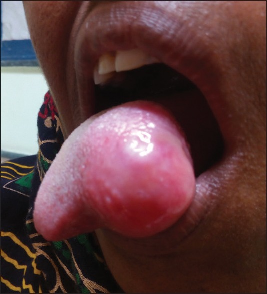
Photo showing globular swelling on the left lateral aspect of tongue
Excised specimen of the swelling was received. Grossly, the specimen was firm to hard and globular. The external surface was shiny and glistening. The cut surface showed myxoid areas. Representative sections were taken and stained with hematoxylin and eosin. Sections examined show stratified squamous epithelium with underlying skeletal muscle, beneath which there were lobules of mature hyaline cartilage separated by fibrovascular septa. Thus, histopathology was suggestive of cartilaginous choristoma [Figures 2 and 3].
Figure 2.

Photomicrograph showing stratified squamous epithelium of tongue (H&E, ×400)
Figure 3.

(a) Photomicrograph showing lobule of mature cartilage in the subepithelial tissue (H&E, ×400). (b) Photomicrograph showing mature chondrocytes (H&E, ×1000)
DISCUSSION
Choristomas of oral soft tissue are rare lesions.[3] Cartilage, salivary gland, bone, thyroid, sebaceous gland, brain tissue and gastric mucosa are identified as the sources of intraoral choristoma.[2] The presence of mature cartilage is termed as cartilaginous choristoma. The age of diagnosis varies greatly from 10 to 80 years. It characteristically presents as painless, firm nodule in young adults, especially in females.[5] In the present case, the presentation was also painless hard lump.
Different authors have described choristomas at different sites. Bhargava et al.[6] described a case of cartilaginous choristoma in tonsil. Malis et al.[7] described it in nasopharynx. Toida et al.[4] reported cartilaginous choristoma of the tongue and. In the tongue, they have a predilection of middle dorsal aspect.[3]
The pathogenesis of cartilaginous choristoma is still unclear. Undifferentiated multipotent mesenchymal cells may be considered as a possible origin of cartilage.[1] Another possible origin is from the embryonic remnants. During fetal development, the incomplete resorption of embryonic cartilaginous tissue of the lingual septum, and depending on the persistence after birth, explains a chondromatous proliferation along the tongue midline.[4] However, choristoma of the tonsil appears to be a developmental anomaly associated with the 2nd pharyngeal arch.[1] Zegarelli[8] proposed that the lesions, that have only chondromatous growth should be termed as cartilaginous choristoma of the tongue. However, cartilaginous choristomas of the tongue are often associated with the presence of other elements, such as bone and adipose tissue. Thus, the occurrence of cartilaginous choristoma of the tongue is extremely rare. In our case, there was the presence of only mature cartilaginous tissue only. Its histological variants have also been described as lipocartilagenous and osteocartilagenous.[3]
Cartilaginous choristoma should be distinguished from cartilaginous metaplasia that usually occurs in the soft-tissue beneath trauma or neoplastic degeneration. These lesions are usually located in laryngotracheal area.[4] Histologically, cartilagenous metaplasia is characterized by diffuse deposits of calcium and cartilaginous cells arranged in various stages of maturation in single or clustered cartilaginous foci.[1] It usually occurs as a result of trauma due to ill-fitting dentures. In the present case, there was no history of dentures.
Differential diagnosis of cartilaginous choristoma includes various benign and malignant conditions. Benign conditions include pleomorphic adenoma, chondroma, neurofibroma, papilloma and ectomesenchymal chondromyxoid tumor.[9] A possibility of granular cell tumor can be considered as it is a common lesion on the tongue.[8]
The present case is distinguished from the other differential diagnosis. There was the absence of epithelial and mesenchymal component which helped in differentiating it from pleomorphic adenoma. The absence of round to oval cells embedded in myxofibrillary matrix in cytology and absence of lobulation and surrounding collagenous stroma histologically differentiated it from chondroma. In ectomesenchymal chondromyxoid tumor, the cells are arranged in cords, strands and sheets in a myxoid and chondromyxoid stroma; however, in choristoma, there was the presence of mature cartilage.
It is also important to distinguish it from malignant cartilaginous neoplasms, including primary chondrosarcoma and metastasis from a primary intraosseous chondrosarcoma.[3,9] These differential diagnoses can be ruled out by histopathological examination. Surgical excision is the treatment of choice. No recurrence has been reported in the literature.[9]
In conclusion, cartilaginous choristomas are benign developmental lesions which can mimic various lesions clinically. However, a complete histopathological examination helps in establishing the correct diagnosis.
Declaration of patient consent
The authors certify that they have obtained all appropriate patient consent forms. In the form the patient(s) has/have given his/her/their consent for his/her/their images and other clinical information to be reported in the journal. The patients understand that their names and initials will not be published and due efforts will be made to conceal their identity, but anonymity cannot be guaranteed.
Financial support and sponsorship
Nil.
Conflicts of interest
There are no conflicts of interest.
REFERENCES
- 1.Kannar V, Prabhakar K, Shalini S. Cartilaginous choristoma of tonsil: A hidden clinical entity. J Oral Maxillofac Pathol. 2013;17:292–3. doi: 10.4103/0973-029X.119779. [DOI] [PMC free article] [PubMed] [Google Scholar]
- 2.Chou LS, Hansen LS, Daniels TE. Choristomas of the oral cavity: A review. Oral Surg Oral Med Oral Pathol. 1991;72:584–93. doi: 10.1016/0030-4220(91)90498-2. [DOI] [PubMed] [Google Scholar]
- 3.Bansal R, Trivedi P, Patel S. Cartilaginous choristoma of the tongue-report of twocases and review of literature. Oral Oncol Extra. 2005;41:25–9. [Google Scholar]
- 4.Toida M, Sugiyama T, Kato Y. Cartilaginous choristoma of the tongue. J Oral Maxillofac Surg. 2003;61:393–6. doi: 10.1053/joms.2003.50065. [DOI] [PubMed] [Google Scholar]
- 5.Makoto T, Takatoshi S, Yukihiro K. Cartilaginous choristoma of the tongue. J Oral Maxillofac Surg. 2002;61:393–6. doi: 10.1053/joms.2003.50065. [DOI] [PubMed] [Google Scholar]
- 6.Bhargava D, Raman R, Khalfan Al Abri R, Bushnurmath B. Heterotopia of the tonsil. J Laryngol Otol. 1996;110:611–2. doi: 10.1017/s0022215100134401. [DOI] [PubMed] [Google Scholar]
- 7.Malis DJ, Breisch EA, Billman GF. Cartilaginous choristoma of the nasopharynx. Clin Anat. 2000;13:263–6. doi: 10.1002/1098-2353(2000)13:4<263::AID-CA6>3.0.CO;2-3. [DOI] [PubMed] [Google Scholar]
- 8.Zegarelli DJ. Chondroma of the tongue. Oral Surg Oral Med Oral Pathol. 1977;43:738–45. doi: 10.1016/0030-4220(77)90059-7. [DOI] [PubMed] [Google Scholar]
- 9.Desmedt M, Weynand B, Reychler H. Cartilaginous choristoma of the oral cavity: A report of two cases. B-ENT. 2007;3:87–91. [PubMed] [Google Scholar]