



ABSTRACT
Assembly of the RNA polymerases in both yeast and humans is proposed to occur in the cytoplasm prior to their nuclear import. Our previous studies identified a cold-sensitive mutation, rpc128-1007, in the yeast gene encoding the second largest Pol III subunit, Rpc128. rpc128-1007 is associated with defective assembly of Pol III complex and, in consequence, decreased level of tRNA synthesis. Here, we show that rpc128-1007 mutant cells remain largely unbudded and larger than wild type cells. Flow cytometry revealed that most rpc128-1007 mutant cells have G1 DNA content, suggesting that this mutation causes pronounced cell cycle delay in the G1 phase. Increased expression of gene encoding Rbs1, the Pol III assembly/import factor, could counteract G1 arrest observed in the rpc128-1007 mutant and restore wild type morphology of mutant cells. Concomitantly, cells lacking Rbs1 show a mild delay in G1 phase exit, indicating that Rbs1 is required for timely cell cycle progression. Using the double rpc128-1007 maf1Δ mutant in which tRNA synthesis is recovered, we confirmed that the Pol III assembly defect associated with rpc128-1007 is a primary cause of cell cycle arrest. Together our results indicate that impairment of Pol III complex assembly is coupled to cell cycle inhibition in the G1 phase.
KEYWORDS: RNA polymerase III, assembly, cell-cycle, G1 arrest, Rbs1, tRNA, transcription, yeast
Introduction
RNA polymerase III (Pol III) is responsible for the synthesis of small non-coding RNAs, mostly tRNAs, which are required for cell growth and proliferation. Although tRNA gene transcription is regulated during cell cycle progression, this regulation varies among organisms. In human and other vertebrates tRNA transcription is high during the S and G2 phases, low during the M (mitosis) and early G1 phases and then increase again at the G1/S transition [1–4]. tRNA synthesis fluctuates also during cell cycle in budding yeast Saccharomyces cerevisiae, although the data reported so far are controversial. A previous study has found that tRNA synthesis is higher in M phase relative to the G1 phase. Moreover, entry into mitosis is correlated with increased Pol III occupancy of tRNA genes[5]. More recent data show increased transcription of tRNA genes during late S phase and reveal an underlying molecular mechanism based on the recruitment of cyclin dependent kinase Cdk1 to Pol III chromatin[6].
Links between Pol III activity and cell cycle regulation were discovered in both budding yeast and mammalian cells using genetic approaches. Mutants with impaired Pol III activity have reduced transcription of tRNA genes as well as cell cycle arrest in the G1 phase [7,8]. Yeast Pol III mutants cells arrested in the G1 phase were large, round and unbudded[7]. Arrest of S. cerevisiae cells in the G1 phase following exposure to the chelating agent o-phenanthroline resulted in markedly reduced tRNA synthesis[9]. Together these results indicate that Pol III activity and, thus, tRNA synthesis vary during the cell cycle.
We have recently described an yeast Pol III mutation, rpc128-1007, which occurs in the C-terminal region of second largest subunit of Pol III, Rpc128. This mutant strain has a severe defect in Pol III complex assembly in addition to the previously documented reduction in tRNA levels [10,11]. Genetic suppressor screening for genes that can suppress the cold-sensitive growth phenotype of the rpc128-1007 mutant identified the gene encoding the ABC10β subunit that is shared by all three RNA polymerases and is involved in polymerase assembly[12]. Pol III assembly defect and cold-sensitive growth phenotype were also suppressed by overproduction of Rbs1, which physically interacts with a subset of Pol III subunits: AC19, AC40 and ABC27/Rpb5[10]. Rbs1 also interacts with the exportin Crm1 and shuttles between the cytoplasm and the nucleus. We thus postulated that Rbs1 protein functions as an assembly/import factor for the Pol III complex[10]. Numerous previous studies concerning the biogenesis of multi-subunit RNA polymerases suggest that the Pol III complex is assembled in the cytoplasm with the help of assembly factors and then transported to the nucleus where it transcribes tRNA genes (reviewed in [13,14]).
Here we provide evidence that the rpc128-1007 mutation causes defects in cell proliferation and cell cycle arrest at the G1 phase. Overproduction of Rbs1 counteracts the rpc128-1007-mediated G1-arrest, indicating that the Rbs1 protein is important for cell cycle progression. We also identified a regulatory link between Pol III assembly and the cell cycle by demonstrating that the Pol III assembly defect is a primary cause of cell cycle arrest in the rpc128-1007 mutant.
Results
The rpc128-1007 mutation promotes Rbs1-dependent inhibition of cell proliferation
To investigate the effects of the rpc128-1007 mutation on cell proliferation, we analyzed the morphology of mutant cells grown under standard conditions in rich medium with glucose (YPD) at 30°C. Both visual observation of yeast harvested during the logarithmic growth phase and forward scattering measurements in a flow cytometer showed a clear increase in cell size among the rpc128-1007 mutant population (Figure 1(a, b)). Moreover, mutant cells arrested their division as unbudded cells (Figure 1(c)). About 45% of mutant cells harvested in the logarithmic growth phase were unbudded, compared to 20% of isogenic wild type cells grown under the same conditions. Upon reaching the stationary phase, 70% of rpc128-1007 mutant cells were unbudded, relative to 50% of wild type cells. Overexpression of RBS1 in the rpc128-1007 mutant resulted in a reduction in cell size (Figure 1(a, b)) and the number of unbudded cells, particularly in the population harvested at the stationary growth phase (Figure 1(c)). In parallel, we verified that 98% and 94.6% of rpc128-1007 transformants grown in non-selective YPD medium to logarithmic or stationary phase, respectively, maintained the RBS1 plasmid. Thus, we confirmed that the observed phenotypic effects of RBS1 overexpression on cell cycle in the rpc128-1007 cells were dependent on the RBS1 gene present on a plasmid.
Figure 1.

Cell proliferation defects and morphological changes in the rpc128-1007 mutant can be partially corrected by RBS1 overexpression.
Control strain (WT), isogenic rpc128-1007 mutant and rpc128-1007 transformed with a multicopy RBS1 plasmid, rpc128-1007 [RBS1], were grown in YPD medium at 30°C and harvested during the logarithmic (panels a-d) or stationary phase (panel c). (a) Morphology of rpc128-1007 mutant cells observed by phase microscopy. (b) Size distribution of cell populations measured using flow cytometry for forward angle scattering (FSC). (c) Percentage of unbudded cells among cells harvested during the logarithmic (log-phase) and stationary phase (stat-phase) was estimated after inspection of at least 100 cells. Bars present the mean value from three independent experiments with standard deviation. (d) Pheromone response assay using 750 µM α-factor. The zone of growth inhibition was measured and the respective values are presented in Table 1.
In an halo assay, rpc128-1007 mutant yeast cells had increased sensitivity to the mating pheromone α-factor (Figure 1(d)). This increased sensitivity of the mutant cells was diminished in the presence of RBS1 overexpression to the levels that were comparable to those in the wild type strain (Table 1). Enhanced sensitivity to α-factor and large cell size have been previously described for mutants in CLN3, the gene encoding G1 cyclin involved in cell cycle progression. Moreover, cln3 mutants have a delay at G1-S transition [15–17].
Table 1.
Increased α-factor sensitivity of the rpc128-1007 mutant.
| Strains | Growth inhibition zone (mm) |
||
|---|---|---|---|
| 500 µM* | 750 µM* | 1 mM* | |
| wt | 12 ± 1 | 14 ± 1 | 16 ± 1 |
| rpc128-1007 | 18 ± 1 | 19 ± 1 | 20 ± 1 |
| rpc128-1007 [RBS1] | 14 ± 1 | 15 ± 1 | 16 ± 1 |
* α -factor concentration
Taken together, the proliferation defects observed in the rpc128-1007 mutant could be partially rescued by RBS1 overproduction, which suggests that impaired Pol III assembly/activity is linked to defects in cell-cycle progression.
rpc128-1007 mutation inhibits cell cycle progression at the G1 phase in a Rbs1-dependent manner
The rpc128-1007 cell morphology suggested that these cells were arrested in the G1 phase of the cell cycle. Flow cytometry of a logarithmically growing asynchronous (AS) population of yeast cells confirmed that most wild type cells were in G2/M phase with duplicated DNA (2C), whereas the rpc128-1007 mutant most of the cells had a G1 DNA content (1C) indicating a cell cycle arrest before the S phase (Figure 2(a), AS panel). To further characterize the cell cycle arrest caused by the rpc128-1007 mutation, yeast cultures were synchronized by incubation with pheromone α-factor for 2 h to arrest the cells in the G1 phase followed by transfer into fresh media without α-factor. Samples were collected at 20 min intervals and analyzed by flow cytometry to measure cell cycle progression (Figure 2(a)). Between 40 and 60 min after α-factor removal, a strong accumulation of G1 phase rpc128-1007 cells was seen. Most of these cells remained unbudded with no increase of DNA content, whereas wild type cells initiated DNA synthesis and progressed toward G2/M phase. Prolonged G1 arrest of the rpc128-1007 mutant was additionally confirmed by the kinetics of cell cycle progression of AS (Figure S1A) or synchronized cells (Figure S1B) following a 140 min incubation in the media lacking α-factor. RBS1 overexpression could partially overcome rpc128-1007-mediated G1 arrest. Forty min after α-factor release, rpc128-1007 [RBS1] cells initiated DNA replication and slowly accumulated in the G2/M phase (Figure 2(a), Fig. S1), suggesting a role for Rbs1 protein in progression of the cell cycle.
Figure 2.
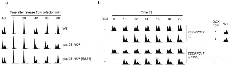
rpc128-1007 inhibition of cell cycle progression at the G1 phase can be partially overcome by RBS1 overexpression.
Flow cytometry histograms of cellular DNA content. The two prominent peaks reflect the 1C and 2C DNA content of cells in G1 and G2/M, respectively. The trough between the peaks reflects the relative number of cells in the S phase. (a) Flow cytometry histograms of wild type (WT), rpc128-1007 mutant and rpc128-1007 transformed with a multicopy RBS1 plasmid, rpc128-1007 [RBS1]. Asynchronous logarithmically growing cells (AS), after synchronization using α-factor (time point 0) and after transfer to fresh medium lacking α-factor at the indicated time points. The differences in the cell cycle are also visible in asynchronous cells. (b) Flow cytometry histograms of the TET:RPC17 mutant harboring the RPC17 gene under control of the TET-off promoter transformed with empty plasmid, TET:RPC17 [-] and RBS1 plasmid, TET:RPC17 [RBS1]. Cells were examined after addition of doxycycline (final concentration, 5μg/ml) to logarithmically growing cultures. A WT strain (BY4741) was used as a control (right panel).
Since RBS1 overexpression partially corrected defective cell cycle progression in rpc128-1007 cells, we checked whether RBS1 overexpression can overcome G1 arrest in another mutant strain that has decreased Pol III activity. Yeast strain carrying the Pol III subunit RPC17 under the control of TET-off promoter displays decreased tRNA levels and pronounced accumulation of cells in the G1 phase in the presence of doxycycline[18]. TET-off RPC17 cells transformed with plasmids carrying the RBS1 gene collected at 2 h intervals (from 10 h to 20 h) after doxycycline addition and analyzed by flow cytometry showed that RBS1 overexpression did not relieve G1 cell cycle arrest in RPC17-depleted cells (Figure 2(b)). This outcome also indicates that the effect of increased RBS1 expression on the cell cycle is specific to rpc128-1007-mediated Pol III assembly defects. This result is consistent with an earlier study showing that rpc128-1007 was the only one among several other Pol III mutants tested for which the growth phenotype could be suppressed by RBS1 overexpression[10].
Cells lacking RBS1 show a mild delay in exit from the G1 phase
To explore the potential role of Rbs1 in cell cycle progression, we next examined the yeast strain in which RBS1 gene was deleted. Since flow cytometry of asynchronously growing cells revealed no changes in the cell cycle progression in these cells (Figure S2), we instead used an α-factor-nocodazole trap assay. Using this assay, we could determine the relative amount of G1 cells that remained sensitive to α-factor and showed mating projections, as compared to large-budded post-G1 cells trapped by nocodazole at G2/M. Wild type and rbs1Δ cells were treated with α-factor for 120 min to induce G1 arrest. At 5-min and 10-min intervals after α-factor release, cells were collected, washed, and then incubated in the medium containing α-factor and nocodazole. Phase microscopy showed that the rbs1Δ cells exhibited a statistically significant mild delay in exit from the G1 phase (Figure 3(a)).
Figure 3.

Deletion of the RBS1 gene causes mild delay in exit from the G1 phase.
(a) Scheme of α-factor-nocodazole trap assay used and percentage of wild type (WT) and rbs1Δ cells remaining in G1 after release from α-factor synchronization as determined by this assay. Two cell populations, shmoo and budded, were observed by phase microscopy; cells showing shmoo were quantified. Bars represent the mean with standard deviation of three independent experiments. p values were calculated using Student’s t-test as ratios of WT to rbs1Δ cells in different time points after release from α-factor and showed statistical significance from 10 min to 65 min (p value <0.01). (b) Scheme of G1 cell synchronization in response to α-factor and percentage of cells in the G1 phase synchronized with α-factor for 90 min. Cells showing shmoo were quantified during this period. The mean with standard deviation was calculated from two independent experiments.
Next we tested whether the observed effect of the rbs1Δ strain on the cell cycle is associated with increased sensitivity to α-factor. When wild type and rbs1Δ strains were synchronized by α-factor for 90 min and cells showing shmoo projections were counted during this period (Figure 3(b)), both strains showed similar sensitivity to pheromone.
Inactivation of Maf1, general repressor of Pol III-mediated transcription, results in elevated tRNA level in rpc128-1007 cells
Since the rpc128-1007 mutation affects Pol III complex assembly and decreases tRNA levels [10,11], in addition to inhibiting cell cycle progression, we sought to understand the primary cause of the observed G1 arrest in the rpc128-1007 mutant. To discriminate between the Pol III assembly defect and inefficient tRNA synthesis, we inactivated Maf1, a general repressor of Pol III in rpc128-1007 cells. The maf1Δ strain was previously shown to have elevated tRNA levels[19]. Thus, if suppression of tRNA transcription is the primary effect of rpc128-1007, then the tRNA levels should vary in the double rpc128-1007 maf1Δ mutant, whereas the assembly defect would persist.
The growth phenotype of the double rpc128-1007 maf1Δ mutant was, as expected, cold-sensitive, with no growth seen on YPD medium at 16°C, which was similar to that seen for the rpc128-1007 single mutant, thus indicating that the Pol III assembly defect was preserved (Figure 4(a)). Moreover, the rpc128-1007 allele could compensate for the growth defect of maf1Δ on medium with a nonfermentable carbon source (YPGly) at 37°C as observed previously[11].
Figure 4.
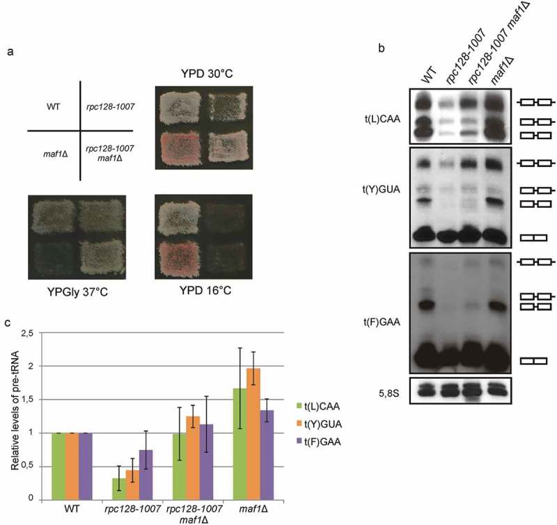
MAF1 gene deletion elevates tRNA levels in rpc128-1007 cells.
(a) Phenotype analysis of wild type (WT), rpc128-1007, maf1Δ and rpc128-1007 maf1Δ strains. Cells were incubated on YPD medium at 16°C or 30°C and on YPGly medium at 37°C. (b) Northern analysis of RNA using probes specific for tRNAs: t(L)CAA, t(Y)GUA and t(F)GAA. : tRNA precursor containing introns and unprocessed 5’, 3’-ends;
 : tRNA precursor with intron and unprocessed 3’-end;
 : end-matured intron-containing pre-tRNA;
 : mature tRNA. Cells were grown to logarithmic phase in YPD medium at 30°C and then shifted to 16°C for 2 h. (c) Amounts of primary transcripts (
 ) were normalized relative to the loading control (5,8S rRNA) and calculated relative to the amounts present in the WT strain, which were set to 1. Bars represent the mean with standard deviation of three independent experiments.
To evaluate the efficiency of tRNA synthesis in the rpc128-1007 maf1Δ double mutant, we used northern hybridization with specific oligonucleotide probes to identify precursors tRNAs: t(L)CAA, t(Y)GUA and t(F)GAA (Figure 4(b)). Cells were grown to logarithmic phase in YPD medium at 30°C and were then shifted to 16°C for 2 h. The double rpc128-1007 maf1Δ mutant had about 2-fold higher levels of newly synthesized tRNA precursors compared to the single rpc128-1007 mutant. The levels of tRNA precursors in the rpc128-1007 maf1Δ were lower than those for the maf1Δ strain and were comparable to that detected in the wild type strain (Figure 4(c)). Thus, inactivation of Maf1 in rpc128-1007 cells indeed increased tRNA levels.
Arrest of the double rpc128-1007 maf1Δ mutant in the G1 phase of cell cycle can be partially overcome by increased RBS1 expression
Next, we sought to determine whether increased tRNA transcription in the rpc128-1007 maf1Δ double mutant restored wild type cell cycle progression kinetics. Unexpectedly, flow cytometry revealed that, like rpc128-1007 cells, rpc128-1007 maf1Δ mutant cells were also arrested in G1 (Figure 5(a)). Quantification of asynchronous cells in the G1 phase confirmed that approximately 50% of rpc128-1007 maf1Δ cells were arrested in G1 as compared to about 25% in the wild type strain (Figure 5(b)). Meanwhile, quantification of cells synchronized with α-factor cells showed that 40–60 min after α-factor removal, about 50%-70% of rpc128-1007 maf1Δ cells and only 10%-30% of wild type cells remained in G1 (Figure 5(c)). We consistently observed an inhibition of the cell cycle at the G1 phase for the single rpc128-1007 mutant and the double rpc128-1007 maf1Δ mutant carrying a plasmid that expresses active MAF1, which were used as controls (Figure 5(a-c)). Plasmid-based MAF1 expression in rpc128-1007 maf1Δ cells compensated for the maf1Δ chromosomal allele, whereas cell cycle defects persisted due to the presence of rpc128-1007 (Figure 5(a), line 3, rpc128-1007 maf1Δ [MAF1]). In addition, RBS1 overdose in rpc128-1007 maf1Δ could partially overcome the G1 arrest (Figure 5(a), the last line, rpc128-1007 maf1Δ [RBS1]). Between 40 and 80 min after removal of α-factor, we observed an increase in the 2C DNA content for rpc128-1007 maf1Δ [RBS1] cells that yielded a similar pattern as that seen for the wild type strain. Quantification of G1-arrested asynchronous and synchronous rpc128-1007 maf1Δ [RBS1] cells confirmed that RBS1 overexpression was associated with a decreased number of G1 phase cells (Figure 5(b,c)).
Figure 5.

Arrest of the double rpc128-1007 maf1Δ mutant in the G1 phase can be partially overcome by increased RBS1 expression.
Wild type (WT), rpc128-1007 mutant, and double rpc128-1007 maf1Δ transformed with empty vectors [-] or multicopy [MAF1] and [RBS1] plasmids were grown to logarithmic phase in YPD medium at 30°C. (a) Flow cytometry histograms showing DNA content of asynchronous cells during logarithmic growth (AS), after synchronization by α-factor (time point 0) and after transfer to fresh medium lacking α-factor at the indicated time points. Note that differences in the cell cycle are also visible in logarithmically growing cells. (b) Number of asynchronous cells in the G1 phase and (c) after synchronization with α-factor and transfer to fresh medium at the indicated time points were calculated from three independent experiments. Bars in panel b and curves in panel c represent the mean with standard deviation.
These results indicated that MAF1 deletion in the rpc128-1007 mutant does not restore normal cell cycle progression despite the increase in tRNA transcription. Thus, we conclude that impairment of Pol III complex assembly, and not decreased tRNA transcription levels, is the primary reason for the G1 arrest observed for the rpc128-1007 mutant.
Discussion
Here we present evidence that the Pol III assembly mutant rpc128-1007 causes defects in cell cycle progression. These mutant cells showed an abnormal morphology manifested as increased cell size and numbers of unbudded cells relative to the isogenic wild type control (Figure 1(a-c)). The kinetics of cell cycle progression assessed by flow cytometry were clearly affected and the mutant cells were arrested in the G1 phase of the cell cycle (Figure 2(a)). Our results are consistent with previously observed features of yeast carrying mutations in the Pol III subunit Rpc53 that are also arrested in the G1 phase[7]. The temperature-sensitive (ts) cell cycle mutant of hamster cells arrested in G1 that also had decreased transcriptional Pol III activity could be complemented by the human homologue of yeast Rpc53 [8,20]. Moreover, cell cycle arrest was reported for some Pol II mutants. The ts mutation in the gene encoding Rpb1, the largest Pol II subunit in budding yeast, impaired cell cycle progression[21]. Similarly, mutation in the largest Pol II subunit in the fission yeast Schizosaccharomyces pombe led to defects in cell cycle progression[22]. The ts mutant in the Rpb1 subunit in a hamster cell line also arrests the cell cycle in the G1 phase[23]. Together, these results suggest that exit from the G1 phase and entry into the S/G2/M phases of the cell cycle requires active RNA polymerases. Furthermore, the detection of similar cell cycle defects in Pol II and Pol III mutants in various organisms from yeast to mammals suggests evolutionary conservation of the mechanisms that link cell cycle control and RNA transcription.
Our data indicated that overproduction of Rbs1 activated Pol III machinery and relieved the cell cycle defect in rpc128-1007 cells. As we showed previously, RBS1 is a strong suppressor of the rpc128-1007 cold-sensitive phenotype and could compensate for the low tRNA levels induced by a Rpc128 point mutation occurring at a crucial site for Pol III complex assembly[10]. Rbs1 overproduction also improves interaction between Pol III subunits in the rpc128-1007 mutant[10]. Our current study shows that RBS1 overexpression relieves proliferation defects of rpc128-1007 and resulted in fewer unbudded cells as well as a reduction in cell size compared to rpc128-1007 alone (Figure 1(a-c)). Increased RBS1 expression overcame the rpc128-1007-mediated G1 arrest, causing more rapid passage into the next cell cycle phase (Figure 2(a)). Concomitantly, cells lacking Rbs1 showed mild delay in exit from the G1 phase (Figure 3(a)). These results, together with previous findings, raise the possibility that Rbs1 plays a role in cell cycle regulation. This hypothesis is supported by literature data. A global proteomics-based study identified Rbs1 as a substrate of cyclin-dependent kinase Cdk1 (also known as Cdc28), the master regulator of cell cycle in yeast[24], which initiates the cell cycle by activating cell cycle-specific transcription factors that activate a transcriptional program during the late G1 phase[25]. Significantly Cdk1 kinase has been reported recently to gate cell cycle dependent tRNA synthesis by regulating Pol III activity. Cdk1 is recruited to tRNA genes in the late S phase and boost their transcription. By phosphorylation of TFIIIB component, Bdp1 protein, Cdk1 stimulates interaction between TFIIIB and TFIIIC and increases Pol III dynamics in vivo[6].
Cdk1 kinase activity is also involved in regulation of Pol II transcription machinery in S. cerevisiae (reviewed in [26]). Cdk1 cooperates with Kin28, another major cyclin-dependent kinase of the cell cycle, to recruit and phosphorylate the C-terminal domain of Rpb1, the largest Pol II subunit, and to promote mRNA capping[27]. Importantly, potential Cdk1 phosphorylation sites were identified in the Rbs1 sequence[28], suggesting that Cdk1-mediated phosphorylation plays a regulatory role in Rbs1 function, although further study is needed to explore this possibility.
In human cells, Pol III transcription is also regulated during the cell cycle by phosphorylation of the Pol III general transcription factor, TFIIIB (reviewed in [29]). TFIIIB is inactivated by phosphorylation during mitosis which corresponds with repressed Pol III activity [4,30,31]. In frogs, TFIIIB can be inhibited in vitro by the cyclin dependent kinase, Cdk1[3]. Polo-like kinase 1 (Plk1), a serine/threonine kinase which plays multiple roles in cell cycle progression[32], was also described to repress Pol III activity during mitosis by phosphorylation of TFIIIB[33]. Regulation of yeast Pol III during cell cycle progression possibly involves phosphorylation of the Pol III subunit Rpc53 by Mck1 kinase which is associated with inhibition of Pol III transcriptional activity[34]. Mck1 has been implicated in many cellular functions, including cell cycle regulation. Mck1 inhibits activity of the Clb2-Cdk1 complex after nuclear division[35]. In mammals, the Mck1 homolog, glycogen synthase kinase-3 (GSK-3) is also implicated in cyclin inhibition, suggesting a conserved function for both yeast and mammalian glycogen synthase kinases in the regulation of the cell cycle [36,37]. The link between Mck1, Pol III transcription and the cell cycle suggest that Mck1-mediated phosphorylation of Rpc53 may inhibit Pol III transcription in a cell cycle-dependent manner.
Here we showed that the assembly defect seen for the Pol III mutant rpc128-1007 is correlated with a delayed exit from the G1 phase (Figure 2). Cell cycle arrest was also observed in the double rpc128-1007 maf1Δ mutant in which tRNA synthesis was restored to the level similar to the wild type control (Figures 4 and 5). Moreover, increased expression of Rbs1, which functions as a Pol III assembly factor[10], overcame G1 arrest in both rpc128-1007 single and rpc128-1007maf1Δ double mutant cells (Figures 2 and 5). From these results we conclude that impaired assembly of the Pol III complex in the rpc128-1007 mutant is a primary cause of cell cycle arrest in the G1 phase. We propose a model in which assembly of the Pol III (and possibly other Pols) complex occurs at the G1/S checkpoint. Rbs1 protein, which binds and facilitates Pol III assembly, promotes exit from the cell cycle G1 phase. Our findings therefore link Pol III assembly to cell cycle control, two fundamental processes that are essential for cell proliferation.
Material and methods
Yeast strains, media and plasmids
The yeast strains used in this study were described previously [10,11]. Yeast strains were grown in YPD (2% glucose, 2% peptone, 1% yeast extract), YPGly (2% glycerol, 2% peptone, 1% yeast extract) or synthetic minimal medium SC-leu or SC-ura (2% glucose, 0.67% yeast nitrogen base supplemented with 20 µg/ml amino acids and nucleotides required for growth, except for leucine or uracil, respectively). Solid media contained 2% agar.
The following yeast/E. coli vectors were used in this study: pFL44L-MAF1 called here [MAF1], a multicopy plasmid (URA3, 2µ) containing the MAF1 gene; pFL44L-RBS1 or YEp181-RBS1, called here [RBS1], multicopy plasmids (URA3, 2µ or LEU2, 2µ, respectively) containing the RBS1 gene, and empty pFL44L or YEp181 vectors, called here [-]. Yeast cells were transformed with a plasmid DNA using the lithium acetate method as described previously[38], and were cultivated at 30°C in SC-leu or SC-ura medium for three days.
Analysis of plasmids loss in non-selective medium
Transformants, rpc128-1007 with the multicopy plasmid [RBS1] were grown in liquid non-selective medium, YPD to logarithmic or stationary phase and plated on solid YPD medium (~100 cells per plate) following incubation for two days at 30°C. Growing colonies were transferred onto fresh YPD plate, incubated one day at 30°C and replicated on selective SC-leu plate. Subsequently, SC-leu negative colonies that lost plasmids were quantified and percentage loss of plasmids was determined.
Pheromone response assay
Cells were grown in YPD untill OD600nm 1.0 was reached, then cultures were diluted to OD600nm 0.3. 100 µl of culture was spread on YPD plate and 5 mm diameter sterile discs of Whatmann paper were placed on the agar plate. 3 µl of α-factor (Sigma) solution of different concentration was dropped on the disc. Plates were incubated for two days at 30ºC and the zone of growth inhibition was measured. The experiment was repeated three times.
Cell cycle synchronization
Cells were incubated in YPD medium to OD600nm 0.5–0.7, then diluted to OD600nm 0.3 and synchronized by α-factor added to a final concentration of 5 μM following incubation at 30ºC in YPD for 120 min. Next, cells were washed two times with YPD and then transferred to fresh YPD medium to release from G1 arrest.
α-factor–nocodazole trap assay
Yeast cells were synchronized in 5 μM α-factor at 106 cells per ml for 120 min in YPD, washed with YPD, and then released from arrest to the fresh YPD medium. To detect onset and kinetics of G1 exit, at 5-min and 10-min intervals 0.5-ml samples were collected. Next, samples were combined with 0.5 ml trapping YPD medium (10 μM α-factor, 30 μg/ml nocodazole) and incubated for 90 min at 30°C, fixed, and examined by phase microscopy to count cells displaying mating projections (G1 cells) or buds (post-G1 cells).
Flow cytometry
Yeast cells attained from 1ml of culture harvested at an optical density of about 0.5 OD600nm were fixed by incubation with 70% ethanol for 60 min at room temperature (or stored several days at 4°C). Next, cells were washed two times and suspended in 1 ml of 50 mM sodium citrate, pH 7.2, then sonicated thoroughly to eliminate aggregates. The suspension of cells was incubated with RNase A (0.25 µg/ml) at 55°C for 60 min and subsequently with proteinase K (1 mg/ml) at 55°C for 60 min. Finally, cells were stained with propidium iodide (160 µg/ml) at room temperature for 60 min, sonicated again and analyzed with a Becton Dickinson FACSCalibur flow cytometer.
DIC microscopy
To analyze the morphology of yeast strains, cells were fixed in 70% (vol/vol) ethanol and examined by differential interference contrast (DIC) microscopy (Carl Zeiss Axio Imager M2, 100x oil objective).
Northern analysis
RNA isolation and northern hybridization was done as described previously [39], using 5 µg of RNA separated by electrophoresis on 10% polyacrylamide, 8 M urea gel. The following DIG-labeled oligonucleotides were used for RNA hybridization: tL(CAA) 5’-TATTCCCACAGTTAACTGCGGTCA-3’; tY(GUA) 5’-GAGAGTCGATTTCTTGC-3’; tF(GAA) 5’-GCGCTCTCCCAACTGAGCT-3’; and 5.8S rRNA, 5’-GCGTTGTTCATCGATGC-3’.
Funding Statement
This work was supported by the Foundation for Polish Science (Parent-Bridge Programme/2010-2/2 for M.C. and MISTRZ 7/2014 for M. B.).
Acknowledgments
We thank Damian Graczyk for critical reading of the manuscript.
Disclosure statement
No potential conflict of interest was reported by the authors.
Supplemental material
Supplemental data for this article can be accessed here.
References
- [1].Gottesfeld JM, Forbes DJ.. Mitotic repression of the transcriptional machinery. Trends Biochem Sci. 1997;22:197–202. [DOI] [PubMed] [Google Scholar]
- [2].Hartl P, Gottesfeld J, Forbes DJ.. Mitotic repression of transcription in vitro. J Cell Biol. 1993;120:613–624. [DOI] [PMC free article] [PubMed] [Google Scholar]
- [3].Gottesfeld JM, Wolf VJ, Dang T, et al. Mitotic repression of RNA polymerase III transcription in vitro mediated by phosphorylation of a TFIIIB component. Science. 1994;263:81–84. [DOI] [PubMed] [Google Scholar]
- [4].White RJ, Gottlieb TM, Downes CS, et al. Cell cycle regulation of RNA polymerase III transcription. Mol Cell Biol. 1995;15:6653–6662. [DOI] [PMC free article] [PubMed] [Google Scholar]
- [5].Chen M, Gartenberg MR. Coordination of tRNA transcription with export at nuclear pore complexes in budding yeast. Genes Dev. 2014;28:959–970. [DOI] [PMC free article] [PubMed] [Google Scholar]
- [6].Herrera MC, Chymkowitch P, Robertson JM, et al. Cdk1 gates cell cycle-dependent tRNA synthesis by regulating RNA polymerase III activity. Nucleic Acids Res. 2018;46:11698–11711. [DOI] [PMC free article] [PubMed] [Google Scholar]
- [7].Mann C, Micouin JY, Chiannilkulchai N, et al. RPC53 encodes a subunit of Saccharomyces cerevisiae RNA polymerase C (III) whose inactivation leads to a predominantly G1 arrest. Mol Cell Biol. 1992;12:4314–4326. [DOI] [PMC free article] [PubMed] [Google Scholar]
- [8].Ittmann M, Ali J, Greco A, et al. The gene complementing a temperature-sensitive cell cycle mutant of BHK cells is the human homologue of the yeast RPC53 gene, which encodes a subunit of RNA polymerase C (III). Cell Growth Differ. 1993;4:503–511. [PubMed] [Google Scholar]
- [9].Johnston GC, Singer RA. RNA synthesis and control of cell division in the yeast S. cerevisiae. Cell. 1978;14:951–958. [DOI] [PubMed] [Google Scholar]
- [10].Cieśla M, Makała E, Płonka M, et al. Rbs1, a new protein implicated in RNA polymerase III biogenesis in yeast Saccharomyces cerevisiae. Mol Cell Biol. 2015;35:1169–1181. [DOI] [PMC free article] [PubMed] [Google Scholar]
- [11].Cieśla M, Towpik J, Graczyk D, et al. Maf1 is involved in coupling carbon metabolism to RNA polymerase III transcription. Mol Cell Biol. 2007;27:7693–7702. [DOI] [PMC free article] [PubMed] [Google Scholar]
- [12].Gadal O, Shpakovski GV, Thuriaux P. Mutants in ABC10beta, a conserved subunit shared by all three yeast RNA polymerases, specifically affect RNA polymerase I assembly. J Biol Chem. 1999;274:8421–8427. [DOI] [PubMed] [Google Scholar]
- [13].Wild T, Cramer P. Biogenesis of multisubunit RNA polymerases. Trends Biochem Sci. 2012;37:99–105. [DOI] [PubMed] [Google Scholar]
- [14].Leśniewska E, Boguta M. Novel layers of RNA polymerase III control affecting tRNA gene transcription in eukaryotes. Open Biol. 2017;7:170001. [DOI] [PMC free article] [PubMed] [Google Scholar]
- [15].Cross FR. DAF1, a mutant gene affecting size control, pheromone arrest, and cell cycle kinetics of Saccharomyces cerevisiae. Mol Cell Biol. 1988;8:4675–4684. [DOI] [PMC free article] [PubMed] [Google Scholar]
- [16].Nash R, Tokiwa G, Anand S, et al. The WHI1+ gene of Saccharomyces cerevisiae tethers cell division to cell size and is a cyclin homolog. Embo J. 1988;7:4335–4346. [DOI] [PMC free article] [PubMed] [Google Scholar]
- [17].Wijnen H, Landman A, The FB. G(1) cyclin Cln3 promotes cell cycle entry via the transcription factor Swi6. Mol Cell Biol. 2002;22:4402–4418. [DOI] [PMC free article] [PubMed] [Google Scholar]
- [18].Gómez-Herreros F, Rodríguez-Galán O, Morillo-Huesca M, et al. Balanced production of ribosome components is required for proper G1/S transition in Saccharomyces cerevisiae. J Biol Chem. 2013;288:31689–31700. [DOI] [PMC free article] [PubMed] [Google Scholar]
- [19].Pluta K, Lefebvre O, Martin NC, et al. Maf1p, a negative effector of RNA polymerase III in Saccharomyces cerevisiae. Mol Cell Biol. 2001;21:5031–5040. [DOI] [PMC free article] [PubMed] [Google Scholar]
- [20].Ittmann M, Greco A, Basilico C. Isolation of the human gene that complements a temperature-sensitive cell cycle mutation in BHK cells. Mol Cell Biol. 1987;7:3386–3393. [DOI] [PMC free article] [PubMed] [Google Scholar]
- [21].Drebot MA, Johnston GC, Friesen JD, et al. An impaired RNA polymerase II activity in Saccharomyces cerevisiae causes cell-cycle inhibition at START. Mol Gen Genet. 1993;241:327–334. [DOI] [PubMed] [Google Scholar]
- [22].Sugaya K, Ajimura M, Tsuji H, et al. Alteration of the largest subunit of RNA polymerase II and its effect on chromosome stability in Schizosaccharomyces pombe. Mol Gen Genet. 1998;258:279–287. [DOI] [PubMed] [Google Scholar]
- [23].Sugaya K, Sasanuma S, Cook PR, et al. A mutation in the largest (catalytic) subunit of RNA polymerase II and its relation to the arrest of the cell cycle in G(1) phase. Gene. 2001;274:77–81. [DOI] [PubMed] [Google Scholar]
- [24].Ubersax JA, Woodbury EL, Quang PN, et al. Targets of the cyclin-dependent kinase Cdk1. Nature. 2003;425:859–864. [DOI] [PubMed] [Google Scholar]
- [25].Enserink JM, Kolodner RD. An overview of Cdk1-controlled targets and processes. Cell Div. 2010;5:11. [DOI] [PMC free article] [PubMed] [Google Scholar]
- [26].Chymkowitch P, Enserink JM. The cell cycle rallies the transcription cycle: cdc28/Cdk1 is a cell cycle-regulated transcriptional CDK. Transcription. 2013;4:3–6. [DOI] [PMC free article] [PubMed] [Google Scholar]
- [27].Chymkowitch P, Eldholm V, Lorenz S, et al. Cdc28 kinase activity regulates the basal transcription machinery at a subset of genes. Proc Natl Acad Sci USA. 2012;109:10450–10455. [DOI] [PMC free article] [PubMed] [Google Scholar]
- [28].Holt LJ, Tuch BB, Villén J, et al. Global analysis of Cdk1 substrate phosphorylation sites provides insights into evolution. Science. 2009;325:1682–1686. [DOI] [PMC free article] [PubMed] [Google Scholar]
- [29].Graczyk D, Cieśla M, Boguta M. Regulation of tRNA synthesis by the general transcription factors of RNA polymerase III - TFIIIB and TFIIIC, and by the MAF1 protein. Biochim Biophys Acta. 2018;1861:320–329. [DOI] [PubMed] [Google Scholar]
- [30].White RJ, Gottlieb TM, Downes CS, et al. Mitotic regulation of a TATA-binding-protein-containing complex. Mol Cell Biol. 1995;15:1983–1992. [DOI] [PMC free article] [PubMed] [Google Scholar]
- [31].Fairley JA, Scott PH, White RJ. TFIIIB is phosphorylated, disrupted and selectively released from tRNA promoters during mitosis in vivo. Embo J. 2003;22:5841–5850. [DOI] [PMC free article] [PubMed] [Google Scholar]
- [32].Barr FA, Silljé HHW, Nigg EA. Polo-like kinases and the orchestration of cell division. Nat Rev Mol Cell Biol. 2004;5:429–441. [DOI] [PubMed] [Google Scholar]
- [33].Fairley JA, Mitchell LE, Berg T, et al. Direct regulation of tRNA and 5S rRNA gene transcription by Polo-like kinase 1. Mol Cell. 2012;45:541–552. [DOI] [PubMed] [Google Scholar]
- [34].Lee J, Moir RD, McIntosh KB, et al. TOR signaling regulates ribosome and tRNA synthesis via LAMMER/Clk and GSK-3 family kinases. Mol Cell. 2012;45:836–843. [DOI] [PMC free article] [PubMed] [Google Scholar]
- [35].McQueen J, van Dyk D, Young B, et al. The Mck1 GSK-3 kinase inhibits the activity of Clb2-Cdk1 post-nuclear division. Cell Cycle. 2012;11:3421–3432. [DOI] [PMC free article] [PubMed] [Google Scholar]
- [36].Xu C, Kim N-G, Gumbiner BM. Regulation of protein stability by GSK3 mediated phosphorylation. Cell Cycle. 2009;8:4032–4039. [DOI] [PMC free article] [PubMed] [Google Scholar]
- [37].Doble BW, Woodgett JR. GSK-3: tricks of the trade for a multi-tasking kinase. J Cell Sci. 2003;116:1175–1186. [DOI] [PMC free article] [PubMed] [Google Scholar]
- [38].Chen DC, Yang BC, Kuo TT. One-step transformation of yeast in stationary phase. Curr Genet. 1992;21:83–84. [DOI] [PubMed] [Google Scholar]
- [39].Foretek D, Wu J, Hopper AK, et al. Control of Saccharomyces cerevisiae pre-tRNA processing by environmental conditions. Rna. 2016;22:339–349. [DOI] [PMC free article] [PubMed] [Google Scholar]
Associated Data
This section collects any data citations, data availability statements, or supplementary materials included in this article.