Abstract Abstract
Two new endophytic species, Colletotrichumjishouensesp. nov. and. C.tongrenensesp. nov. were isolated from Nothapodytespittosporoides in Guizhou and Hunan provinces, China. Detailed descriptions and illustrations of these new taxa are provided and morphological comparisons with similar taxa are explored. Phylogenetic analysis with combined sequence data (ITS, GAPDH, ACT and TUB2) demonstrated that both species formed distinct clades in this genus. This is the first record of Colletotrichum species from N.pittosporoides in China.
Keywords: Ascomycota , Multi-loci, Phylogeny, Morphology, Taxonomy
Introduction
Nothapodytespittosporoides (Oliv.) Sleum (Icacinacceae) has been used as Traditional Chinese Medicine (TCM) and is mainly distributed in southern China (Fang 1981). It is quickly gaining attention as the characteristic compounds of camptothecin and its derivatives (CIDs) in N.pittosporoides (Dong et al. 2015) are used as anti-cancer drugs in the world market (Demain and Vaishnav 2011). It is recognised that endophytes reside in the internal tissues of living plants and potentially have the capability to produce the same functional compounds as their hosts (Stierle et al. 1993, 1995; Kusari et al. 2008; Bhalkar et al. 2016; Uzma et al. 2018). The endophytic fungi in N.pittosporoides were therefore studied for their secondary metabolites with pharmaceutical potential.
Endophytic fungi were isolated from different parts of Nothapodytespittosporoides (Zhou et al. 2017; Qiao et al. 2018) collected from different sites. A high diversity of fungi were found, of which several species of Colletotrichum were isolated and identified.
Colletotrichum species are globally distributed and occur in various plants as endophytes (Tibpromma et al. 2018). Colletotrichum is the sole genus in the family Glomerellaceae (Glomerellales, Sordariomycetes, Wijayawardene et al. 2018) and was introduced by Corda (1831) with the type species C.lineola (Jayawardena et al. 2016, 2017, Wijayawardene et al. 2017). Recently, several studies have analysed this genus and these are summarised in Hyde et al. (2014), who accepted 163 names. Since this review, about 30 more species have been introduced (Baroncelli et al. 2017; Douanla-meli et al. 2017; Jayawardena et al. 2017; Silva et al. 2018).
In this study, we introduce two novel species, C.jishouense sp. nov. and C.tongrenense sp. nov. isolated as endophytes from N.pittosporoides. These species are based on both morphological features and molecular sequence data evidence.
Material and methods
Sample collection
Fresh healthy plant samples (leaves, stems and roots) of Nothapodytespittosporoides were collected in Tongren City, Guizhou Province and Jishou City, Hunan Province, China. Materials were kept in zip-lock bags on ice. Fungal isolation was carried out within 24 hours of collection.
Isolation and cultivation of fungal endophytes
Each part of the plant was surface sterilised to eliminate epiphytic microorganisms. The samples were washed thoroughly in running tap water, followed by immersion in 70% (v/v) ethanol for 3 min to sterilise the surfaces, then rinsed with sterilised distilled water for 1 min. Samples were dried on sterilised filter paper and then placed in 3% hydrogen peroxide for 7 min, washed in sterilised distilled water and dried on a sterilised filter paper again. Each plant tissue was then cut into small cubes (0.5 × 0.5 cm) using a sterilised blade. The cubes were placed on potato dextrose agar (PDA) medium in Petri dishes containing with antibiotic (100 mg/l chloramphenicol) and incubated at 25 °C until fungal growth emerged from the plant segments. The endophytic fungi were isolated and sub-cultured on fresh PDA plates at 25 °C in darkness. Fungal isolates were stored on PDA and covered with sterilised water at 4 °C.
The type specimens are deposited in Guizhou Agricultural College (GACP), Guiyang, China. Ex-type living cultures are deposited at Guizhou Medical University Culture Collection (GMBC). Mycobank numbers are provided.
DNA extraction, PCR amplification, and sequencing
Genomic DNA was extracted from fresh fungal mycelia using the BIOMIGA Fungus Genomic DNA Extraction Kit (GD2416, Biomiga, USA), following the manufacturer’s instructions. DNA samples were stored at -20 °C until used for polymerase chain reaction (PCR). Four loci, rDNA regions of internal transcribed spacers (ITS), partial β-tubulin (TUB2), actin (ACT) and glyceraldehyde-3-phosphate dehydrogenase (GAPDH) genes were amplified by PCR with primers ITS1 (Gardes and Bruns 1993) + ITS4 (White et al. 1990), Bt-2a + Bt-2b (Glass and Donaldson 1995), ACT-512F + ACT-783R (Carbone and Kohn 1999) and GDF1 + GDR1 (Guerber et al. 2003), respectively. The components of a 50 µl volume PCR mixture were used as follows: 2.0 µl of DNA template, 1 µl of each forward and reverse primer, 25 µl of 2 × Easy TaqPCR Super Mix (mixture of Easy Taq TM DNA Polymerase, dNTPs and optimised buffer, Beijing Trans Gen Biotech Co., Chaoyang District, Beijing, China) and 19 µl sterilised water. PCR thermal cycle programmes for ITS and ACT gene amplification were provided as: initial denaturation at 95 °C for 3 min, followed by 35 cycles of denaturation at 95 °C for 30 s, annealing at 52 °C for 50 s, elongation at 72 °C for 45 s and final extension at 72 °C for 10 min. The PCR thermal cycle programme for GAPDH gene amplification was provided as: initial denaturation at 95 °C for 3 min, followed by 35 cycles of denaturation at 94 °C for 30 s, annealing at 60 °C for 30 s, elongation at 72 °C for 45 s and final extension at 72 °C for 10 min. The PCR thermal cycle programme for TUB2 gene amplification was provided as: initial denaturation 95 °C for 3 min, followed by 35 cycles of denaturation at 94 °C for 30 s, annealing at 55 °C for 45 s, elongation at 72 °C for 45 s and final extension at 72 °C for 10 min. The quality of PCR products were checked with 1.5% agarose gel electrophoresis stained with ethidium bromide. PCR products were sent for sequencing to Sangon Co., Shanghai, China.
Sequence alignment and phylogenetic analyses
Sequence data of the four loci were blasted in the GenBank database and all top hits, including the corresponding type sequences, were retrieved (Table 1). Multiple sequence alignments for ITS, TUB2, ACT and GAPDH were constructed and carried out using the MAFFT v.7.110 online programme (http://mafft.cbrc.jp/alignment/server/, Katoh and Standley 2013) with the default settings. Four datasets of ITS, TUB2, ACT and GAPDH of Colletotrichum spp. were combined and manually adjusted using BioEdit v.7.0.5.3 (Hall 1999), then assembled using SequenceMatrix1.7.8 (Vaidya et al. 2011). The final alignments contained 1593 characters with gaps, ITS with 522 sites, TUB2 with 510 sites, ACT with 269 sites and GAPDH with 292 sites. Fifty-four taxa and 1593 sites were used for phylogenetic analyses. Gaps were treated as missing data in maximum likelihood (ML), Bayesian Inference (BI) and parsimony trees. The phylogeny website tools “ALTER” (Glez-Peña et al. 2010) were used to convert the alignment file from Fasta to PhyLip file for RAxML analysis and Nexus for MrBayes. All loci were tested based on single maximum likelihood (ML) trees and Bayesian Inference (BI) methods.
Table 1.
Taxa used for phylogenetic analyses in the study.
| Species name | Isolate No.b | GenBank Accession No. | |||
|---|---|---|---|---|---|
| ITS | GAPDH | ACT | TUB | ||
| Colletotrichum agaves | AR3920 | DQ286221 | –a | – | – |
| C. anthrisci | CBS 125334* | GU227845 | GU228237 | GU227943 | GU228139 |
| C. aracearum | LC1041 | KX853167 | KX893586 | KX893578 | KX893582 |
| C. arxii | CBS 132511 | KF687716 | KF687843 | KF687802 | KF687881 |
| C. brevisporum | BCC 38876* | JN050238 | JN050227 | JN050216 | JN050244 |
| C. chlorophyte | IMI 103806* | GU227894 | GU228286 | GU227992 | GU228188 |
| C. citricola | SXC151* | KC293576 | KC293736 | KC293616 | KC293656 |
| C. citri-maximae | AGMy0254* | KX943582 | KX943578 | KX943567 | KX943586 |
| C. cliviae | CBS 125375* | JX519223 | JX546611 | JX519240 | JX519249 |
| C. coccodes | CBS 369.75 | HM171679 | HM171673 | HM171667 | JX546873 |
| C. colombiense | CBS 129818* | JQ005174 | JQ005261 | JQ005522 | JQ005608 |
| C. conoides | CAUG17* | KP890168 | KP890162 | KP890144 | KP890174 |
| C. constrictum | CBS 128504* | JQ005238 | JQ005325 | JQ005586 | JQ005672 |
| C. cordylinicola | ICMP18579* | JX010226 | JX009975 | HM470235 | JX010440 |
| C. dematium | CBS 125.25* | GU227819 | GU228211 | GU227917 | GU228113 |
| C. dracaenophilum | CBS 118199 | JX519222 | JX546707 | JX519238 | JX519247 |
| C. euphorbiae | CPC 21823 | KF777146 | KF777131 | KF777125 | KF777247 |
| C. excelsum-altitudum | CGMCC 3.15130* | HM751815 | KC843502 | KC843548 | JX625211 |
| C. fructi | CBS 346.37* | GU227844 | GU228236 | GU227942 | GU228138 |
| C. fuscum | CBS 133701* | KM105174 | KM105524 | KM105384 | KM105454 |
| C. fusiforme | MFLU 13-0291* | KT290266 | KT290255 | KT290251 | KT290256 |
| C. gigasporum | CBS 133266 | KF687715 | KF687822 | – | KF687866 |
| C. godetiae | CBS 133.44* | JQ948402 | JQ948733 | JQ949723 | JQ950053 |
| C. grevilleae | CBS 132879* | KC297078 | KC297010 | KC296941 | KC297102 |
| C. hymenocallidicola | MFLUCC 12–0531* | KT290264 | KT290263 | – | – |
| C. jishouense | GZU_HJ2_G2 | MH482931 | MH681657 | MH708134 | MH727472 |
| C. jishouense | GZU_HJ2_G3 | MH482929 | MH681658 | MH708135 | MH727473 |
| C. jishouense | GZU_HJ2_G4 | MH482932 | MH681659 | MH708136 | MH727474 |
| C. jishouense | GZU_HJ3_J5 | MH482930 | MH492706 | MH708137 | – |
| C. kahawae | C1266.1 | JX010231 | JX010012 | JX009452 | JX010444 |
| C. ledebouriae | CPC 25671* | KX228254 | – | KX228357 | – |
| C. liaoningense | CAUOS2* | KP890104 | KP890135 | KP890097 | KP890111 |
| C. lindemuthianum | CBS 144.31* | JQ005779 | JX546712 | JQ005842 | JQ005863 |
| C. magnisporum | CBS 398.84 | KF687718 | KF687842 | KF687803 | KF687882 |
| C. malvarum | CBS 521.97* | KF178480 | KF178504 | KF178577 | KF178601 |
| C. neosansevieriae | CPC 25127* | KR476747 | KR476791 | KR476790 | KR476797 |
| C. nymphaeae | CBS 515.78 | JQ948197 | JQ948527 | JQ949518 | JQ949848 |
| C. orchidophilum | CBS 632.80* | JQ948151 | JQ948481 | JQ949472 | JQ949802 |
| C. pisicola | CBS 724.97* | KM105172 | KM105522 | KM105382 | KM105452 |
| C. pseudoacutatum | CBS 436.77* | JQ948480 | JQ948811 | JQ949801 | JQ950131 |
| C. pseudomajus | CBS 571.88 | KF687722 | KF687826 | KF687801 | KF687883 |
| C. radices | CBS 529.93 | KF687719 | KF687825 | KF687785 | KF687869 |
| C. rhombiforme | CBS 129953* | JQ948457 | JQ948788 | JQ949778 | JQ950108 |
| C. sansevieriae | MAFF 239721* | NR_152313 | – | – | – |
| C. spinosum | CBS 515.97* | KF178474 | KF178498 | KF178571 | KF178595 |
| C. tanaceti | CBS 132693* | JX218228 | JX218243 | JX218238 | JX218233 |
| C. trichellum | CBS 217.64* | GU227812 | GU228204 | GU227910 | GU228106 |
| C. tongrenense | GZU_TRJ1-37 | MH482933 | MH705332 | MH717074 | MH729805 |
| C. tropicicola | L58 | JN050240 | JN050229 | JN050218 | JN050246 |
| C. truncatum | CBS 151.35 | GU227862 | GU228254 | GU227960 | GU228156 |
| C. vietnamense | CBS 125478 | KF687721 | KF687832 | KF687792 | KF687877 |
| C. yunnanense | CBS 132135* | JX546804 | JX546706 | – | JX519248 |
| Monilochaetes infuscans | CBS 869.96 | JQ005780 | JX546612 | JQ005843 | JQ005864 |
Notes: New strains are in bold. * ex-type strains. a No data in GenBank. bBCC: BIOTEC Culture Collection, National Center for Genetic Engineering and Biotechnology (BIOTEC), Khlong Luang, Pathumthani, Thailand; CBS: Culture collection of the Centraalbureau voor Schimmelcultures, Fungal Biodiversity Centre, Utrecht, The Netherlands; CGMCC: China General Microbiological Culture Collection; CPC: Working collection of Pedro W. Crous, housed at CBS; IMI: Culture collection of CABI Europe UK Centre, Egham, UK; LC: Working collection of Lei Cai, housed at CAS, China; MAFF: MAFF GenBank Project, Ministry of Agriculture, Forestry and Fisheries, Tsukuba, Japan; MFLUCC: Mae Fah Luang University Culture Collection, Chiang Rai, Thailand; MFLU: Herbarium of Mae Fah Luang University, Chiang Rai, Thailand; ICMP: International Collection of Microorganisms from Plants, Auckland, New Zealand.
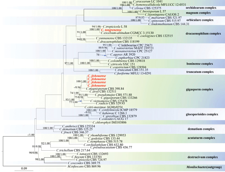
Maximum Likelihood (ML) analysis was performed on the website of CIPRES Science Gateway v.3.3 (http://www.phylo.org/portal2/, Miller et al. 2010) using RAxML-HPC Blackbox version 8.2.10. All free model parameters were estimated by RAxML and ML estimate of 25 per site rate categories. Final ML searches were conducted using the GTRGAMMA model. Bootstrap Support values (BS) equal to or greater than 60% are given above each node (Fig. 1).
Figure 1.
Phylogram generated from Maximum Likelihood (RAxML) analysis based on combined ITS, ACT, TUB2 and GAPDH DNA sequence data of Colletotrichum. Bayesian Posterior Probabilities (BSPP) greater than 0.90 and Maximum Likelihood Bootstrap Support values (MLBS) greater than 70% are shown above branches. New isolates are in red. The tree is rooted with MonilochaetesinfuscansCBS 869.96.
For Bayesian Inference (BI), a Markov Chain Monte Carlo (MCMC) algorithm was used to generate phylogenetic trees with Bayesian probabilities using MrBayes 3.2.6 (Ronquist et al. 2012) for the combined sequence datasets. MrModeltest v.2.3 (Nylander 2004) was used to carry out the statistical selection of the best-fit model of nucleotide substitution. GTR+G model was selected for ITS, a GTR+I+G model for TUB2, a HKY+I+G model for ACT and GAPDH were incorporated into the analysis. Models of nucleotide substitution for each gene determined by MrModeltest v. 2.3 were included for each set of gene sequence data. Two runs were executed simultaneously for 1,000,000 generations and sampled every 100 generations. Of the trees, 25% were discarded as burn-in and the remaining trees were used to calculate the posterior probabilities. Convergence was assumed when the standard deviation of split sequences was less than 0.01. Phylogenetic trees were visualised using FigTree v1.4.0 (http://tree.bio.ed.ac.uk/software/figtree/, Rambaut 2012). The final alignment was deposited in Treebase (http://www.treebase.org, submission number 23622).
Morphological analysis
Isolates were grown on PDA, water agar (WA) with bamboo and corn malt agar medium (CMA) for examination of morphological characters. Colonies were examined after 7, 14 and 21 d at 25 °C in darkness. The morphological characters of mycelia, conidiophores, conidiogenous cells and conidia were observed and photographed using a Nikon NI-SS microscope and processed with Adobe Photoshop CS3 Extended version 10.0 software (Adobe Systems, USA).
Results
Sample collection and isolation
Four hundred and forty endophytic fungi were isolated from different parts of Nothapodytespittosporoides in Jishou, Hunan Province and Tongren, Guizhou Province, belonging to twenty-four genera based on ITS sequences analysis. Colletotrichum was a common genus amongst the isolates. Herein, five endophytic taxa were isolated and identified as Colletotrichum of which GZU_HJ2_G2, GZU_HJ2_G3 and GZU_HJ2_G4 were isolated from roots and GZU_HJ3_J5 from stems of N.pittosporoides in Jishou, Hunan Province. GZU_TRJ1-37 was isolated from stems of N.pittosporoides in Tongren, Guizhou Province.
Phylogenetic analyses
Phylogenetic analysis of four loci (ITS, GAPDH, ACT and TUB2) sequence datasets included 54 taxa, 1,593 positions including gaps (ITS: 1–522, TUB2: 523–1032, ACT: 1033–1301, GAPDH: 1302–1593) and Monilochaetesinfuscans (CBS 869.96) was selected as the outgroup taxon. The 50% majority rule consensus Bayesian phylogram presented in Fig. 1 and the topology is recovered with the RAxML tree. Values of the Bayesian PP ≥ 0.70 from MCMC analyses and bootstrap support values of RAxML ≥ 90% are given on the branches.
Representatives of complexes and species in Colletotrichum (Noireung et al. 2012; Tao et al. 2013; Liu et al. 2014; Jayawardena et al. 2016; Douanla-meli et al. 2017) are included in the phylogenetic analyses (Fig. 1). Four isolates, GZU_HJ2_G2, GZU_HJ2_G3, GZU_HJ2_G4 and GZU_HJ3_J5, were identified as distinct new species and are described as Colletotrichumjishouense sp. nov., and as C.tongrenense sp. nov., based on their morphology and molecular phylogeny.
Taxonomy
Colletotrichum jishouense
SX. Zhou, JC. Kang & K.D. Hyde sp. nov.
828723
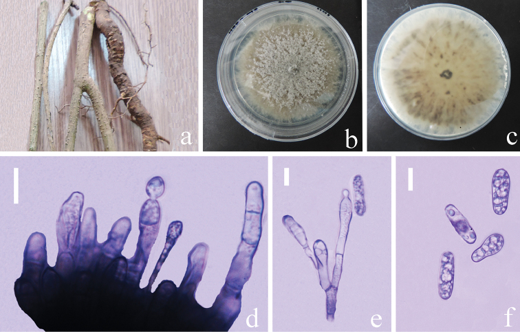
Figure 2.
Colletotrichumjishouense (GACP GZU_HJ2_G3, holotype) a stems and roots of Nothapodytespittosporoidesb,c colonies on PDAd conidiophores in cotton blue e conidiophores with conidia in cotton blue f conidia in cotton blue. Scale bars: 10 µm (d), 5 µm (e, f).
Etymology.
‘Jishouense’ referring to Jishou City, site of collection of type species.
Description.
Endophytic fungus in root of Nothapodytespittosporoides. Sexual morph: Undetermined. Asexual morph: Vegetative hyphae 0.5–1.2 µm diam. (n=10), hyaline, smooth-walled, septate, branched. Chlamydospores not observed. Conidiophores formed on a basal cushion, hyaline to pale brown, clavate or cylindrical, septate and irregularly branched. Conidiogenous cells 4–11 × 2–3 μm (x‒= 6.7 ± 3.0 × 2.6 ± 0.4 μm, n=20), L/W ratio= 2.5, hyaline, smooth-walled, clavate to mostly ampulliform or cylindrical. Conidia hyaline, smooth-walled, aseptate, straight, cylindrical, some clavate, the apex and base rounded, 5–14 × 3–5 μm (x‒ = 10.8 ± 1.8 × 3.7 ± 0.5 μm, n = 40), L/W ratio= 2.9. Appressoria not observed.
Culture characteristics.
Colonies on PDA, reaching 55–60 mm diam. in 14 days at 25 °C in darkness, circular, mycelium superficial and partially immersed, more or less planar, brown in the medium but covered with abundant, pale and lanose to cottony aerial mycelium, reverse greenish pale brown, margin entire and irregular.
Material examined.
CHINA, Hunan Province, Jishou City (28°55'24"N, 109°10'24"E), isolated from healthy roots of Nothapodytespittosporoides, 27 May 2016, S.X. Zhou (Holotype GACP GZU_HJ2_G3 dried culture), ex-type living culture, GMBC0209, living culture, GZU_HJ2_G2, living culture, GZU_HJ2_G4.
China, Hunan Province, Jishou City (28°55'24"N, 109°10'24"E), isolated from healthy stem of Nothapodytespittosporoides, 27 May 2016, S.X. Zhou, living culture, GZU_HJ3_J5.
Notes.
Colletotrichumjishouense belongs in the gigasporum species complex. C.jishouense has shorter and narrower conidiogenous cells and conidia than all the related species in the C.gigasporum complex (Liu et al. 2014). Phylogenetically, our four new isolates clustered together with C.magnisporum (CBS 398.84). The pairwise dissimilarities of DNA sequences between C.jishouense and C.magnisporum were 2 bp, 20 bp, 5 bp and 9 bp in ITS, TUB2, ACT and GAPDH, respectively. They are phylogenetically distinct species and, therefore, C.jishouense sp. nov. is introduced.
Colletotrichum tongrenense
S.X. Zhou, J.C. Kang & K.D. Hyde sp. nov.
828725
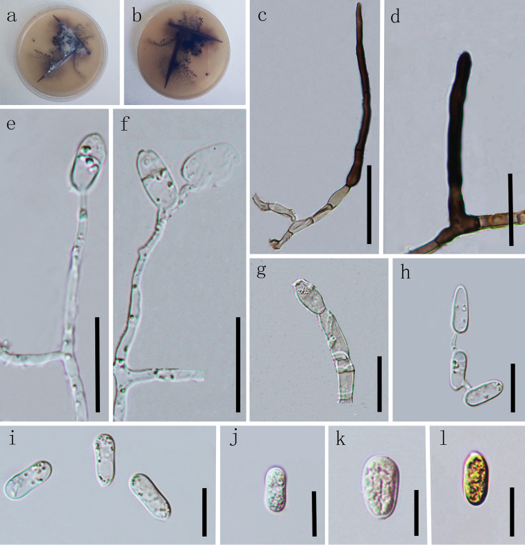
Figure 3.
Colletotrichumtongrenense (GACP GZU_TRJ1-37, holotype) a, b colonies on WAc–g Conidiophores h–l Conidia. Scale bars: 40 µm (c), 20 µm (d, g), 10 µm (e, f), 10 µm (h–l).
Etymology.
‘tongrenense’ referring Tongren City, site of collection of type species.
Description.
Endophytic in leaves and stems of Nothapodytespittosporoides. Sexual morph: Undetermined. Asexual morph: On WA, vegetative hyphae 1.4–6 µm diam. (n=10), smooth-walled, septate, branched, hyaline. Chlamydospores not observed. Setae unbranched, septate, tapering to rounded at apical end, pale brown to dark brown, smooth-walled, 45–90 µm long, 5.9–6.2 μm wide at widest part, 2.6–5.8 µm wide at bottom, 1.5–1.6 µm wide at apex. Conidiophores pale brown, septate, branched. Conidiogenous cells pale, hyaline, smooth-walled, erect, clavate or cylindrical, 2–11 × 1–2 μm (x‒ = 6.3 ± 4.4 × 1.7 ± 0.4 μm, n = 20), L/W ratio= 3.7. Conidia hyaline, aseptate, smooth-walled, variable in size and shape, thick-walled, ellipsoidal to subglobose, the apex and base rounded, slightly constricted in the middle, 11–14 × 5–7 μm (x‒ = 13.1 ± 1.0 × 5.5 ± 0.6 μm, n = 40), L/W ratio= 2.4.
Culture characteristics.
Cultures on WA at 25 °C in darkness, reaching 15–18 mm diam. in 21 days, white to grey, asymmetrical surface, reverse dark grey to black.
Colonies on PDA at 25 °C reaching 45–55 mm diam. in 12 days in darkness, circular, more or less planar, surface dark brown, covered with abundant, pale grey, lanose to cottony aerial mycelium, margin smooth, entire and pale white. Reverse dark grey, margin pale white.
Cultures on CMA, 10–15 mm diam. in 21 days, covered with dark brown aerial mycelium, sparse, reverse light brown, margin irregular.
Material examined.
CHINA, Guizhou province, Tongren (27°35'37"N, 109°10'58"E, elevation 332.8 m), isolated from healthy stems of Nothapodytespittosporoides, 27 May 2016, S.X. Zhou and L.J. Qiao (Holotype GACP GZU-TRJ1-37 dried culture), ex-type living culture, GMBC0209.
Notes.
Colletotrichumtongrenense belongs to the C.dracaenophilum species complex (Damm et al. 2019). Morphologically, C.tongrenense resembles C.tropicicola and C.excelsum-altitudum in conidia characters, but it can be distinguished from C.tropicicola in having setae and longer conidia (15–19 µm vs 11–14 µm) (Noireung et al. 2012). C.tongrenense is distinguished from C.excelsum-altitudum (Tao et al. 2013) in having smaller conidiophores (2–11 × 1–2 μm vs 8.5–25 × 4–5 μm). Phylogenetically, the new isolate GZU_TRJ1-37 clusters together with C.tropicicola with good bootstrap support (94% MLBS, 1.00 PP) (Fig. 1) and the phylogenetic analysis supports it as a distinct species. There are 6, 4, 2 and 5 base pairs differences in ITS, TUB2, ACT and GAPDH gene regions, respectively, between the new isolate and the type strain of C.tropicicola, which confirms that they are separate species. Therefore, it is introduced as a novel species.
Discussion
Colletotrichum appears to have a wide host range and a geographic distribution (Yang et al. 2009, Hyde et al. 2014, Jayawardena et al. 2016). This study reports on five endophytic Colletotrichum isolates which were isolated from Nothapodytespittosporoides. Two new species were introduced, named C.jishouense and C.tongrenense, respectively, based on morphological characters and multilocus (ITS, TUB2, ACT and GAPDH) phylogenetic analyses. The C.gigasporum species complex is associated with various host plants as pathogens and endophytes and also isolated from air and stored grain, indicating that the members are not host-specific and apparently have different life styles (Than et al. 2008, Yang et al. 2009, Liu et al. 2014, Jayawardena et al. 2016). The C.dracaenophilum species complex contains a few apparently host-specific species and these species seem to be uncommon (Damm et al. 2019). The complex includes C.coelogynes, C.dracaenophilum, C.excelsum-altitudinum, C.tropicicola and C.yunnanense. A further strain, C.tongrenense was identified to the C.dracaenophilum species complex in the study, based on the multilocus phylogeny and morphological features. Amongst them, C.excelsum-altitudinum was described from healthy leaves of Bletillaochracea (Orchidaceae) in Guizhou, China (Tao et al. 2013.), C.tropicicola were described from leaves of Citrusmaxima and Paphiopedilum sp. in Thailand and a further strain from C. sp. in Mexico (Noireung et al. 2012, Damm et al. 2019). The C.coelogynes strain CBS 132504 is an endophytic Colletotrichum isolate from both Dendrobium spp. in China (Yuan et al. 2009, Gao and Guo, unpublished data). C.yunnanense was described from healthy leaves of Buxus sp. in Yunnan, China (Liu et al. 2007).
Morphological features and genes sequence data are recognised as a basis for describing new species, but sometimes morphological features of Colletotrichum are not stable and may change under different growth conditions (Liu et al. 2014). DNA sequence comparison and multi-gene phylogenetic analyses can provide sufficient evidence to show distinct taxa (Jeewon and Hyde 2016). However, single gene data, including ITS, are usually insufficient for species identification in most of the Colletotrichum species complexes (Hyde et al. 2009). Multi-locus phylogenies are therefore necessary to describe Colletotrichum species (Jayawardena et al. 2016).
The composition of endophytic microorganisms may depend on the plant age, tissue, host type and time of isolation (Rosenblueth and Martinez-Romero 2006). The new species, Colletotrichumtongrenense lives in stems and C.jishouense lives in roots and stems of Nothapodytespittosporoides. Nothing is known about their infection strategies on the host. It is also the first report of Colletotrichum species from N.pittosporoides. This study enriches the host diversity of Colletotrichum.
Supplementary Material
Acknowledgements
This work was funded by grants of the National Natural Science Foundation of China (NSFC Grants nos. 31670027 &31460011 & 30870009). Sixuan Zhou thanks Dr. Shaun Pennycook, Prof. Jiangming Lv, Yongzhong Lu and Jianfei Gao for their help.
Citation
Zhou S, Qiao L, Jayawardena RS, Hyde KD, Ma X, Wen T, Kang J (2019) Two new endophytic Colletotrichum species from Nothapodytes pittosporoides in China. MycoKeys 49: 1–14. https://doi.org/10.3897/mycokeys.49.31904
References
- Bhalkar BN, Patil SM, Govindwar SP. (2016) Camptothecine production by mixed fermentation of two endophytic fungi from Nothapodytesnimmoniana Fungal Biology120: 873–883. 10.1016/j.funbio.2016.04.003 [DOI] [PubMed]
- Baroncelli R, Talhinhas P, Pensec F, Sukno SA, Floch GL, Thon MR. (2017) The Colletotrichumacutatum species complex as a model system to study evolution and host specialization in plant pathogens. Frontiers in Microbiology 8: 2001. 10.3389/fmicb.2017.02001 [DOI] [PMC free article] [PubMed]
- Carbone I, Kohn LM. (1999) A method for designing primer sets for speciation studies in filamentous ascomycetes. Mycologia 91: 553–556. 10.2307/3761358 [DOI] [Google Scholar]
- Corda ACI. (1831) Die Pilze Deutschlands. In: Sturm J. (Ed.) Deutschlands Flora in Abbildungen nach der Natur mit Beschreibungen.Sturm, Nürnberg 3 (12), 33–64.
- Damm U, Sato T, Alizadeh A, Groenewald JZ, Crous PW. (2019) The Colletotrichumdracaenophilum, C.magnum and C.orchidearum species complexes. Studies in Mycology 92(5): 1–46. 10.1016/j.simyco.2018.04.001 [DOI] [PMC free article] [PubMed] [Google Scholar]
- Demain AL, Vaishnav P. (2011) Natural products for cancer chemotherapy. Microbial Biotechnology 4(6): 687–699. 10.1111/j.1751-7915.2010.00221.x [DOI] [PMC free article] [PubMed] [Google Scholar]
- Douanla-meli C, Unger JG. (2017) Phylogenetic study of the Colletotrichum species on imported citrus fruits uncovers a low diversity and a new species in the Colletotrichumgigasporum complex. Fungal Biology 121(10): 858–868. 10.1016/j.funbio.2017.06.003 [DOI] [PubMed] [Google Scholar]
- Fang WP. (1981) Flora Republicae. Popularis Sinicae 46. Science Press, Beijing, Tomus, 49 pp. [Google Scholar]
- Gardes M, Bruns TD. (1993) ITS primers with enhanced specificity for basidiomycetes – application to the identification of mycorrhizae and rusts. Molecular Ecology 2: 113–118. 10.1111/j.1365-294X.1993.tb00005.x [DOI] [PubMed] [Google Scholar]
- Glass NL, Donaldson GC. (1995) Development of primer sets designed for use with the PCR to amplify conserved genes from filamentous ascomycetes. Applied Environmental Microbiology 61: 1323–1330. [DOI] [PMC free article] [PubMed] [Google Scholar]
- Glezpeña D, Gómezblanco D, Reboirojato M, Fdezriverola F, Posada D. (2010) ALTER: program-oriented conversion of DNA and protein alignments. Nucleic Acids Research 38: 14–18. 10.1093/nar/gkq321 [DOI] [PMC free article] [PubMed] [Google Scholar]
- Guerber JC, Liu B, Correll JC, Johnston PR. (2003) Characterization of diversity in Colletotrichumacutatum sensu lato by sequence analysis of two gene introns, mtDNA and intron RFLPs, and mating compatibility. Mycologia 95(5): 872–895. 10.1080/15572536.2004.11833047 [DOI] [PubMed] [Google Scholar]
- Guo DY, Ling TJ, Cai XH. (2015) Chemical constituents of Nothapodytespittosporoides (Icacinaceae). Biochemical Systematics and Ecology 61: 293–296. 10.1016/j.bse.2015.06.039 [DOI] [Google Scholar]
- Hall TA. (1999) BioEdit: a user-friendly biological sequence alignment editor and analysis program for Windows 95/98/NT. Nucleic Acids Symposium Series 41: 95–98. [Google Scholar]
- Hyde KD, Cai L, Cannon PF, Crouch JA, Crous PW, Damm U, Goodwin PH, Chen H, Johnston PR, Jones EBG, Liu ZY, McKenzie EHC, Moriwaki J, Noireung P, Pennycook SR, Pfenning LH, Prihastuti H, Sato T, Shivas RG, Tan YP, Taylor PWJ, Weir BS, Yang YL, Zhang JZ. (2009) Colletotrichum names in current use. Fungal Diversity 39: 147–182.
- Hyde KD, Nilsson RH, Alias SA, Ariyawansa HA, Blair JE, Cai L, de Cock AWAM, Dissanayake AJ, Glockling SL, Goonasekara ID, Gorczak M, Hahn M, Jayawardena RS, van Kan JAL, Laurence MH, Lévesque CA, Li XH, Liu JK, Maharachchikumbura SSN, Manamgoda DS, Martin FN, McKenzie EHC, McTaggart AR, Mortimer PE, Nair PVR, Pawłowska J, Rintoul TL, Shivas RG, Spies CFJ, Summerell BA, Taylor PWJ, Terhem RB, Udayanga D, Vaghefi N, Walther G, Wilk M, Wrzosek M, Xu JC, Yan JY, Zhou N. (2014) One stop shop: backbones trees for important phytopathogenic genera: I. Fungal Diversity 67: 21–125. 10.1007/s13225-014-0298-1 [DOI] [Google Scholar]
- Index Fungorum (2017) http://www.indexfungorum.org/names/Names.asp
- Jayawardena RS, Camporesi E, Elgorban AM, Bahkali AH, Yan J, Hyde KD. (2017) A new species of Colletotrichum from Sonchus sp. in Italy. Phytotaxa 314(1): 55–63. 10.11646/phytotaxa.314.1.3 [DOI] [Google Scholar]
- Jayawardena RS, Hyde KD, Damm U, Cai L, Liu M, Li XH, Zhang W, Zhao WS, Yan JY. (2016) Notes on currently accepted species of Colletotrichum. Mycosphere 7: 1192–1260. 10.5943/mycosphere/si/2c/9 [DOI] [Google Scholar]
- Jeewon R, Hyde KD. (2016) Establishing species boundaries and new taxa among fungi: recommendations to resolve taxonomic ambiguities. Mycosphere 7(11): 1669–1677. 10.5943/mycosphere/7/11/4 [DOI] [Google Scholar]
- Kusari S, Lamshöft M, Zühlke S, Spiteller M. (2008) An endophytic fungus from Hypericumperforatum that produces hypericin. Journal of Natural Products 71: 159–16. 10.1021/np070669k [DOI] [PubMed] [Google Scholar]
- Katoh K, Standley DM. (2013) MAFFT multiple sequence alignment software version 7: improvements in performance and usability. Molecular Biology and Evolution 30(4): 772–780. 10.1093/molbev/mst010 [DOI] [PMC free article] [PubMed] [Google Scholar]
- Liu F, Cai L, Crous PW, Damm U. (2014) The Colletotrichumgigasporum species complex. Persoonia 33: 83–97. 10.3767/003158514X684447 [DOI] [PMC free article] [PubMed] [Google Scholar]
- Liu X, Xie X, Duan J. (2007) Colletotrichumyunnanense sp. nov., a new endophytic species from Buxus sp. Mycotaxon 100: 137–144. [Google Scholar]
- Miller MA, Pfeiffer W, Schwartz T. (2010) Creating the CIPRES Science Gateway for inference of large phylogenetic trees. 2010 Gateway Computing Environments Workshop (GCE), IEEE, 2010. 10.1109/GCE.2010.5676129 [DOI]
- Noireung P, Phoulivong S, Liu F, Cai L, McKenzie EHC, Chukeatirote E, Jones EBG, Bahkali AH, Hyde KD. (2012) Novel species of Colletotrichum revealed by morphology and molecular analysis. Cryptogamie, Mycologie, 33(3): 347–362. 10.7872/crym.v33.iss3.2012.347 [DOI] [Google Scholar]
- Nylander JAA. (2004) MrModeltest v2. Program distributed by the author. Evolutionary Biology Centre, Uppsala University, Uppsala.
- Qiao LJ, Zhou SX, Wen TC, Kang JC, Lei BX. (2018) Diversity of endophytic fungi from Nothapodytespittosporoides in Guizhou Province. Mycosystema 37(1): 43–51. [Google Scholar]
- Rambaut A. (2012) FigTree version 1.4. http://tree.bio.ed.ac.uk/software/figtree
- Ronquist F, Teslenko M, van der Mark P, Ayres DL, Darling A, Höhna S, Larget B, Liu L, Suchard MA, Huelsenbeck JP. (2012) MrBayes3.2: efficient Bayesian phylogenetic inference and model choice across a large model space. Systematic Biology 61(3): 539–542. 10.1093/sysbio/sys029 [DOI] [PMC free article] [PubMed] [Google Scholar]
- Rosenblueth M, Martinez-Romero E. (2006) Bacterial endophytes and their interactions with hosts. Acta Pharmacologica Sinica 19: 827–837. 10.1094/MPMI-19-0827 [DOI] [PubMed] [Google Scholar]
- Silva MD, Cruz ES, Veloso TGR, Miranda L, Pereiram OL, Bocayuva MF, Kasuya MCM. (2018) Colletorichumserranegrense sp. nov., a new endophytic species from the roots of the endangered Brazilian epiphytic orchid Cattleyajongheana. Phytotaxa 351(2): 163–170. 10.11646/phytotaxa.351.2.4 [DOI] [Google Scholar]
- Stamatakis A. (2014) RAxML version 8: a tool for phylogenetic analysis and post-analysis of large phylogenies. Bioinformatics 30(9): 1312–1313. 10.1093/bioinformatics/btu033 [DOI] [PMC free article] [PubMed] [Google Scholar]
- Stierle A, Strobel G, Stierle D. (1993) Taxol and taxane production by Taxomycesandreanae, an endophytic fungus of Pacific yew. Science 260: 214–216. 10.1126/science.8097061 [DOI] [PubMed] [Google Scholar]
- Stierle A, Strobel G, Stierle D, Grothaus P, Bignami G. (1995) The search forataxol-producing microorganism among the endophytic fungi of the Pacific yew, Taxusbrevifolia. Journal of Nature Products 58: 1315–1324. 10.1021/np50123a002 [DOI] [PubMed] [Google Scholar]
- Tao G, Liu ZY, Liu F, Gao YH, Cai L. (2013) Endophytic Colletotrichum species from Bletillaochracea (Orchidaceae), with descriptions of seven new species. Fungal Diversity 61(1): 139–164. 10.1007/s13225-013-0254-5 [DOI] [Google Scholar]
- Than PP, Prihastuti H, Phoulivong S, Taylor PWJ, Hyde KD. (2008) Chillianthracnose disease caused by Colletotrichum species. Journal of Zhejiang University-Science B 9: 764–788. 10.1631/jzus.B0860007 [DOI] [PMC free article] [PubMed] [Google Scholar]
- Tibpromma S, Hyde KD, Bhat JD, Mortimer PE, Xu JC, Promputtha I, Doilom M, Yang JB, Tang AMC, Karunarathna SC. (2018) Identification of endophytic fungi from leaves of Pandanaceae based on their morphotypes and DNA sequence data from southern Thailand. Mycokeys 33: 25–67. 10.3897/mycokeys.33.23670 [DOI] [PMC free article] [PubMed] [Google Scholar]
- Uzma F, Mohan CD, Hashem A, Konappa NM, Rangappa S, Kamath PV, Singh BP, Mudili V, Gupta VK, Siddaiah CN. (2018) Endophytic fungi-alternative sources of cytotoxic compounds: a review. Frontiers in Pharmacology, 9: 309. 10.3389/fphar.2018.00309 [DOI] [PMC free article] [PubMed]
- Vaidya G, Lohman DJ, Meier R. (2011) SequenceMatrix: concatenation software for the fast assembly of multi-gene datasets with character set and codon information. Cladistics 27: 171–180. 10.1111/j.1096-0031.2010.00329.x [DOI] [PubMed] [Google Scholar]
- White TJ, Bruns T, Lee S, Taylor JW. (1990) Amplification and direct sequencing of fungal ribosomal RNA genes for phylogenetics. PCR protocols: a guide to methods and applications. Academic Press, San Diego,California 18: 315–322. 10.1016/B978-0-12-372180-8.50042-1 [DOI] [Google Scholar]
- Wijayawardene NN, Hyde KD, Lumbsch T, Liu JK, Maharachchikumbura SSN, Ekanayaka AH, Tian Q, Phookamsak R. (2018) Outline of Ascomycota – 2017. Fungal Diversity 88: 167–263. 10.1007/s13225-018-0394-8 [DOI] [Google Scholar]
- Wijayawardene NN, Hyde KD, Rajeshkumar KC, Hawksworth DL, Madrid H, Kirk PM, Braun U. (2017) Notes for genera: ascomycota. Fungal Diversity 86(1): 1–594. 10.1007/s13225-017-0386-0 [DOI] [Google Scholar]
- Wijayawardene DNN, Song Y, Bhat DJ, McKenzie EHC, Chukeatirote E, Wang Y, Hyde KD. (2013) Wojnowiciaviburni sp. nov. from China and its phylogenetic placement. Sydowia 65: 181–190. [Google Scholar]
- Yang YL, Liu ZY, Cai L, Hyde KD, Yu ZN, McKenzie EHC. (2009) Colletotrichumanthracnose of Amaryllidaceae. Fungal Diversity 39: 123–146. [Google Scholar]
- Yuan ZL, Chen YC, Yang Y. (2009) Diverse non-mycorrhizal fungal endophytes inhabiting an epiphytic, medicinal orchid (Dendrobiumnobile): estimation and characterization. World Journal of Microbiology & Biotechnology 25(2): 295–303. hhttps://doi.org/ 10.1007/s11274-008-9893-1 [DOI]
- Zhou SX, Qiao LJ, Kang JC, Hyde KD, Ma XY. (2017) A new species of Monilochaetes from Nothapodytespittosporoides. Phytotaxa 326(2): 129–136. 10.11646/phytotaxa.326.2.4 [DOI] [Google Scholar]
Associated Data
This section collects any data citations, data availability statements, or supplementary materials included in this article.


