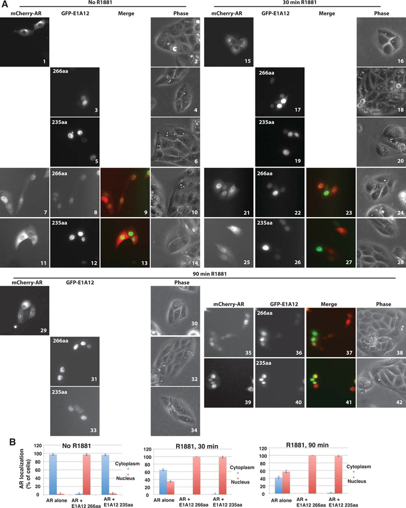
Figure 3. E1A12 promotes AR nuclear entry independently of androgens.

(A) Saos-2 cells were transfected with GFP-E1A12 266aa, 235aa, or mCherry-AR alone, or a combination thereof. At 8 h after transfection, the culture medium was removed and cells were washed with PBS. The DMEM medium supplemented with 10% CSS was then added to the culture plates. Cells were imaged at 24 h after transfection. Immediately after the initial imaging, the androgen analog R1881 was added to the cell cultures to the final concentration of 1 nM. The cells were then imaged at the indicated times after R1881 addition. Representative images are shown. (B) Quantification of AR subcellular localization. Cells exhibiting exclusive or predominant nuclear AR signal are grouped as cells with nuclear AR; likewise, cells with exclusive or predominant cytoplasmic AR signal are shown as cells with AR in the cytoplasm. For each transfection experiment, over 100 transfected cells in five random microscopic fields were quantified. The average percent values of the five fields along with SEM are shown.
