Abstract
The developing cardiovascular system of zebrafish is a sensitive target for many environmental pollutants, including dioxin-like compounds and pesticides. Some polychlorinated biphenyls (PCBs) can compromise the cardiovascular endothelial function by activating oxidative stress-sensitive signaling pathways. Therefore, we exposed zebrafish embryos to PCB126 or to several redox-modulating chemicals to study their ability to modulate the dysmorphogenesis produced by PCB126. PCB126 produced a concentration-dependent induction of pericardial edema and circulatory failure, and a concentration-dependent reduction of cardiac output and body length at 80 hours post fertilization (hpf). Among several modulators tested, the effects of PCB126 could be both positively and negatively modulated by different compounds; co-treatment with α-tocopherol (vitamin E liposoluble) prevented the adverse effects of PCB126 in pericardial edema, whereas co-treatment with sodium nitroprusside (a vasodilator compound) significantly worsened PCB126 effects. Gene expression analysis showed an up-regulation of cyp1a, hsp70, and gstp1, indicative of PCB126 interaction with the aryl hydrocarbon receptor (AhR), while the transcription of antioxidant genes (sod1, sod2; cat and gpx1a) was not affected. Further studies are necessary to understand the role of oxidative stress in the developmental toxicity of low concentrations of PCB126 (25 nM). Our results give insights into the use of zebrafish embryos for exploring mechanisms underlying the oxidative potential of environmental pollutants.
Keywords: cardiovascular toxicity, phenotype, zebrafish embryo, redox modulators, qPCR
1. Introduction
Congenital heart defects (CHD) constitute the largest group of congenital anomalies [1]. Although the etiology of the majority of CHD remains unknown, it is likely to be multifactorial, with roles for both genetic and environmental causes. There are several epidemiological studies linking maternal exposure to environmental pollutants with occurrence of a wide range of CHD [2] and experimental studies demonstrate that the developing cardiovascular system is a sensitive target of many environmental pollutants, including dioxins, dioxin-like polychlorinated biphenils (DLCs), and some pesticides [3]. Moreover, accumulating evidence indicates that exposure to environmental chemicals contribute to cardiovascular disease risk, incidence, and severity [4].
Fish are among the most sensitive vertebrates to DLC-induced teratogenicity [5]. Although fish are more sensitive to these effects than mammals are, the developmental effects observed are similar to other vertebrates (mammals and birds) and phenotypically resemble some birth defects in humans [6]. The hallmark endpoints after DLC exposure in fish consists of circulatory failure, edema, craniofacial malformation, and growth retardation leading to lethality [7,8]. 3,3’,4,4’,5-Pentachlorobiphenyl (PCB126) is the most representative coplanar congener of dioxin-like PCBs and has similar structure and biological effects to 2,3,7,8-tetrachlorodibenzo-p-dioxin (TCDD). PCB-mediated dysfunction in the vascular endothelium has been linked to increased oxidative stress mediated through the activation of a cytochrome P450 oxidase, CYP1A, following the activation of the Ah receptor (AhR) [9]. While AhR activation seems to be a prerequisite for DLCs toxicity [10], the identity of the trigger gene or genes regulating teratogenesis remains unknown.
Zebrafish (Danio rerio) has received increasing attention as an animal model for understanding chemical effects during development and to study diseases [11]. Particularly, its heart resembles that of a human embryo at three weeks of gestation and effects on the cardiovascular system can be easily assessed in living zebrafish using the microscope [12]. Additionally, unlike mammals, organs and tissues of zebrafish embryo do not depend on the cardiac output for oxygen delivery. Embryos rely on oxygen diffusion through the skin from the swimming medium up to 14 days post-fertilization (dpf) [13], thereby allowing a detailed analysis of animals with severe cardiovascular defects.
Previously studies have been shown that PCB126 exposure in zebrafish at high concentration (32 to 128 µg/L) produces oxidative stress [14,15]. However, there are contradictory data concerning whether that is true at the low concentrations at which the phenotypic effect is still present [16]. The purpose of the current study was to investigate if oxidative stress is a component of the developmental toxicity of PCB126 in zebrafish at a low concentration level with established phenotypic cardiovascular effects. For that reason, we have first characterized the cardiovascular effects of PCB126 in zebrafish embryos and subsequently evaluated the potential for a set of redox modulator chemicals to reverse the effects of PCB126.
2. Results
2.1. Concentration-Dependent Cardiovascular Toxicity of PCB126
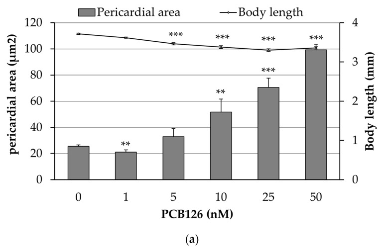
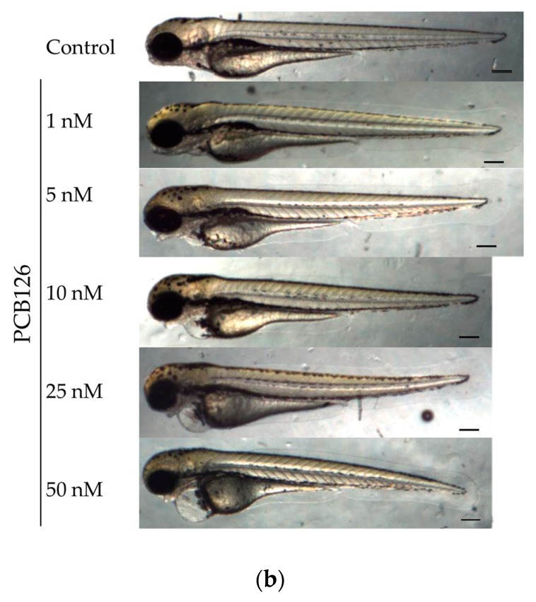
Embryos were exposed to increasing concentrations of PCB126 (1, 5, 10, 25, and 50 nM) and vehicle (embryo medium with 0.01% acetone) in order to characterize the concentration-dependent cardiovascular toxicity of the compound. As Figure 1 shows, exposure to PCB126 produced a concentration-dependent increase in the pericardial sac area and a reduction on the body length of the larvae with a correlation coefficient of r = −0.78. Moreover, the heart of PCB126 exposed embryos (25 nM) failed to loop correctly (observed as a linear heart tube), and at high concentrations (50 nM) embryos also showed ventricular standstill (Supplementary video file 1).
Figure 1.
Concentration-dependent morphological effects of PCB126 exposure: (a) Mean pericardial sac area and body length of embryos exposed to increased concentration of PCB126. Values are mean ± SEM. Asterisks represent statistical significance at * p < 0.05, ** p < 0.01, and *** p < 0.001; n = 4 pools of five embryos (two biological replicates). (b) Representative images of embryos at 80 hpf exposed to increasing concentrations of PCB126 (Scale bar = 200 µm).
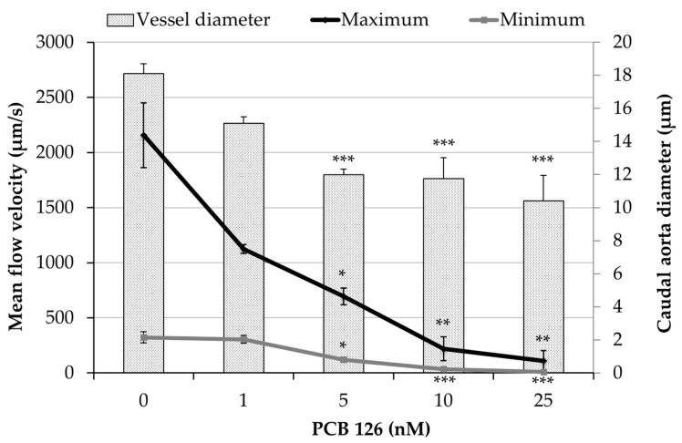
To determine whether PCB126 exposure disrupted peripheral blood circulation, we examined the maximum and minimum caudal aortic blood flow. As Figure 2 shows, exposure to PCB126 produced a concentration dependent decrease in peripheral blood velocity as well as a reduction in the diameter of caudal aortic vessel that resulted in a significantly decreased mean flow from concentration 1nM of PCB126 (Table 1). The reduction on peripheral blood flow circulation also came with a reduction in red blood cells passing through the caudal aortic vessel (Supplementary video file 2). Embryos exposed to PCB126 showed a significantly reduced cardiac output but an unaltered heart rate at 3 dpf (Table 1).
Figure 2.
Effects of PCB126 on peripheral blood flow. Maximum and minimum caudal aortic flow velocity after exposure of embryos to PCB126. Vessel diameter plotted on secondary axis. Values are mean ± SEM (n = 6 larvae). Asterisks represent statistical significance at * p < 0.05, ** p < 0.01, and *** p < 0.001.
Table 1.
PCB126 exposure decreased peripheral mean blood flow velocity from 1 nM, caused no change in heart rate but significantly decreased cardiac output. The highest concentration tested (50 nM) could not be measured due to the high reduction of blood cells. Values are mean ± SEM on n = 6 larvae. Asterisks represent statistical significance at * p < 0.05, ** p < 0.01, and *** p < 0.001.
| Treatment | Maximum Blood Flow (nL/min) | Minimum Blood Flow (nL/min) | Heart Rate (beats/min) | Cardiac Output (nL/min) |
|---|---|---|---|---|
| Control | 34.7 ± 6.8 | 5.2 ± 1.1 | 215.7 ± 3.4 | 47.0 ± 6.1 |
| PCB 126 1nM | 12.1 ± 0.7 * | 3.2 ± 0.3 * | 219.4 ± 5.7 | 35.1 ± 4.4 |
| PCB 126 5 nM | 4.7 ± 0.6 * | 0.8 ± 0.1 * | 215.0 ± 10.7 | 22.8 ± 1.4 ** |
| PCB 126 10 nM | 2.7 ± 1.6 * | 0.4 ± 0.3 * | 226.8 ± 17.0 | 14.1 ± 4.7 *** |
| PCB 126 25 nM | 1.7 ± 1.6 * | 0.1 ± 0.1 * | 209.2 ± 5.8 | 22.0 ± 5.9 ** |
2.2. Influence of Redox-Modulators on the Cardiovascular Toxicity Induced by PCB126
Embryos were co-exposed to several redox-modulating chemicals (Table 2) to determine whether a more reduced or oxidized redox level would alter the gross defects observed after PCB126 exposure (pericardial effusion severity or body length). The concentration of PCB126 used was 25 nM, concentration that was found to produce a fully and uniform phenotype in zebrafish using our protocol. Each redox-modulating chemical was first tested independently at increasing concentrations and the maximum tolerable concentration (MTC) was established and subsequently used in the co-exposure experiments.
Table 2.
Overview of the chemicals used as modulators in the cardiac toxicity produced by PCB126.
| Substance | CAS Number | MTC 1 (µM) | Description |
|---|---|---|---|
| N-Acetyl-L-cysteine (NAC) | 616-91-1 | 100 | Glutathione precursor |
| Diethyl maleat (DEM) | 141-05-9 | 0.05 | Glutathione depletor |
| (±)-α-Lipoic acid | 1077-28-7 | 10 | Antioxidant and free radical scavenger |
| (±)-α-Tocopherol | 10191-41-0 | 100 | Antioxidant and peroxyl radical scavenger |
| (±)-6-Hydroxy-2,5,7,8-tetramethylchromane-2-carboxylic acid (TROLOX) | 53188-07-1 | 15 | Water-soluble analogue of alpha-tocopherol |
| Quercetin | 117-35-9 | 12 | Flavonoid (mitochondrial ATPase and phosphodiesterase inhibitor) |
| Nω-Nitro-L-arginine methyl ester hydrochloride (L-NAME) | 51298-62-5 | 100 | Inhibitor of nitric oxide synthase |
| Sodium nitroprusside dihydrate (SNP) | 13755-38-9 | 250 | Nitric oxide donor |
| Indomethacin | 53-86-1 | 30 | Non-steroidal anti-inflammatory compound |
Note: 1 Maximum tolerable concentration.
We attempted to modulate deformities by decreasing glutathione (GSH) levels with dimethyl maleate (DEM) and increasing GSH pools with N-acetyl cysteine (NAC). Co-treatment with NAC (100 µM) did not prevent deformities and co-treatment with DEM (0.05 µM) failed to worsen deformities (Table 3). We also examined whether several antioxidants could modulate deformities produced by PCB126. Co-treatment with lipoic acid (10 µM) did not ameliorate pericardial effusion and growth of the PCB126 treated larvae at 3 dpf (Table 3), but produced a non-significant increase in pericardial area compared to PCB126. A dual role of lipoic acid as antioxidant/pro-oxidant molecule has already been described and could be related to such a result [17].
Table 3.
Pericardial sac area and body length of larvae at 3 dpf exposed to PCB126 and co-exposed to several redox-modulating chemicals. Data are presented as mean ± SEM on n = 3–5 pools of three embryos. Different letters on top of the bars indicate statistically significant differences with p < 0.05.
| Treatment | Pericardial Sac Area (% Control Vehicle area) |
Body Length (% Control Vehicle Length) |
|---|---|---|
| Vehicle control | 100 ± 4.1 a | 100 ± 1.1 a |
| DEM 50nM | 128.7 ± 3.9 a | 98.4 ± 0.5 a |
| PCB126 25 nM | 329.5 ± 20.6 b | 94.1 ± 0.9 b |
| PCB126 25 nM + DEM 50 nM | 302.5 ± 10.8 b | 93.7 ± 0.4 b |
| Vehicle control | 100 ± 2.9 a | 100 ± 1.2 a |
| NAC 100 µM | 88.3 ± 8.2 a | 101.2 ± 1.4 a |
| PCB126 25 nM | 205.0 ± 21.5 b | 94.1 ± 0.5 b |
| PCB126 25 nM + NAC 100 µM | 216.5 ± 24.4 b | 92.8 ± 0.9 b |
| Vehicle control | 100 ± 4.9 a | 100 ± 0.8 a |
| Lipoic acid 10 µM | 102.9 ± 2.9 a | 98.4 ± 1.0 a |
| PCB126 25 nM | 271.6 ± 10.4 b | 93.9 ± 0.1 b |
| PCB126 25 nM + lipoic acid 10 µM | 318.9 ± 39.4 b | 88.9 ± 0.8 b |
| Vehicle control | 100 ± 3.1 a | 100 ± 0.1 a |
| Quercetin 12 µM | 118 ± 6.4 a | 98.5 ± 0.6 a |
| PCB126 25 nM | 350.5 ± 13.0 b | 91.5 ± 0.5 b |
| PCB126 25 nM + quercetin 12 µM | 340.3 ± 26.4 b | 91.6 ± 1.0 b |
| Vehicle control | 100 ± 10.1 a | 100 ± 0.3 a |
| Indomethacin 30 µM | 114.9 ± 1.7 a | 96.5 ± 0.7 a |
| PCB126 25 nM | 223.9 ± 19.2 b | 92.3 ± 0.5 b |
| PCB126 25 nM + indomethacin 30 µM | 246.9 ± 22.8 b | 89.6 ± 1.3 b |
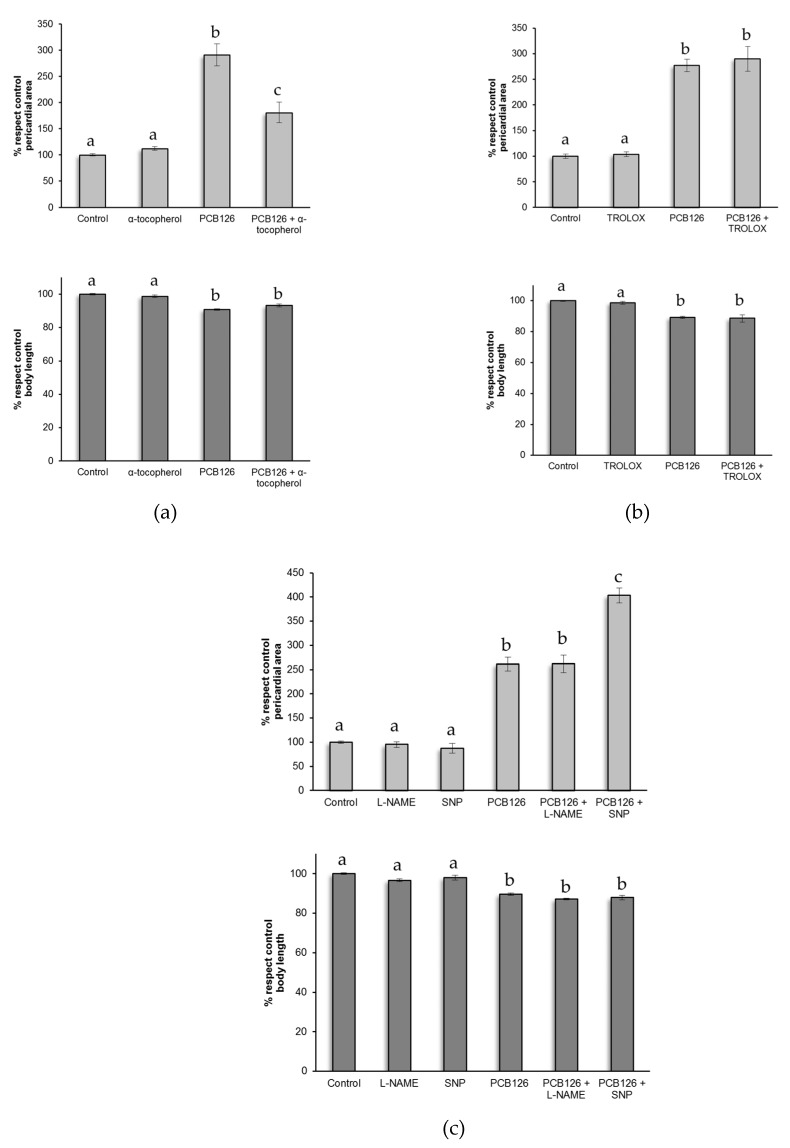
Co-treatment with α-tocopherol (vitamin E liposoluble) at 100 µM ameliorated pericardial sac area, but did not prevent PCB126 induction of pericardial effusion and shortening of body length (Figure 3a). On the other hand, pericardial effusion and shortening of the body length was not prevented by co-treatment with a soluble analogue of vitamin E (Trolox) at 15 µM (Figure 3b). Studies showed that coplanar PCBs have proinflammatory effects on vascular endothelial cells [18] through endothelial nitric oxide synthase (eNOS) signalling. Therefore, we attempted to modulate this effect by co-exposing embryos to indomethacin, a non-steroidal anti-inflammatory compound, and quercetin, a flavonoid with anti-inflammatory properties. Embryos co-exposed either to indomethacin (30 µM) or quercetin (12 µM) still exhibited pericardial effusion and shortening of the body length at 3 dpf compared to control group (Table 4). As shown in Figure 3c, co-exposure to the nitric oxide synthase inhibitor, Nω-Nitro-L-arginine methyl ester hydrochloride (L-NAME, 100 µM), did not result in any significant change in pericardial sac area and body length at 3 dpf. However, co-treatment with a nitric oxide donor, sodium nitroprusside (SNP, 250 µM), worsened the pericardial sac area but did not significantly shorten body length.
Figure 3.
Pericardial sac area and body length of larvae at 80 hpf exposed to PCB126 and co-exposed to (a) α-tocopherol (100 µM), (b) Trolox (15 µM), (c) L-NAME (100 µM), and SNP (250 µM). Data are presented as mean ± SEM on n = 6–9 pools of three embryos. Different letters on top of the bars indicate statistically significant differences with p < 0.05.
Table 4.
Expression of genes involved in the redox status of the embryo after exposure to PCB126 (25 nM) and after deformity development at 3 dpf. Relative expression was calculated respect the housekeeping gene (gapdh) and control (0.01% acetone). Data are represented as mean ± SEM of n = 3 pools of 15 embryos from different biological replicates. Asterisk indicates significant induction compared to the vehicle control (p < 0.05).
| Gene | Fold Change |
|---|---|
| cyp1a | 199.6 ± 62.1 * |
| hsp70 | 1.72 ± 0.2 * |
| gpx1a | 1.21 ± 0.2 |
| sod1 | 1.03 ± 0.07 |
| sod2 | 1.02 ± 0.06 |
| cat | 0.91 ± 0.09 |
| gstp1 | 1.74 ± 0.5 * |
2.3. Gene Expression Analysis of Oxidative Stress Related Genes
To determine the redox status of zebrafish embryo exposed to PCB126, gene expression of several antioxidant genes was analyzed by quantitative RT-PCR. Alteration of the redox status was examined in embryos treated with PCB126 at a concentration of 25 nM and after dysmorphogenesis at 3 dpf. The targeted genes involved in the defense against oxidative stress (gpx1a, sod1, sod2, cat, and gstp1), cyp1a, related to the AhR machinery, and hsp70 involved in the cellular stress response. Cyp1a expression increased by nearly 200-fold in embryos exposed to PCB126. We also observed a significant fold change induction of about 1.7 for hsp70 and gstp1 genes. For the remaining genes (gpx1a, sod1, sod2, and cat), no alteration in expression was observed (Table 4).
3. Discussion
The results of this study demonstrate that PCB126 developmental exposure elicits a cardiovascular concentration-dependent toxicity in zebrafish embryos. PCB126 exposure leads to a concentration-dependent increase in pericardial sac area, which is in accordance with previous reports by Jönsson et al. [7], where craniofacial malformations, heart malformations (hypotrophy and reduced looping), slower and weaker heartbeats, impaired circulation, and immobility were also described. Our study also shows that PCB126 exposure leads to a concentration-dependent shortened body length, being the LOAEC 5 nM of PCB126 (Figure 1). This reduction in body length could suggests an impairment of the growth of the larva, but it is also an effect often observed when there is a cardiovascular defect [19], and, in this case, a significant reduction in maximum blood flow at concentrations not affecting body length was detected. An increased pericardial sac area was moderately correlated (r = −0.78) with reduced body length.
Chemicals that are AhR agonists are known to reduce blood flow in trunk vessels of zebrafish embryos at 72 hpf or later [20]. In our study, exposure to PCB126, a known AhR agonist, significantly reduced peripheral blood circulation and aortic caudal vessel diameter in a concentration-dependent way from concentration of 5 nM (Figure 2). Particularly, reduction in blood vessel diameter suggests impairment in endothelial cell function. Considering that, hemodynamic changes play an important role in blood vessel formation and changes in blood flow can lead to severe vascular distortion [21]. Grimes et al. [22] have already strongly suggested a link between hemodynamic forces and myocardial dysmorphogenesis produced by PCB126. In line with this, the observed significant reduction on peripheral blood flow circulation came together with a reduction in red blood cells passing through the caudal aortic vessel (Supplementary video file 2), and cardiac malformation (no heart looping). However, it is not possible to discern whether the PCB126-heart phenotype we observed is secondary to the endocardial disruption and hemodynamic impairment or not. It should be noted that measurement of peripheral blood flow provides more accurate information about the blood pumped by the heart than measuring cardiac output. This is because the method used to measure cardiac output does not take into account a possible blood reflux between chambers. In any case, PCB126 exposure produced a significant decrease in cardiac output from concentration of 5 nM (Table 3). This reduction in cardiac output occurring at the same time was not related to an altered heart rate but to the abnormal morphogenesis of the heart ventricle observed in PCB126 exposed embryos. Antkiewicz et al. [10] also observed a significantly decreased stroke volume and cardiac output, with no effect on heart rate after TCDD treatment. In our case, some treated embryos at high concentrations showed reflux of blood between chambers or a contractile ventricle chamber without blood passing through it (Supplementary video file 2) that made measurement of cardiac output difficult.
As mentioned before, PCB126 is a strong agonist for AhR, a ligand-activated cytosolic transcription factor that induces the expression of a battery of genes involved in xenobiotic metabolism. The activity of these genes contributes to the formation of reactive oxygen species that can subsequently lead to cellular oxidative stress, lipid peroxidation, and DNA fragmentation [23]. Some studies indicate that an increase in cellular oxidative stress and an imbalance in antioxidant status are critical events in PCB-mediated induction of inflammatory genes and endothelial cell dysfunction [18]. Therefore, in this study we sought to determine the role of redox imbalance in mediating the cardiac toxicity caused by PCB126 in the developing zebrafish. For that reason, embryos were exposed to several redox modulating chemicals to demonstrate if oxidative stress is a component of the developmental toxicity of PCB126. Nine redox modulating chemicals were tested in the zebrafish that are known to modulate different redox mechanisms: increase or decrease in GSH pools (NAC [24] and DEM [25], respectively), free radical scavengers (Lipoic acid [26], vitamin E [14], Trolox [26]), anti-inflammatory agents (indomethacin [27], quercetin [28]), inhibitor of NO synthase (L-NAME [29]), and a nitric oxide donor (SNP [30]). Among all the redox modulating chemicals tested we have observed that PCB126 toxicity could only be modulated by vitamin E. We could observe only a partial amelioration in the pericardial sac area and no significant effect in body length of larvae, in contrast with what was observed in the study of Na et al. [14]. This difference could be due to differences in experimental protocol. On the other hand, our results demonstrated that SNP, a nitric oxide donor, worsened pericardial effusion induced by PCB126 exposure. Pelster et al. [29] demonstrated that the vasodilator effect of NO contributes to changes in local blood flow, and modifications of shear stress. Endothelial cells are able to convert mechanical stimuli into intracellular signals that affect cellular functions (e.g., permeability or remodeling) [31]. Some studies have shown that PCB126 decreases NO production and alters the expression of eNOS in human umbilical vein endothelial cells [32,33] and increases endothelial production of the inflammatory mediator IL-6 [18]. Additionally, it has been suggested that NO can induce COX activity and subsequent production of proinflammatory prostaglandins [34]. Cyclooxygenase-2 (Cox-2) enzymes have been proposed to be involved in some DLC-mediated toxicities [35]. Particularly, PCB126 showed a concentration dependent increase in COX-2 expression [36]. Our results suggest that NO signalling pathway is involved in PCB126 induced endothelial cell permeability dysfunction, although more studies with inhibitors or gene knockdown specific for zebrafish COX-2 are required for further understanding the roles of zebrafish nitric oxide and COX-2 involvement in the cardiotoxicity elicited by PCB126.
It is well established that PCB126 binds to the AhR and induces the expression of a battery of genes (the AhR gene battery), including cyp1a. PCB126 has been shown to markedly contribute to cellular oxidative stress in endothelial cells in vitro by inducing cyp1a activity and decreasing vitamin E content [37]. Accordingly in our study, embryos exposed to PCB126 during 24 h showed a strong induction of cyp1a at 3 dpf (Table 4). Our results are in line with previous reports [7] demonstrating the largest fold change between basal and induced expression on day 3 (236 ± 34 fold the control). Besides, our analysis shows significant fold induction for HSP70 gene after PCB126 exposure. Heat shock proteins (HSPs) are a highly conserved, ubiquitously expressed family of stress response proteins, which are expressed at low levels under normal physiological conditions. HSPs can function as molecular chaperones, facilitating protein folding, preventing protein aggregation, or targeting improperly folded proteins to specific degradative pathways [38]. Moreover, it has been demonstrated that HSP activity is required for physiological responses, such as endothelial cell migration and blood vessel repair specifically, as hsp70 morphants of zebrafish have impaired vessel development and sprouting [39]. On the other hand, the expression of genes related to the NRF2 pathway was not altered after exposure to 25 nM of PCB126 at 3 dpf, but there was the upregulation of gstp1 by PCB126. There are contradictory data concerning the role of NRF2 in regulation of phase II enzymes in zebrafish [16,40,41]. Moreover, some studies have been shown that knockdown of gstp2 did not serve as a protective function against the cardiac toxicity caused by PCB126 [42].
Although the production of PCBs has been strictly regulated in most industrialized countries since the late 1970s, they are still prevalent in the environment due to their high persistence and resistance to bio-degradation. The most abundant PCB congener in serum is usually PCB153, a congener that does not display “dioxin-like” activity. As an approach to compare “dioxin-like” compound concentrations in serum or breast milk with the concentrations used in this study, we have calculated the corresponding toxic equivalent (TEQ). The total TEQ concentrations detected in these biological samples were in the pg/L range, being the highest level in blood around 142.4 pg/L [43]. In contrast, our study is in the range of 326-1630 ng/L. However, how the effect of concentrations in zebrafish embryos can be related to appropriate effect levels in mammalian models is still a major issue for future research. In our study, nominal concentrations were used and may represent a limitation. Under short-term exposures, a steady state may only be reached for test substances with lower hydrophobicity and rarely reached for more hydrophobic substances [44], leading to an underestimation of the apparent short-term toxicity of PCB126. Therefore, the potential health risk for the developing human to “dioxin-like” compounds still needs further investigation.
4. Materials and Methods
4.1. Zebrafish Maintenance and Egg Production
Adult female and male zebrafish were obtained from a commercial supplier (Pisciber, Barcelona) and housed separately in a closed flow-through system in standardized dilution water (ISO, 1996; 2 mM CaCl2·2 H2O; 0.5 mM MgSO4·7 H2O; 0.75 mM NaHCO3; 0.07 mM KCl). Fish were maintained at 26 ± 1 °C under a 14:10 light:dark cycle. The study was approved by the Ethic Committee for Animal Experimentation of the University of Barcelona (398/14, Mai 2014) and by the Department of Environment and Housing of the Generalitat de Catalunya with the license number DAAM 9967 (Mai, 2018).
Embryos were collected after natural spawning of adult zebrafish, as previously described [45]. Embryos used in the experiments were held in 0.3× Danieau’s solution (5.8 mM NaCl; 0.23 mM KCl; 0.13 mM MgSO4·7 H2O; 0.2 mM Ca(NO3)2; 5 mM HEPES; pH 7.4) at 27 °C and at 14:10 light:dark cycle. All exposures were performed in glass crystallization dishes.
4.2. Exposure to PCB126
Groups of 25 embryos at approximately 8 h post-fertilization (hpf) were exposed to increasing concentrations of PCB126 (Dr. Ehrenstorfer GmbH, Augsburg, Germany) or carrier solvent (0.01% acetone, v/v) in 20 mL of 0.3× Danieau’s solution. After 24 h, the exposure test solution was removed and replaced with fresh 0.3× Danieau’s solution. Embryos were held, with daily changes of the medium, until 3 dpf.
4.3. Exposure to Redox Modulating Chemicals
Embryos were exposed to chemical modulators at 4 hpf and during the whole test until 3 dpf, renewal of medium was done every 24 h. All redox modulating chemicals were dissolved in 0.3× Danieau’s solution, except lipoic acid, α-tocopherol, and indomethacin that were initially prepared in 100% DMSO and subsequently diluted in embryo medium (0.05% DMSO, v/v). Trolox stock solution was prepared at 2 mM in 0.3× Danieau’s solution and filtered (0.22-μm syringe filter) (GE Healthcare Whatman®, Chicago, Illinois, USA). The concentration was adjusted using absorption at 290 nm and extinction coefficient of 2350/M/cm. A preliminary range finding test was performed in order to determine the maximum tolerable concentration (Table 2), defined as the maximum concentration tested that does not produce a significant increase in mortality or dysmorphogenesis. At 3 dpf, induction of the pericardial sac area and body shortening was compared between embryos treated with PCB126 alone and embryos co-treated with PCB126 chemical modulator.
4.4. Heart Rate
Cardiovascular function of embryos was evaluated by time-lapse recording using a high-speed digital camera (Exilim, CASIO). Sequential images of the heart (480 frames per second –fps–) were obtained with the embryo positioned on its side from the lateral position and with a duration of 5 s. To avoid the movement of zebrafish embryo during recoding, embryos were anaesthetized by adding tricaine to embryo medium and mounted in 3% methylcellulose on double depression slides. The final concentration of tricaine was 0.004% (w/v), which has shown to not affect heart rate in zebrafish embryos [46].
To quantify heartbeat in the zebrafish, video recording were analyzed with ImageJ [47]. A plot of dynamic pixels was obtained by selecting an area of the heart ventricle with high deviation of pixel intensity by means of the z projection function. The frequency as frames/beat was obtained by analyzing the waveform of dynamic pixels by Short-term Fourier Transform using Statgraphics software (Statpoint Technologies Inc., Virginia, Washington D.C., USA). Then, the heart beat frequency (beats/min) was calculated taking into account the speed of video recording (480 fps).
4.5. Stroke Volume and Cardiac Output
To ensure that a comparable plane of focus was examined for ventricular analysis, zebrafish hearts were imaged in a standardized lateral right position, with the ventricle clearly visible in the plane of focus throughout the cardiac cycle. The atrium usually lay outside the plane of focus. Ventricular performance of embryos was analyzed by identifying frames that captured ventricular end systole and end diastole in the sequential still frames (as shown in [48]). From the ventricle, image major and minor axes measurements were extracted and exported to an Excel spreadsheet. Calculation of ventricle volume during systole and diastole was based on the formula for the volume of a prolate spheroid (V = 4πab2/3), where “a” represents the major axe radius and “b” the minor axe radius of the ventricle image. The stroke volume (SV), the volume of blood ejected by the ventricle in one heart beat was calculated using the Equation (1).
| SV (nl) = (end-diastolic volume – end-systolic volume) | (1) |
Cardiac output (CO), the volume of blood ejected by the heart in one minute, was calculated using Equation (2).
| CO (nl/min) = SV (nl) × heart rate (beats/min) | (2) |
4.6. Peripheral Blood Flow
To identify a reproducible location for analysis of blood circulation, the dorsal aorta adjacent to the cloaca was chosen as a defined imaging landmark. Video recordings of the dorsal aorta at 240 fps were processed with ImageJ. A scan line was placed parallel on the blood flux on the stack of images, and a series of lines were obtained whose slopes were inversely proportional to cellular velocity. OrientationJ plugin [49] of ImageJ was used in order to calculate cellular velocity from the scanned lines.
4.7. Pericardial Sac Area and Body Length
To quantify the degree of increase in the pericardial sac area and body shortening, lateral images were obtained for at least 10 larvae mounted in 3% methylcellulose. By tracing the boundaries of the pericardial space with ImageJ software, the pixel area was obtained. For body length, a line starting at the anterior-most point of the head and ending at the tail end was measured.
4.8. Quantitative RT-PCR
Total RNA was extracted from a pool of 15 embryos per sample homogenized zebrafish larvae (3 dpf) using TRIZOL® reagent (Life Technologies S.A., Carlsbad, California, USA) according to the manufacturer’s instructions. RNA quantity and quality was analyzed spectrophotometrically using a NanoDrop ND1000 (NanoDrop Technologies, Wilmington, Delaware, USA). RNA samples were stored at −80 °C until use. cDNA was produced from 2 µg of total DNAse treated RNA samples using RevertAid™ Reverse Transcriptase (MBI Fermentas, Thermo Fisher Scientific, Inc., Waltham, Massachusetts, USA) in 20 µL.
Real-time PCR reactions were carried out using the SensiMix SYBR Hi-ROX One-Step Kit (Bioline Reagents Ltd., London, UK) and following manufacturer’s instructions. Amplifications were performed in 384-well plates on an ABI 7900HT (AppliedBiosystems, Life Technologies S.A., Carlsbad, California, USA) real-time PCR machine at the Scientific and Technologic Center of UB (CCITUB) under the following thermal cycle: initial denaturation for 10 min at 95 °C, 40 cycles of denaturation for 10 s at 95 °C, annealing for 20 s at 55 °C, and elongation for 20 s at 72 °C. A final denaturation was performed for 30 s at 95 °C. This was followed by generation of a melting curve, starting from 60 °C to 95 °C. Temperature was raised in 0.5 °C increments, holding each temperature for 7 s. Primer sequences were obtained from literature or designed using Primer3 program [50]. Primer sequences and accession numbers of all investigated genes are listed in Supplementary Table S1. Gapdh gene was used as a housekeeping gene for qPCR normalization.
4.9. Statistics
Statistical analysis was performed with SPSS 15.0 (IBM, Chicago, USA). One-way analysis of variance (ANOVA) followed by post hoc multi-comparison with Bonferroni’s test was used to analyze homogeneous data of the continuous variables. Significance threshold was established at p < 0.05.
The comparative Ct method [51] was used to determine average fold induction of mRNA by comparing the Ct of the target gene to that of the reference gen (Gapdh). The fold change obtained for each biological replicate pool of 15 embryos was averaged for treatments. Statistical analysis was performed with REST© software [52].
5. Conclusions
PCB126 exposure leads to a cardiovascular failure manifested by the development of pericardial effusion, body shortening, altered peripheral blood flow, and cardiac performance in zebrafish larvae. It is likely that PCB126 disrupts endothelial cell permeability and it seems that altered hemodynamic forces could be implicated. However, the precise nature of this cellular dysfunction and how it leads to abnormal heart morphology remains to be elucidated. The results further suggest that redox modulation did not serve as a protective function against cardiovascular effects caused by PCB126 in developing zebrafish. Oxidative stress may not be the primary factor contributing to the deformities caused by low concentration of PCB126.
Abbreviations
| CHD | Congenital heart defects |
| DLC | Dioxin-like polychlorinated biphenil |
| TCDD | 2,3,7,8-tetrachlorodibenzo-p-dioxin |
| PCB126 | 3,3’,4,4’,5-Pentachlorobiphenyl |
| AhR | Aryl hydrocarbon receptor |
| MTC | Maximum tolerable concentration |
| GSH | Glutathione |
| DEM | Dimethyl maleate |
| NAC | N-Acetyl-L-cysteine |
| TROLOX | (±)-6-Hydroxy-2,5,7,8-tetramethylchromane-2-carboxylic acid |
| L-NAME | Nω-Nitro-L-arginine methyl ester hydrochloride |
| SNP | Sodium nitroprusside dihydrate |
Supplementary Materials
Supplementary materials can be found at https://www.mdpi.com/1422-0067/20/5/1065/s1.
Author Contributions
Conceptualization, J.M.L. and J.G.-C.; data curation, E.T.; formal analysis, E.T.; funding acquisition, J.M.L. and J.G.-C; investigation, E.T.; methodology, E.T. and E.P.; resources, E.P.; software, E.P.; supervision, M.B., J.M.L., and J.G.-C.; validation, J.G.-C.; writing—original draft, E.T.; writing—review and editing, M.B., J.G.-C., and E.T.
Funding
This research work was supported by funds from Fundació Bosch i Gimpera, project number FBG-300155, and University of Barcelona fundings for Open access publishing. We thank the staff of the zebrafish facility (CCiTUB) for their excellent technical support. Also, the work of E.T. was supported by the APIF 2010 fellowship, granted by the Universitat de Barcelona and a grant from the Spanish ministry of Education for the mobility of European Doctorate in 2010. We want to thank Dani Perez for his support in the lab and Nils Klüver for the practical information on gene expression analysis. We thank the anonymous reviewers for their critical and helpful comments.
Conflicts of Interest
The authors declare no conflict of interest.
References
- 1.EUROCAT, 2010. European Surveillance of Congenital Anomalies. Available online: http://www.eurocat-network.eu/AccessPrevalenceData/PrevalenceTables.
- 2.Dadvand P., Rankin J., Rushton S., Pless-Mulloli T. Association Between Maternal Exposure to Ambient Air Pollution and Congenital Heart Disease: A Register-based Spatiotemporal Analysis. Am. J. Epidemiol. 2011;173:171–182. doi: 10.1093/aje/kwq342. [DOI] [PMC free article] [PubMed] [Google Scholar]
- 3.Kopf P.G., Walker M.K. Overview of Developmental Heart Defects by Dioxins, PCBs, and Pesticides. J. Environ. Sci. Health Part C. 2009;27:6–85. doi: 10.1080/10590500903310195. [DOI] [PubMed] [Google Scholar]
- 4.Mastin J.P. Environmental cardiovascular disease. Cardiovasc. Toxicol. 2005;5:91–94. doi: 10.1385/CT:5:2:091. [DOI] [PubMed] [Google Scholar]
- 5.Peterson R.E., Theobald H.M., Kimmel G.L. Developmental and Reproductive Toxicity of Dioxins and Related Compounds: Cross-Species Comparisons. Crit. Rev. Toxicol. 1993;23:283–335. doi: 10.3109/10408449309105013. [DOI] [PubMed] [Google Scholar]
- 6.Puga A. Perspectives on the potential involvement of the AH receptor-dioxin axis in cardiovascular disease. Toxicol. Sci. 2011;120:256–261. doi: 10.1093/toxsci/kfq393. [DOI] [PMC free article] [PubMed] [Google Scholar]
- 7.Jönsson M.E., Jenny M.J., Woodin B.R., Hahn M.E., Stegeman J.J. Role of AHR2 in the Expression of Novel Cytochrome P450 1 Family Genes, Cell Cycle Genes, and Morphological Defects in Developing Zebra Fish Exposed to 3,3′,4,4′,5-Pentachlorobiphenyl or 2,3,7,8-Tetrachlorodibenzo-p-dioxin. Toxicol. Sci. 2007;100:180–193. doi: 10.1093/toxsci/kfm207. [DOI] [PubMed] [Google Scholar]
- 8.Di Paolo C., Groh K.J., Zennegg M., Vermeirssen E.L.M., Murk A.J., Eggen R.I.L., Hollert H., Werner I., Schirmer K. Early life exposure to PCB126 results in delayed mortality and growth impairment in the zebrafish larvae. Aquat. Toxicol. 2015;169:168–178. doi: 10.1016/j.aquatox.2015.10.014. [DOI] [PubMed] [Google Scholar]
- 9.Slim R., Toborek M., Robertson L.W., Hennig B. Antioxidant protection against PCB-mediated endothelial cell activation. Toxicol. Sci. 1999;52:232–239. doi: 10.1093/toxsci/52.2.232. [DOI] [PubMed] [Google Scholar]
- 10.Antkiewicz D.S., Peterson R.E., Heideman W. Blocking Expression of AHR2 and ARNT1 in Zebrafish Larvae Protects Against Cardiac Toxicity of 2,3,7,8-Tetrachlorodibenzo-p-dioxin. Toxicol. Sci. 2006;94:175–182. doi: 10.1093/toxsci/kfl093. [DOI] [PubMed] [Google Scholar]
- 11.Goldsmith P. Zebrafish as a pharmacological tool: The how, why and when. Curr. Opin. Pharmacol. 2004;4:504–512. doi: 10.1016/j.coph.2004.04.005. [DOI] [PubMed] [Google Scholar]
- 12.Chico T.J.A., Ingham P.W., Crossman D.C. Modeling Cardiovascular Disease in the Zebrafish. Trends Cardiovasc. Med. 2008;18:150–155. doi: 10.1016/j.tcm.2008.04.002. [DOI] [PubMed] [Google Scholar]
- 13.Jacob E., Drexel M., Schwerte T., Pelster B. Influence of hypoxia and of hypoxemia on the development of cardiac activity in zebrafish larvae. Am. J. Physiol. Regul. Integr. Comp. Physiol. 2002;283:R911–R917. doi: 10.1152/ajpregu.00673.2001. [DOI] [PubMed] [Google Scholar]
- 14.Na Y.-R., Seok S.-H., Baek M.-W., Lee H.-Y., Kim D.-J., Park S.-H., Lee H.K., Park J.-H. Protective effects of vitamin E against 3,3′,4,4′,5-pentachlorobiphenyl (PCB126) induced toxicity in zebrafish embryos. Ecotoxicol. Environ. Saf. 2009;72:714–910. doi: 10.1016/j.ecoenv.2008.09.015. [DOI] [PubMed] [Google Scholar]
- 15.Liu H., Nie F.-H., Lin H.-Y., Ma Y., Ju X.-H., Chen J.-J., Gooneratne R. Developmental toxicity, oxidative stress, and related gene expression induced by dioxin-like PCB 126 in zebrafish (Danio rerio) Environ. Toxicol. 2016;31:295–303. doi: 10.1002/tox.22044. [DOI] [PubMed] [Google Scholar]
- 16.Rousseau M.E., Sant K.E., Borden L.R., Franks D.G., Hahn M.E., Timme-Laragy A.R. Regulation of Ahr signaling by Nrf2 during development: Effects of Nrf2a deficiency on PCB126 embryotoxicity in zebrafish (Danio rerio) Aquat. Toxicol. 2015;167:157–171. doi: 10.1016/j.aquatox.2015.08.002. [DOI] [PMC free article] [PubMed] [Google Scholar]
- 17.Moini H., Packer L., Saris N.-E.L. Antioxidant and prooxidant activities of alpha-lipoic acid and dihydrolipoic acid. Toxicol. Appl. Pharmacol. 2002;182:84–90. doi: 10.1006/taap.2002.9437. [DOI] [PubMed] [Google Scholar]
- 18.Hennig B., Meerarani P., Slim R., Toborek M., Daugherty A., Silverstone A.E., Robertson L.W. Proinflammatory properties of coplanar PCBs: In vitro and in vivo evidence. Toxicol. Appl. Pharmacol. 2002;181:174–183. doi: 10.1006/taap.2002.9408. [DOI] [PubMed] [Google Scholar]
- 19.Billiard S.M., Querbach K., Hodson P.V. Toxicity of retene to early life stages of two freshwater fish species. Environ. Toxicol. Chem. 1999;18:2070–2077. doi: 10.1002/etc.5620180927. [DOI] [Google Scholar]
- 20.Carney S.A., Peterson R.E., Heideman W. 2,3,7,8-Tetrachlorodibenzo-p-dioxin activation of the aryl hydrocarbon receptor/aryl hydrocarbon receptor nuclear translocator pathway causes developmental toxicity through a CYP1A-independent mechanism in zebrafish. Mol. Pharmacol. 2004;66:512–521. doi: 10.1124/mol.66.3. [DOI] [PubMed] [Google Scholar]
- 21.Yashiro K., Shiratori H., Hamada H. Haemodynamics determined by a genetic programme govern asymmetric development of the aortic arch. Nature. 2007;450:285–288. doi: 10.1038/nature06254. [DOI] [PubMed] [Google Scholar]
- 22.Grimes A.C., Erwin K.N., Stadt H.A., Hunter G.L., Gefroh H.A., Tsai H.J., Kirby M.L. PCB126 exposure disrupts zebrafish ventricular and branchial but not early neural crest development. Toxicol. Sci. 2008;106:193–205. doi: 10.1093/toxsci/kfn154. [DOI] [PMC free article] [PubMed] [Google Scholar]
- 23.Barouki R., Morel Y. Repression of cytochrome P450 1A1 gene expression by oxidative stress: Mechanisms and biological implications. Biochem. Pharmacol. 2001;61:511–516. doi: 10.1016/S0006-2952(00)00543-8. [DOI] [PubMed] [Google Scholar]
- 24.Timme-Laragy A.R., Van Tiem L.A., Linney E.A., Di Giulio R.T. Antioxidant Responses and NRF2 in Synergistic Developmental Toxicity of PAHs in Zebrafish. Toxicol. Sci. 2009;109:217–227. doi: 10.1093/toxsci/kfp038. [DOI] [PMC free article] [PubMed] [Google Scholar]
- 25.Gallagher E.P., Hasspieler B.M., Di Giulio R.T. Effects of buthionine sulfoximine and diethyl maleate on glutathione turnover in the channel catfish. Biochem. Pharmacol. 1992;43:2209–2215. doi: 10.1016/0006-2952(92)90180-Q. [DOI] [PubMed] [Google Scholar]
- 26.Reimers M.J., La Du J.K., Periera C.B., Giovanini J., Tanguay R.L. Ethanol-dependent toxicity in zebrafish is partially attenuated by antioxidants. Neurotoxicol. Teratol. 2006;28:497–508. doi: 10.1016/j.ntt.2006.05.007. [DOI] [PubMed] [Google Scholar]
- 27.Teraoka H., Kubota A., Dong W., Kawai Y., Yamazaki K., Mori C., Harada Y., Petersona R.E., Hiraga T. Role of the cyclooxygenase 2-thromboxane pathway in 2,3,7,8-tetrachlorodibenzo-p-dioxin-induced decrease in mesencephalic vein blood flow in the zebrafish embryo. Toxicol. Appl. Pharmacol. 2009;234:33–40. doi: 10.1016/j.taap.2008.09.021. [DOI] [PubMed] [Google Scholar]
- 28.Ramadass P., Meerarani P., Toborek M., Robertson L.W., Hennig B. Dietary flavonoids modulate PCB-induced oxidative stress, CYP1A1 induction, and AhR-DNA binding activity in vascular endothelial cells. Toxicol. Sci. 2003;76:212–219. doi: 10.1093/toxsci/kfg227. [DOI] [PubMed] [Google Scholar]
- 29.Parng C., Ton C., Lin Y.-X., Roy N.M., McGrath P. A zebrafish assay for identifying neuroprotectants in vivo. Neurotoxicol. Teratol. 2006;28:509–516. doi: 10.1016/j.ntt.2006.04.003. [DOI] [PubMed] [Google Scholar]
- 30.Pelster B., Grillitsch S., Schwerte T. NO as a mediator during the early development of the cardiovascular system in the zebrafish. Comp. Biochem. Physiol. Part A Mol. Integr. Physiol. 2005;142:215–220. doi: 10.1016/j.cbpb.2005.05.036. [DOI] [PubMed] [Google Scholar]
- 31.Li Y.-S.J., Haga J.H., Chien S. Molecular basis of the effects of shear stress on vascular endothelial cells. J. Biomech. 2005;38:1949–1971. doi: 10.1016/j.jbiomech.2004.09.030. [DOI] [PubMed] [Google Scholar]
- 32.Omori N., Fukata H., Sato K., Yamazaki K., Aida-Yasuoka K., Takigami H., Kuriyama M., Ichinose M., Mori C. Polychlorinated biphenyls alter the expression of endothelial nitric oxide synthase mRNA in human umbilical vein endothelial cells. Hum. Exp. Toxicol. 2007;26:811–816. doi: 10.1177/0960327107072394. [DOI] [PubMed] [Google Scholar]
- 33.Andersson H., Garscha U., Brittebo E. Effects of PCB126 and 17β-oestradiol on endothelium-derived vasoactive factors in human endothelial cells. Toxicology. 2001;285:46–56. doi: 10.1016/j.tox.2011.04.003. [DOI] [PubMed] [Google Scholar]
- 34.Vassalle C., Domenici C., Lubrano V., L’Abbate A. Interaction between Nitric Oxide and Cyclooxygenase Pathways in Endothelial Cells. J. Vasc. Res. 2003;40:491–499. doi: 10.1159/000074550. [DOI] [PubMed] [Google Scholar]
- 35.Teraoka H., Okuno Y., Nijoukubo D., Yamakoshi A., Peterson R.E., Stegeman J.J., Kitazawa T., Hiraga T., Kubota A. Involvement of COX2–thromboxane Pathway in TCDD-Induced Precardiac Edema in Developing Zebrafish. Aquat. Toxicol. 2014;154:19–26. doi: 10.1016/j.aquatox.2014.04.025. [DOI] [PMC free article] [PubMed] [Google Scholar]
- 36.Jönsson M.E., Kubota A., Timme-Laragy A.R., Woodin B., Stegeman J.J. Ahr2-dependence of PCB126 effects on the swim bladder in relation to expression of CYP1 and cox-2 genes in developing zebrafish. Toxicol. Appl. Pharmacol. 2012;265:166–174. doi: 10.1016/j.taap.2012.09.023. [DOI] [PMC free article] [PubMed] [Google Scholar]
- 37.Toborek M., Barger S.W., Mattson M.P., Espandiari P., Robertson L.W., Hennig B. Exposure to polychlorinated biphenyls causes endothelial cell dysfunction. J. Biochem. Toxicol. 1995;10:219–226. doi: 10.1002/jbt.2570100406. [DOI] [PubMed] [Google Scholar]
- 38.Hartl F.U. Molecular chaperones in cellular protein folding. Nature. 1996;381:571–580. doi: 10.1038/381571a0. [DOI] [PubMed] [Google Scholar]
- 39.Bruns A.F., Yuldasheva N., Latham A.M., Bao L., Pellet-Many C., Frankel P., Stephen S.L., Howell G.J., Wheatcroft S.B., Kearney M.T., et al. A heat-shock protein axis regulates VEGFR2 proteolysis, blood vessel development and repair. PLoS ONE. 2012;7:e48539. doi: 10.1371/journal.pone.0048539. [DOI] [PMC free article] [PubMed] [Google Scholar]
- 40.Kobayashi M., Itoh K., Suzuki T., Osanai H., Nishikawa K., Katoh Y., Takagi Y., Yamamoto M. Identification of the interactive interface and phylogenic conservation of the Nrf2-Keap1 system. Genes Cells. 2002;7:807–820. doi: 10.1046/j.1365-2443.2002.00561.x. [DOI] [PubMed] [Google Scholar]
- 41.Nakajima H., Nakajima-Takagi Y., Tsujita T., Akiyama S.-I., Wakasa T., Mukaigasa K., Kaneko H., Tamaru Y., Yamamoto M., Kobayashi M. Tissue-restricted expression of Nrf2 and its target genes in zebrafish with gene-specific variations in the induction profiles. PLoS ONE. 2011;6:e26884. doi: 10.1371/journal.pone.0026884. [DOI] [PMC free article] [PubMed] [Google Scholar]
- 42.Garner L.V.T., Di Giulio R.T. Glutathione transferase pi class 2 (GSTp2) protects against the cardiac deformities caused by exposure to PAHs but not PCB-126 in zebrafish embryos. Comp. Biochem. Physiol. C Toxicol. Pharmacol. 2012;155:573–579. doi: 10.1016/j.cbpc.2012.01.007. [DOI] [PMC free article] [PubMed] [Google Scholar]
- 43.Todaka T., Hirakawa H., Kajiwara J., Hori T., Tobiishi K., Yasutake D., Onozuka D., Sasaki S., Miyashita C., Yoshioka E., et al. Relationship between the concentrations of polychlorinated dibenzo-p-dioxins, polychlorinated dibenzofurans, and polychlorinated biphenyls in maternal blood and those in breast milk. Chemosphere. 2010;78:185–192. doi: 10.1016/j.chemosphere.2009.09.047. [DOI] [PubMed] [Google Scholar]
- 44.Kühnert A., Vogs C., Atenburger R., Küster E. The Internal Concentration of Organic Substances in Fish Embryos—A Toxicokinetic Approach. Environ. Toxicol. Chem. 2013;32:1819–1827. doi: 10.1002/etc.2239. [DOI] [PubMed] [Google Scholar]
- 45.Teixidó E., Piqué E., Gómez-Catalán J., Llobet J.M. Assessment of developmental delay in the zebrafish embryo teratogenicity assay. Toxicol. In Vitro. 2013;27:469–478. doi: 10.1016/j.tiv.2012.07.010. [DOI] [PubMed] [Google Scholar]
- 46.Langheinrich U., Vacun G., Wagner T. Zebrafish embryos express an orthologue of HERG and are sensitive toward a range of QT-prolonging drugs inducing severe arrhythmia. Toxicol. Appl. Pharmacol. 2003;193:370–382. doi: 10.1016/j.taap.2003.07.012. [DOI] [PubMed] [Google Scholar]
- 47.Schneider C.A., Rasband W.S., Eliceiri K.W. NIH Image to ImageJ: 25 years of image analysis. Nat. Methods. 2012;9:671–675. doi: 10.1038/nmeth.2089. [DOI] [PMC free article] [PubMed] [Google Scholar]
- 48.Teixidó E., Piqué E., Boix N., Llobet J.M. Zebrafish as a model for developmental toxicity assessment. Recent Adv. Pharm. Sci. 2015;661:65–83. [Google Scholar]
- 49.Rezakhaniha R., Agianniotis A., Schrauwen J.T.C., Griffa A., Sage D., Bouten C.V.C., van de Vosse F.N., Unser M., Stergiopulos N. Experimental investigation of collagen waviness and orientation in the arterial adventitia using confocal laser scanning microscopy. Biomech. Model. Mechanobiol. 2012;11:461–473. doi: 10.1007/s10237-011-0325-z. [DOI] [PubMed] [Google Scholar]
- 50.Rozen S., Skaletsky H. Primer3 on the WWW for general users and for biologist programmers. Methods Mol. Biol. 2000;132:365–386. doi: 10.1385/1-59259-192-2:365. [DOI] [PubMed] [Google Scholar]
- 51.Livak K.J., Schmittgen T.D. Analysis of relative gene expression data using real-time quantitative PCR and the 2(-Delta Delta C(T)) Method. Methods. 2001;25:402–408. doi: 10.1006/meth.2001.1262. [DOI] [PubMed] [Google Scholar]
- 52.Pfaffl M.W., Horgan G.W., Dempfle L. Relative expression software tool (REST) for group-wise comparison and statistical analysis of relative expression results in real-time PCR. Nucleic Acids Res. 2002;30:e36. doi: 10.1093/nar/30.9.e36. [DOI] [PMC free article] [PubMed] [Google Scholar]
Associated Data
This section collects any data citations, data availability statements, or supplementary materials included in this article.


