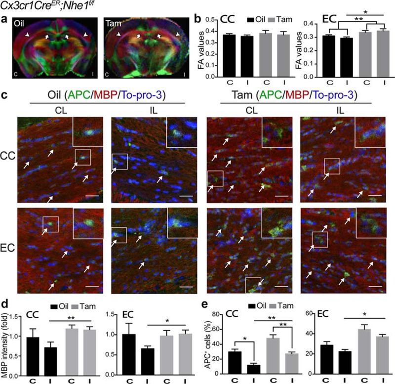
FIGURE 6.

Tam-treated Cx3cr1-CreER;Nhe1f/f mice enhanced white matter repair at the subacute phase post-ischemia. (a) Representative Directionally Encoded Color (DEC) maps of ex vivo oil- and Tam-treated Cx3cr1-CreER;Nhe1f/f brains at 14 days Rp. C, contralateral hemisphere; I, ipsi-lateral hemisphere. Arrow: corpus callosum (CC). Arrowhead: external capsule (EC). (b) Quantitative analysis of fractional anisotropy (FA) values in CC and EC of oil- or Tam-treated Cx3cr1-CreER;Nhe1f/f brains at 14 days Rp. C, contralateral hemisphere; I, ipsilateral hemisphere. Data are mean ± SEM. N = 4. *p < .05, **p < .01. (c) Representative immunofluorescent images of APC, MBP, and To-pro-3 staining in CC and EC areas in the CL and IL of oil- and Tam-treated Cx3cr1-CreER;Nhe1f/f brains at 14 days Rp. (d) Quantitative analysis of MBP intensity (fold change) in CC and EC areas in the CL (C) and IL (I) of oil- and Tam-treated Cx3cr1-CreER;Nhe1f/f brains at 14 days Rp. Data are mean ± SEM. N = 4. *p < .05, **p < .01. (e) Quantitative analysis of APC+ cell percentages in CC and EC areas in the CL (C) and IL (I) of oil- and Tam-treated Cx3cr1-CreER; Nhe1f/f brains at 14 days Rp. Data are mean ± SEM. N = 4. *p < .05, **p < .01 [Color figure can be viewed at wileyonlinelibrary.com]
