

Abstract
The development of a reliable electrochemical sensor using a hybrid nanocomposite consisting of ionic liquid (1-butyl-3-methylimidazolium hexafluorophosphate) functionalized graphene oxide (GrO-IL) and gold nanoparticles (AuNPs) stabilized by chitosan (Chit) was described. The new sensor, labelled as GrO-IL-AuNPs-Chit/CSE, exhibited an improved electrocatalytic response to cancer drugs such as purine antimetabolites (6-thioguanine, 6-mercaptopurine, and azathioprine) in a wide concentration range with a low detection limit (20–40 nmol·L−1, S/N = 3), and satisfactory recoveries (97.1–103.0%). The sensor has been also successfully used for cyclic voltammetric study of a salmon sperm double-stranded DNA degradation and DNA-6-mercaptopurine interaction in aqueous solutions (pH 7.4).
1. Introduction
The family of purines and purine analogs is the most common class of nitrogen-containing heterocyclic compounds which play crucial role in a wide variety of functions of living organisms. In particular, purine bases like adenine (9H-purin-6-amine, Ade) and guanine (2-amino-9H-purin-6(1H)-one, Gua) are components of nucleosides, the building blocks of DNA and RNA [1]. The concentration changes of Gua and Ade and their ratio in the DNA could be considered as important indicators in the clinical diagnosis and treatment of various diseases [2]. The purine antimetabolites such as 6-thioguanine (2-amino-1H-purine-6(7H)-thione, TG), 6-mercaptopurine (3,7-dihydro-purine-6-thione, MP), and azathioprine (6-([(1-methyl-4-nitro-1H-imidazol-5-yl)-sulphonyl]-7H-purine, AZTP) are commonly used in oral chemotherapy for the treatment of leukemia and became increasably popular in therapy of inflammatory diseases (ulcerative colitis, dermatitis, and some other pathologies) [3–5]. The cytostatic effect of all these compounds is associated with impaired synthesis of nucleic acids (DNA and RNA). The action mechanism of thiopurine drugs is probably due to the fact that they may interfere with the DNA synthesis and inhibit the proliferation of quickly growing cells, especially cells of the immune system, after the metabolic conversion to thiopurine nucleotides that substitute regular Ade and Gua nucleotides [6]. Not coincidentally, the chemistry of purine antimetabolites is still one of the most important areas of scientific research. Among the known publications on this topic, a lot of analytical techniques such as HPLC/LC [7–16], CE [17–19], FIA [20, 21], and SIA [22] have been proposed for the determination of these substances in pharmaceutical and clinical samples. The spectrophotometric, spectrofluorimetric, or chemiluminescence detection was based on the oxidation of an imidazole ring in their molecules by typical oxidizing agents [23–28]. The redox activity of purine antimetabolites determined the possibility of their electrochemical detection by using (bio)sensors based on carbon nanotubes-modified materials [29–38]. In recent years, a few studies have been reported, in which graphene- (Gr-) or graphene oxide- (GrO-) based nanocomposites were proposed as electrochemical sensing materials for thiopurines [39–43]. For instance, AZTP sensors were fabricated by electrodeposition of Gr-Chit composite onto the GCE surface [39] and by modification of a graphite electrode by Gr nanosheets decorated with Ag nanoparticles [40]. The development of a pencil graphite electrode modified with poly(neutral red)-electrochemically reduced GrO composite was described for sensing TG in biological and pharmaceutical samples [41]. In another paper [42], the voltammetric behaviour of TG was comparatively investigated on GrO- and reduced GrO-modified carbon paste electrodes. The voltammetric determination of MP at Co(III) tris-phenanthroline complex immobilized on a GCE modified with GrO-decorated DNA was described [43]. In addition, reduced GrO-modified electrode was fabricated to investigate the electrochemical oxidation of nucleic acids [44]. Recently, we proposed the electrochemical sensor based on the GrO-IL nanocomposite immobilized into a Chit biopolymeric matrix [45]. The major aim of the present study was to prepare a more sensitive and reliable electrode material, considering integration of the unique properties of GrO-IL and electrocatalytic activity of gold nanopieces- (AuNPs-) Chit bioconjugate. According to our knowledge, this concept has not been explored to achieve better electrochemical properties. The analytical perspectives and applicability of the new sensor for the voltammetric analysis of thiopurine anticancer drugs and for probing the double-stranded deoxyribonucleic acid (ds-DNA) damage were evaluated.
2. Experimental
2.1. Reagents and Solutions
GrO (powder), medium molecular weight сhitosan (75–85% deacetylation), HAuCl4·3H2O (99%), 1-butyl-3-methylimidazolium hexafluorophosphate ([BMIM]PF6), purines and thiopurine drugs (guanine, adenine, azathioprine, 6-thioguanine, and 6-mercaptopurine monohydrate), double-stranded DNA (ds-DNA) from salmon sperm (31149-11G-F), and other chemicals were purchased from Sigma-Aldrich Chemical Co. All compounds were of analytical grade and used as received. Stock solutions of each purine (1.0 mmol·L−1) were prepared by dissolving in 5.0 mmol·L−1 NaOH. Stock solution of native ds-DNA was prepared by dissolving 0.1 g of the sample in 100 mL water. All stock solutions were stored at +4°C in the dark at least one week. Working standard solutions were freshly prepared by stepwise diluting the respective stock solutions with a 0.1 mol·L−1 phosphate buffer solution containing 1.0 mol·L−1 KCl. A chitosan solution (0.5%) was prepared by dissolving 50 mg of Chit in 10 mL 1.0% (v/v) acetic acid. The solutions were deoxygenated by passing nitrogen gas. Hyperpure water was used throughout the experiment.
2.2. Apparatus
All electrochemical experiments were performed using an Ecotest-VA analyser (Econix-Expert, Russia) interfaced to a computer system with MDEV software. A three-electrode system was used, where a modified carbositall Dick electrode (CSE, Volta, Russia, diameter of 3 mm) served as the working electrode, an Ag/AgCl (3 mol·L−1 KCl) served as the reference electrode, and a platinum wire served as the auxiliary electrode. All potentials reported were referred to the Ag/AgCl electrode at 25 ± 1°C. The computer program Origin 2017 based on the Levenberg–Marquardt algorithm was used for signal processing and peak analysis. pH values were tested by using a pH-meter Model OP-110 (Radelkis, Hungary). The ultrasonic bath (Elmasonic One, Germany, 35 kHz ultrasound) was used in all ultrasonic experiments. The source of UV light was a PRO-4 lamp with a power of 4 W. UV-Vis absorbance spectra of the prepared chitosan-stabilized gold solutions were obtained using a JENWAY 6705 spectrophotometer (Bibby Scientific Ltd.). The measurements were carried out using a quartz cell of thickness 1 cm in the wavelength range of 200–700 nm. The surface morphology of GrO and the prepared composites was examined by using a scanning electron microscope (SEM, Carl Zeiss NVision 40, Germany).
2.3. Sensor Design
The fabrication process for the sensor is depicted in Scheme 1.
Scheme 1.

Schematic illustration of the fabrication process for GrO-IL-AuNPs-Chit/CSE.
The hybrid nanocomposite GrO-IL-AuNPs-Chit was prepared in three steps. In the first step, the biogenic synthesis of AuNPs stabilized by Chit was carried out by UV-induced reduction of Au(III) to Au(0) in an acidic solution of Chit, as follows [46]: 1 mL of 2.0 mmol·L−1 HAuCl4 was added to 5 mL of 1.0 wt.% sodium citrate solution, and the mixture was heated to 50°C with constant stirring. Next, 2 mL of this mixture was added to 2.0 mL of the Chit solution (5 mg·mL−1), and the resulting solution was subjected to UV exposure until its colour turned rose-red. The time of UV irradiation was 50 min. Figure 1 displays the UV-Vis spectra of a HAuCl4–Chit solution before and after UV irradiation.
Figure 1.
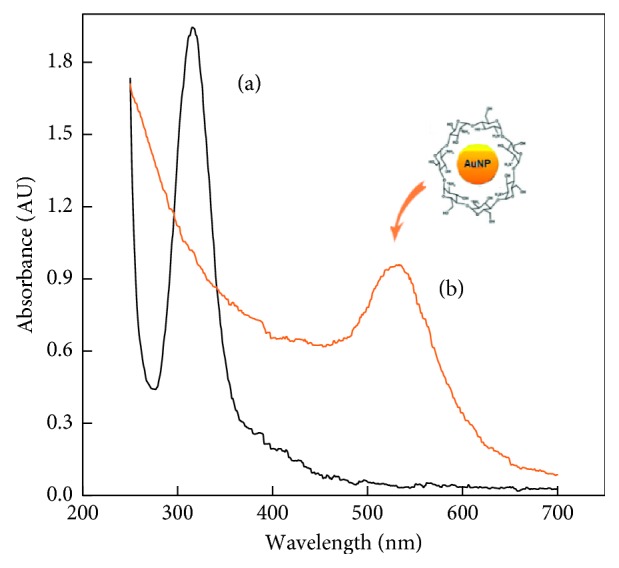
Absorbance spectra of a HAuCl4-Chit solution obtained before (a) and after (b) UV irradiation.
The final spectra indicated that intensive gold reduction occurred during the UV irradiation, as it evidenced by the disappearance of the 315 nm band and the appearance of a new relatively broad band with a maximum of 530 nm, which is due to surface plasmon resonance for metal particles having an average size of about 5 nm [47, 48]. The resulting bioconjugate was labelled as AuNPs-Chit.
In order to improve the electroanalytical properties of GrO, it was modified by a room temperature imidazolium ionic liquid ([BMIM]PF6) [49, 50]. The procedure for the preparation of the GrO-IL nanocomposite have been previously reported in [45]. GrO nanosheets (10 mg) dispersed in 2.0 mL of ethanol were mixed with the ionic liquid (50 μL). Afterwards, the mixture was ultrasonically treated for 1 h, centrifuged, and dried to yield a black-brownish composite labelled as GrO-IL. Then, 10 mg of this composite was mixed with 1.0 mL of AuNPs-Chit bioconjugate solution by using ultrasonication for 30 min, generating the heterogeneous suspension labelled as GrO-IL-AuNPs-Chit. Figure 2 displays the SEM images of GrO-IL, AuNPs-Chit, and hybrid GrO-IL-AuNPs-Chit (c) nanocomposites. The morphology of the hybrid nanocomposite indicated that it was relatively smooth and GrO-IL has been uniformly integrated with AuNPs-Chit.
Figure 2.
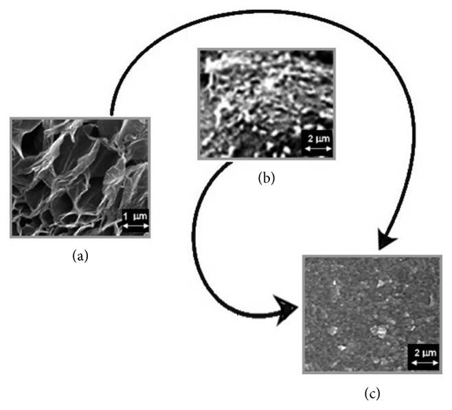
SEM images of the CSE surface covered by GrO-IL nanocomposite (a), AuNPs-Chit bioconjugate (b), and hybrid GrO-IL-AuNPs-Chit nanocomposite (c).
Finally, assembly of the GO-IL-AuNPs-Chit-modified CSE was carried out as described previously [45]. Before modification, the surface of a pure СSE was mechanically polished with 0.3 and 0.05 μm alumina powders. Then, the electrode was thoroughly cleaned with ethanol and deionized water, in order to remove residual alumina particles. The modification of the pretreated CSE was carried by drop casting 5 μL of the GrO-IL-AuNPs-Chit suspension onto its surface followed by subsequent air drying at room temperature for 1 h. Then, in order to stabilize the formed layer, the resulting film was applied with 2 μL of 0.02 mol·L−1 NaOH solution followed by a drying step for 24 h. The electrochemical activation of the fabricated sensor denoted as GrO-IL-AuNPs-Chit/CSE was performed with cyclic voltammetry technique (0.0 – +1.4; 50 mV·s−1, 20 cycles) in PBS (0.1 mol·L−1, pH 7.4). The CSEs modified with only GrO-Chit and GrO-IL-Chit were also prepared through similar procedure for comparison.
2.4. Preparation of DNA Samples
Thermally denatured ds-DNA was prepared according to the literature [51]. Ultrasonic irradiation of ds-DNA was carried out in a sonication bath in 0.1 mol·L−1 phosphate buffer solution (PBS, pH 7.4) for 15 min. Acidic denaturation of ds-DNA was done by treatment in 0.5 mol·L−1·НСlO4 as described early [52].
3. Results and Discussion
3.1. Comparative Electrochemical Characterisation of Various Decorated CSEs
The prepared sensing nanomaterials were characterized by cyclic voltammetry (CV) in 5.0 mmol·L−1 K3[Fe(CN)6] solution containing 1.0 mol·L−1 KCl at a scan rate v from 0.01 to 0.30 V·s−1. As could be seen from the CVs presented in Figure 3, GrO-Chit/CSE (curve a) showed the smallest quasireversible voltammetric response and the peak-to-peak separation potential ΔEp=|Epa − Epc|=160 mV.
Figure 3.
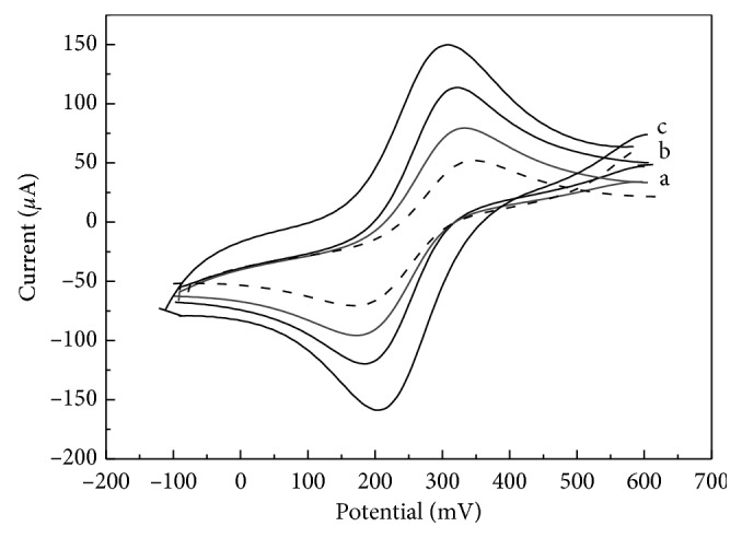
The CVs recorded at GrO-Chit/CSE (a), GrO-IL-Chit/CSE (b), and GrO-IL-AuNPs-Chit/CSE (c) in 5.0 mmol·L−1 K3[Fe(CN)6] containing 1.0 mol·L−1 KCl (v = 0.1 V·s−1). The voltammogram recorded at a bare CSE is indicated by a dotted line.
In case of CSE covered with GrO-IL-Chit composite film, an obvious increase in the voltammetric response was observed (Figure 3(b)). It should be noted that intercalating IL in GrO can enhance the distance between the layers of GrO, resulting a higher electroactive surface area of the sensor (Aact = 0.144 ± 0.005 cm2) compared to pure GrO (Aact = 0.116 ± 0.003 cm2). After CSE modification with the GrO-IL-AuNPs-Chit composite film, the redox peak currents further increased and ΔEp value obviously decreased up to 97 mV (Figure 3(c)). This may be due to the largest effective surface area of GrO-IL-AuNPs-Chit/CSE (Aact = 0.203 ± 0.005 cm2) as well as to the excellent electrocatalytic activity of AuNPs.
Thus, it may be concluded that the GrO-IL-AuNPs-Chit hybrid composite showed advantages for the application as the sensing support material for the sensor.
3.2. Voltammetric Behaviour and Quantitation of Thiopurines at GrO-IL-AuNPs-Chit/CSE
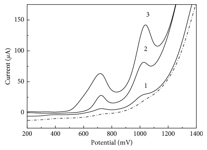
The CV behaviour of thiopurines at GrO-IL-AuNPs-Chit/CSE was investigated in 0.1 M PBS (pH 7.4) in comparison with GrO-IL-Chit/GCE. It is possible to see from Figure 4 that both sensors provided similar electrochemical behaviour. 6-TG exhibited only anodic peaks, indicating that its oxidation is an irreversible one. In contrast, the well-defined anodic and cathodic peaks were recorded for MP. AZTP, an imidazolyl derivative of MP, exhibited one additional oxidation peak at about 0 V, which can be related to the formation of nitroso derivative of AZTP [45]. The second anodic peak may be due to electrochemical reactions involving the imidazole ring oxidation, like in case of TG.
Figure 4.

CVs recorded in 100 μmol·L−1 solutions of TG, AZTP, and MP (PBS, pH 7.4; v = 0.1 V·s−1) at GrO-IL-Chit/GCE (A) and GrO-IL-AuNPs-Chit/CSE (B).
The presence of AuNPs in the new composite film highly promoted the voltammetric response toward all investigated compounds (Figure 4, curves B): the oxidation potentials shifted negatively with enhanced peak currents compared to GrO-IL-Chit/GCE (curves A). The above-described effects might be mainly explained not only to the synergistic effect of GrO-IL and AuNPs but also to the fact that thio-containing compounds are capable to bind to AuNPs-containing surfaces by formation of Au-S bonds. Table 1 summarizes the analytical performance of the proposed sensor for the selected thiopurines.
Table 1.
Main analytical results obtained for the voltammetric detection of the selected thiopurines using. GrO-IL-AuNPs-Chit/CSE (PBS, pH 7.4).
| Analyte | E p, V | Regression equation ΔI (Ipa − Iblank), μA·vs. c (μmol·L−1) | Linear range (µmol·L−1) | LOD (μmol·L−1) (S/N = 3) | Recovery, % |
|---|---|---|---|---|---|
| TG | +0.90 ± 0.02 | ΔI∗ = 4.215c + 0.045 (R2 = 0.9989) | 0–10 | 0.02 | 97.1–102.0 |
| ΔI = 1.528c + 0.083 (R2 = 0.9998) | 10–150 | 99.7–101.5 | |||
|
| |||||
| AZTP | +1.11 ± 0.01 | ΔI∗ = 2.660c + 0.056 (R2 = 0.9991) | 0–10 | 0.04 | 96.5–103.0 |
| ΔI = 1.859c + 0.091 (R2 = 0.9991) | 10–100 | 98.2–101.6 | |||
|
| |||||
| MP | −0.54 ± 0.01 | ΔI∗ = 2.790c + 0.036 (R2 = 0.9990) | 0–20 | 0.03 | 97.4–102.3 |
| ΔI = 0.938c + 0.065 (R2 = 0.9994) | 20–200 | 99.5–100.7 | |||
∗Accumulation time (tacc): 120 s.
The data show that GrO-IL-AuNPs-Chit/CSE provided very high detection sensitivity and wide linear ranges (two linear sections) with relatively low limits of detection (LODs). It characterized by good recoveries (Table 1) and storage stability for at least one month. Comparative evaluation of the developed sensor and Gr- (GrO-) based sensors found in the literature is given in Table 2. As it can be seen, the new sensor is characterized by higher sensitivity and a low detection limit for all three thiopurines.
Table 2.
Comparison of the Gr- (or GrO-) based sensors proposed for the determination of thiopurines by using adsorptive stripping voltammetry.
| Compound | Sensor | Sensitivity (μA/μmol·L−1) | Linear range (μmol·L−1) | LOD (μmol·L−1) | t acc (s) | Reference |
|---|---|---|---|---|---|---|
| AZTP | Gr-Chit/GCE | 0.46 | 0.1–2.0 | 0.05 | 120 | [39] |
| Ag-Gr/GE | 4.74 | 0.7–100 | 0.07 | 50 | [40] | |
| GrO-IL-AuNPs-Chit/CSE | 2.66 | 0.0–10 | 0.04 | 120 | This work | |
|
| ||||||
| TG | RGrO/CPE | 0.23 | 0.4–50 | 0.07 | 40 | [41] |
| Poly(neutral red)-ERGrO/PGE | 0.08 | 0.7–475 | 0.12 | 150 | [42] | |
| GrO-IL-AuNPs-Chit/CSE | 4.22 | 0.0–10 | 0.02 | 120 | This work | |
|
| ||||||
| MP | [Co(phen)3]3+/GrO-DNA/GCE | 0.29 | 0.05–2.0 | 0.02 | [43] | |
| GrO-IL-AuNPs-Chit/CSE | 2.79 | 0.0–20 | 0.03 | 120 | This work | |
GE: graphite electrode; GCE: glassy carbon electrode; CPE: carbon paste electrode; PGE: pencil graphite electrode.
3.3. Voltammetric Detection of ds-DNA at GrO-IL-AuNPs-Chit/CSE
It is well known that degradation of DNA in living organisms leads to mutations and the development of diseases. In this connection, evaluation of the intensity of this process is of great importance, in particular for environmental monitoring of genotoxic compounds [53]. Nanomaterial-modified electrodes can provide very simple and sensing platforms for DNA electroanalysis [54, 55]. The developed GrO-IL-AuNPs-Chit/CSE was found to have excellent adsorption ability and electrocatalytic activity towards the irreversible oxidation of the fish sperm ds-DNA in aqueous solutions (pH 7.4). Therefore, the given sensor was used to study degraded ds-DNA samples by means of adsorptive voltammetry approach. The accumulation of ds-DNA was performed in a stirred solution containing 10.0 μg·mL−1 of the nucleic acid at open circuit potential for 180 s. After washing the electrode for 10 s with a buffer solution, the anodic voltammograms were recorded from +0.2 V to +1.4 V at the scan rate of 100 mV·s−1. As can be observed from Figure 5, the large difference in the oxidation signals is produced by the thermally degraded ds-DNA, ultrasonically irradiated ds-DNA, and acid treated ds-DNA samples. Voltammetric measurements in the solutions of both ultrasonically irradiated and acid-treated ds-DNA showed two well-defined oxidation peaks located around 0.7 V and 1.0 V (Figure 5, curves 2 and 3). These peaks can be attributed to the oxidation of DNA's purine bases (Gua and Ade)—residues of partial depurination of ds-DNA molecules. A noticeable decrease in anode peaks obtained in the thermally denatured ds-DNA solution could be explained by the inaccessibility of electroactive centers for the electron transfer. In this case ds-DNA acted like ss-DNA. The LOD for the thermally, ultrasonically, and perchloric acidic denatured ds-DNA was 0.5 μg·mL−1, 0.3 μg·mL−1, and 0.1 μg·mL−1, respectively.
Figure 5.
Adsorptive stripping voltammograms of thermally denatured ds-DNA (1), ultrasonically irradiated ds-DNA (2), and acid-treated ds-DNA (3) at GrO-IL-AuNPs-Chit/CSE after accumulation for 180 s under open circuit. The native DNA voltammogram is indicated by a dotted line.
3.4. ds-DNA-MP Interaction Study
DNA is the pharmacological goal of many drugs. The interaction of DNA with small molecules represents a fundamental issue in life science and pharmaceutical screening, and it has been the subject of several investigations [55–58]. In order to investigate the possible interaction of the fish sperm ds-DNA with MP, GO-IL-AuNPs-Chit/CSE was immersed into 10 mL of the deoxygenated PBS (pH 7.4) containing 100 μg·mL−1 MP and kept for 240 s under open circuit, for the MP immobilisation onto the electrode surface. Next, the interaction of MP with a 100 μg·mL−1 solution of native ds-DNA (pH 7.4) was performed during different contact time periods ranging from 1 to 30 min at 37°C. The monitoring of the process was explored by measuring the changes of the MP voltammetric signals at −0.54 V and +0.38 V (the native ds-DNA was found to be electrochemically inactive in this potential range). As can be seen from Figure 6, the MP peak currents greatly decreased after the contact with ds-DNA. This effect can be corresponded to the probable intercalating of MP between purine base pairs of ds-DNA. The decrease in the MP signal was calculated as about 44% (n=5) by using 30 min of interaction time. The obtained results indicate that the interaction process of MP with ds-DNA is mainly the intercalation mode.
Figure 6.
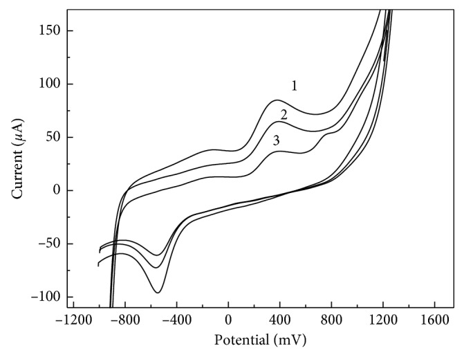
CVs recorded at GrO-IL-AuNPs-Chit/CSE during the interaction between MP and native ds-DNA. Interaction time in min: 3 (1), 10 (2), and 30 (3). Concentration of ds-DNA was 100 μg·mL−1; the supporting electrolyte is 0.1 mol·L−1 PBS, pH 7.4; v = 0.1 V·s−1.
4. Conclusion
In this work, a novel electroactive material was proposed for the fabrication of a nonenzymatic electrochemical sensor. This material is a hybrid nanocomposite that combines a large GrO-IL surface area and highly conductive AuNPs stabilized with Chit functional groups. Cyclic voltammetric results confirmed that the developed sensor clearly exhibited the most electrochemical activity towards the electrooxidation of purine antimetabolites (6-thioguanine, 6-mercaptopurine, and azathioprine). The results demonstrated that it can be used for the determination of these compounds in a wide linear concentration range (up to 100–200 μmol·L−1) with a high sensitivity (2.66–4.22 μA/μmol·L−1), a low detection limit (20–40 nmol·L−1, S/N = 3), and satisfactory recoveries (97.1–103.0%). Besides, the given sensor may be applied to study the double-stranded DNA damage and its interaction with anticancer drug 6-mercaptopurine in phosphate buffer solutions (pH 7.4) by using simple CV technique.
Data Availability
The data used to support the findings of this study are included within the article.
Disclosure
This work was carried out within the framework of the state assignment for the IGIC RAS in the field of fundamental scientific research.
Conflicts of Interest
The authors declare that they have no conflicts of interest.
References
- 1.Ulbricht T. L. V. Purines, Pyrimidines and Nucleotides and the Chemistry of Nucleic Acids. Amsterdam, Netherlands: Elsevier; 1964. [Google Scholar]
- 2.Elion G. B. The purine path to chemotherapy. Science. 1989;244(4900):41–47. doi: 10.1126/science.2649979. [DOI] [PubMed] [Google Scholar]
- 3.Thurston D. E. Chemistry and Pharmacology of Anticancer Drugs. Boca Raton, Florida, USA: CRC Press. Taylor and Francis Group; 2007. [Google Scholar]
- 4.Elgemeie G. H. Thioguanine, mercaptopurine: their analogs and nucleosides as antimetabolites. Current Pharmaceutical Design. 2003;9(31):2627–2642. doi: 10.2174/1381612033453677. [DOI] [PubMed] [Google Scholar]
- 5.Duley J. A., Florin T. H. J. Thiopurine therapies. Therapeutic Drug Monitoring. 2005;27(5):647–654. doi: 10.1097/01.ftd.0000169061.52715.3e. [DOI] [PubMed] [Google Scholar]
- 6.Cara C. J., Pena A. S., Sans M., et al. Reviewing the mechanism of action of thiopurine drugs: towards a new paradigm in clinical practice. Medical Science Monitor. 2004;210(11):RA247–RA254. [PubMed] [Google Scholar]
- 7.Os E. C., McKinney J. A., Zins B. J., et al. Simultaneous determination of azathioprine and 6-mercaptopurine by high-performance liquid chromatography. Journal of Chromatography. B, Biomedical Sciences and Applications. 1996;679:147–154. doi: 10.1016/0378-4347(96)00007-2. [DOI] [PubMed] [Google Scholar]
- 8.Erb N., Haverland U., Harms D., Escherich G., Jankaschaub G. High-performance liquid chromatographic assay of metabolites of thioguanine and mercaptopurine in capillary blood. Journal of Chromatography B. 2003;796(1):87–94. doi: 10.1016/j.jchromb.2003.08.006. [DOI] [PubMed] [Google Scholar]
- 9.Olesen K.-M., Hansen S. H., Sidenius U., Schmiegelow K. Determination of leukocyte DNA 6-thioguanine nucleotide levels by high-performance liquid chromatography with fluorescence detection. Journal of Chromatography B. 2008;864(1-2):149–155. doi: 10.1016/j.jchromb.2008.02.007. [DOI] [PubMed] [Google Scholar]
- 10.Kusmierek K., Chwatko G., Głowacki R., Bald E. Determination of endogenous thiols and thiol drugs in urine by HPLC with ultraviolet detection. Journal of Chromatography B. 2009;887(28):3300–3308. doi: 10.1016/j.jchromb.2009.03.038. [DOI] [PubMed] [Google Scholar]
- 11.Zakrzewski R. Development and validation of a reversed-phase HPLC method with post-column iodine-azide reaction for the determination of thioguanine. Journal of Analytical Chemistry. 2009;64(12):1235–1241. doi: 10.1134/s1061934809120065. [DOI] [Google Scholar]
- 12.Hawwa A. F., Millership J. S., Collier P. S., McElnay J. C. Development and validation of an HPLC method for the rapid and simultaneous determination of 6-mercaptopurine and four of its metabolites in plasma and red blood cells. Journal of Pharmaceutical and Biomedical Analysis. 2009;49(2):401–409. doi: 10.1016/j.jpba.2008.10.045. [DOI] [PubMed] [Google Scholar]
- 13.Nussbaumer S., Bonnabry P., Veuthey J.-L., Fleury-Souverain S. Analysis of anticancer drugs: a review. Talanta. 2011;85(5):2265–2289. doi: 10.1016/j.talanta.2011.08.034. [DOI] [PubMed] [Google Scholar]
- 14.Jacobsen J. H., Schmiegelow K., Nersting J. Liquid chromatography-tandem mass spectrometry quantification of 6-thioguanine in DNA using endogenous guanine as internal standard. Journal of Chromatography B. 2012;881-882:115–118. doi: 10.1016/j.jchromb.2011.11.032. [DOI] [PubMed] [Google Scholar]
- 15.Karadas-Bakirhan N., Sarakbi A., Vandeput M., Ozkan S. A., Kauffmann J.-M. Liquid chromatography with amperometric detection at a silver based detector for the determination of thiocompounds: application to the assay of thiopurine antimetabolites in urine. Analytical Chemistry. 2015;87(13):6730–6735. doi: 10.1021/acs.analchem.5b00879. [DOI] [PubMed] [Google Scholar]
- 16.Sun Y., Yao T., Guo X., Peng Y., Zheng J. Simultaneous assessment of endogenous thiol compounds by LC-MS/MS. Journal of Chromatography B. 2016;1029-1030:213–221. doi: 10.1016/j.jchromb.2016.06.024. [DOI] [PubMed] [Google Scholar]
- 17.Lochman P., Adam T., Friedecký D., Hlídková E., Škopková Z. High-throughput capillary electrophoretic method for determination of total aminothiols in plasma and urine. Electrophoresis. 2003;24(7-8):1200–1207. doi: 10.1002/elps.200390154. [DOI] [PubMed] [Google Scholar]
- 18.Wang C.-C., Chiou S.-S., Wu S.-M. Determination of mercaptopurine and its four metabolites by large-volume sample stacking with polarity switching in capillary electrophoresis. Electrophoresis. 2005;26(13):2637–2642. doi: 10.1002/elps.200500003. [DOI] [PubMed] [Google Scholar]
- 19.Huang Y., Zhao S., Shi M., Liang H. A microchip electrophoresis strategy with online labeling and chemiluminescence detection for simultaneous quantification of thiol drugs. Journal of Pharmaceutical and Biomedical Analysis. 2011;55(5):889–894. doi: 10.1016/j.jpba.2011.03.007. [DOI] [PubMed] [Google Scholar]
- 20.Wang L., Ling B., Chen H., Liang A., Qian B., Fu J. Flow injection chemiluminescence determination of 6-mercaptopurine based on a new system of potassium permanganate-thioacetamide-sodium hexametaphosphate. Luminescence. 2010;25(6):431–435. doi: 10.1002/bio.1171. [DOI] [PubMed] [Google Scholar]
- 21.Wang J., Zhao P., Han S. Direct determination of azathioprine in human fluids and pharmaceutical formulation using flow injection chemiluminescence analysis. Journal of the Chinese Chemical Society. 2012;59(2):239–244. doi: 10.1002/jccs.201100369. [DOI] [Google Scholar]
- 22.Shpigun L. K., Andryukhina E. Yu., Protasov A. S. Development and validation of sequential injection amperometric system for analysis of thiopurine antimetabolic drugs. Journal of Flow Injection Analysis. 2017;34(2):154–159. [Google Scholar]
- 23.Chalmers A. H. A spectrophotometric method for the estimation of urinary azathioprine, 6-mercaptopurine, and 6-thiouric acid. Biochemical Medicine. 1975;12(3):234–241. doi: 10.1016/0006-2944(75)90125-8. [DOI] [PubMed] [Google Scholar]
- 24.Bhaskar M., Manohara Y., Gayasuddin M., Balaraju M., Kumar T. B. Spectrophotometric determination of azathioprine in bulk and pharmaceutical dosage forms. International Journal of ChemTech Research. 2010;2(1):376–378. [Google Scholar]
- 25.Wang L., Zhang Z. The study of oxidization fluorescence sensor with molecular imprinting polymer and its application for 6-mercaptopurine (6-MP) determination. Talanta. 2008;76(4):768–771. doi: 10.1016/j.talanta.2008.04.024. [DOI] [PubMed] [Google Scholar]
- 26.Shen X., Jiang L., Liang H., Lu X., Zhang L., Liu X. Determination of 6-mercaptopurine based on the fluorescence enhancement of Au nanoparticles. Talanta. 2006;69(2):456–462. doi: 10.1016/j.talanta.2005.10.017. [DOI] [PubMed] [Google Scholar]
- 27.Gao M. X., Xu J. L., Li Y. F., Huang C. Z. A rapid and sensitive spectrofluorometric method for 6-mercaptopurine using CdTe quantum dots. Analytical Methods. 2013;5(3):673–677. doi: 10.1039/c2ay25971k. [DOI] [Google Scholar]
- 28.Yang P., Chen Y., Zhu Q., Wang F., Wang L., Li Y. Sensitive chemiluminescence method for the determination of glutathione, L-cysteine and 6-mercaptopurine. Microchimica Acta. 2008;163(3-4):263–269. doi: 10.1007/s00604-008-0006-5. [DOI] [Google Scholar]
- 29.Cao X., Lin L., Zhou Y. Y., et al. Amperometric determination of 6-mercaptopurine on functionalized multi-wall carbon nanotubes modified electrode by liquid chromatography coupled with microdialysis and its application to pharmacokinetics in rabbit. Talanta. 2003;60(5):1063–1070. doi: 10.1016/s0039-9140(03)00187-5. [DOI] [PubMed] [Google Scholar]
- 30.Lu B.-Y., Li H., Deng H., Xu Z., Li W.-S., Chen H.-Y. Voltammetric determination of 6-mercaptopurine using [Co(phen)3]3+/MWNT modified graphite electrode. Journal of Electroanalytical Chemistry. 2008;621(1):97–102. doi: 10.1016/j.jelechem.2008.04.018. [DOI] [Google Scholar]
- 31.Ensafi A. A., Karimi-Maleh H. Modified multiwall carbon nanotubes paste electrode as a sensor for simultaneous determination of 6-thioguanine and folic acid using ferrocenedicarboxylic acid as a mediator. Journal of Electroanalytical Chemistry. 2010;640(1-2):75–83. doi: 10.1016/j.jelechem.2010.01.010. [DOI] [Google Scholar]
- 32.Beitollahi H., Raoof J.-B., Hosseinzadeh R. Electroanalysis and simultaneous determination of 6-thioguanine in the presence of uric acid and folic acid using a modified carbon nanotube paste electrode. Analytical Sciences. 2011;27(10):991–997. doi: 10.2116/analsci.27.991. [DOI] [PubMed] [Google Scholar]
- 33.Zhou P., He L., Gan G., Ni S., Li H., Li W. Fabrication and evaluation of [Co(phen)2L]3+-modified DNA-MWCNT and SDS-MWCNT electrodes for electrochemical detection of 6-mercaptopurine. Journal of Electroanalytical Chemistry. 2012;665:63–69. doi: 10.1016/j.jelechem.2011.11.026. [DOI] [Google Scholar]
- 34.Shahrokhian S., Ghorbani-Bidkorbeh F., Mohammadi A., Dinarvand R. Electrochemical determinations of 6-mercaptopurine on the surface of a carbon nanotube-paste electrode modified with a cobalt salophen complex. Journal of Solid State Electrochemistry. 2012;16(4):1643–1650. doi: 10.1007/s10008-011-1575-5. [DOI] [Google Scholar]
- 35.Keyvanfard M., Khosravi V., Karimi-Maleh H., Alizad K., Rezaei B. Voltammetric determination of 6-mercaptopurine using a multiwall carbon nanotubes paste electrode in the presence of isoprenaline as a mediator. Journal of Molecular Liquids. 2013;177:182–189. doi: 10.1016/j.molliq.2012.10.020. [DOI] [Google Scholar]
- 36.Sharma V., Jelen F., Trnkova L. Functionalized solid electrodes for electrochemical biosensing of purine nucleobases and their analogues: a review. Sensors. 2015;15(1):1564–1600. doi: 10.3390/s150101564. [DOI] [PMC free article] [PubMed] [Google Scholar]
- 37.Karimi-Maleh H., Shojaei A. F., Tabatabaeian K., Karimi F., Shakeri S., Moradi R. Simultaneous determination of 6-mercaptopruine, 6-thioguanine and dasatinib as three important anticancer drugs using nanostructure voltammetric sensor employing Pt/MWCNTs and 1-butyl-3-methylimidazolium hexafluoro phosphate. Biosensors and Bioelectronics. 2016;86:879–884. doi: 10.1016/j.bios.2016.07.086. [DOI] [PubMed] [Google Scholar]
- 38.Mazloum-Ardakani M., Sheikh-Mohseni M. A., Salavati-Niasari M. A Ruthenium complex/carbon nanotube based electrode as the first electrochemical sensor for simultaneous sensing of D-penicillamine, 6-thioguanine and catecholamines. Electroanalysis. 2016;28(6):1370–1376. doi: 10.1002/elan.201500597. [DOI] [Google Scholar]
- 39.Hatami Z., Jalali F. Voltammetric determination of immunosuppressive agent, azathioprine, by using a graphene-chitosan modified-glassy carbon electrode. Russian Journal of Electrochemistry. 2015;51(1):70–76. doi: 10.1134/s1023193515010097. [DOI] [Google Scholar]
- 40.Asadian E., Iraji zad A., Shahrokhian S. Voltammetric studies of azathioprine on the surface of graphite electrode modified with graphene nanosheets decorated with Ag nanoparticles. Materials Science and Engineering: C. 2016;58:1098–1104. doi: 10.1016/j.msec.2015.09.022. [DOI] [PubMed] [Google Scholar]
- 41.Prasad B. B., Singh R., Kumar A. Development of imprinted polyneutral red/electrochemically reduced graphene oxide composite for ultra-trace sensing of 6-thioguanine. Carbon. 2016;102:86–96. doi: 10.1016/j.carbon.2016.02.031. [DOI] [Google Scholar]
- 42.Smarzewska S., Pokora J., Leniart A., Festinger N., Ciesielski W. Carbon paste electrodes modified with graphene oxides—comparative electrochemical studies of thioguanine. Electroanalysis. 2016;28(7):1562–1569. doi: 10.1002/elan.201501101. [DOI] [Google Scholar]
- 43.Yan Zh., Li H. Voltammetric determination of 6-mercaptopurine at Co(III) tris-phenanthroline complex and DNA decorated with graphene oxide modified glassy carbon electrode. International Journal of Electrochemical Science. 2015;10:8714–8726. [Google Scholar]
- 44.Báez D., Pardo H., Laborda I., Marco J., Yáñez C., Bollo S. Reduced graphene oxides: influence of the reduction method on the electrocatalytic effect towards nucleic acid oxidation. Nanomaterials. 2017;7(7):168–183. doi: 10.3390/nano7070168. [DOI] [PMC free article] [PubMed] [Google Scholar]
- 45.Shpigun L. K., Andryukhina E. Y. Electrochemical sensor based on nanocomposite of ionic liquid modified graphene oxide—chitosan and its application for flow injection detection of anticancer thiopurine drugs. Electroanalysis. 2018;30(10):2356–2365. doi: 10.1002/elan.201800358. [DOI] [Google Scholar]
- 46.Gracheva T. A., Kuzmicheva T. A., Perevezentsev V. N. Kinetics and mechanisms of UV induced formation gold nanoparticles in solutions of chitosan doped with HAuCl4. Journal Technical Physics (Russian) 2017;87(8):1216–1220. [Google Scholar]
- 47.Huang H., Yuan Q., Yang X. Preparation and characterization of metal-chitosan nanocomposites. Colloids and Surfaces B: Biointerfaces. 2004;39(1-2):31–37. doi: 10.1016/j.colsurfb.2004.08.014. [DOI] [PubMed] [Google Scholar]
- 48.Wei D., Qian W. Facile synthesis of Ag and Au nanoparticles utilizing chitosan as a mediator agent. Colloids and Surfaces B: Biointerfaces. 2008;62(1):136–142. doi: 10.1016/j.colsurfb.2007.09.030. [DOI] [PubMed] [Google Scholar]
- 49.Valentini F., Roscioli D., Carbone M., et al. Oxidized graphene in ionic liquids for assembling chemically modified electrodes: a structural and electrochemical characterization study. Analytical Chemistry. 2012;84(13):5823–5831. doi: 10.1021/ac301285e. [DOI] [PubMed] [Google Scholar]
- 50.Wang C.-H., Wu C.-H., Wu J.-W., et al. The effects of ionic liquid on the electrochemical sensing performance of graphene- and carbon nanotube-based electrodes. Analyst. 2013;138(2):576–582. doi: 10.1039/c2an36263e. [DOI] [PubMed] [Google Scholar]
- 51.Jelen F., Fojta M., Paleček E. Voltammetry of native double-stranded, denatured and degraded DNAs. Journal of Electroanalytical Chemistry. 1997;427(1-2):49–56. doi: 10.1016/s0022-0728(96)05030-9. [DOI] [Google Scholar]
- 52.Brett C. M. A., Oliveira Brett A. M., Serrano S. H. P. On the adsorption and electrochemical oxidation of DNA at glassy carbon electrodes. Journal of Electroanalytical Chemistry. 1994;366(1-2):225–231. doi: 10.1016/0022-0728(93)02994-S. [DOI] [Google Scholar]
- 53.Cavalieri E., Saeed M., Zahid M., et al. Mechanism of DNA depurination by carcinogens in relation to cancer initiation. IUBMB Life. 2012;64(2):169–179. doi: 10.1002/iub.586. [DOI] [PMC free article] [PubMed] [Google Scholar]
- 54.Kato D., Niva O. Carbon-based electrode materials for DNA electroanalysis. Analytical Sciences. 2013;29(4):385–392. doi: 10.2116/analsci.29.385. [DOI] [PubMed] [Google Scholar]
- 55.Báez D. F., Bollo S. A comparative study of electrochemical performances of carbon nanomaterial-modified electrodes for DNA detection. Nanotubes or graphene? Journal of Solid State Electrochemistry. 2015;20(4):1059–1064. doi: 10.1007/s10008-015-2997-2. [DOI] [Google Scholar]
- 56.Eksin E., Muti M., Erdem A. Chitosan/ionic liquid composite electrode for electrochemical monitoring of the surface-confined interaction between mitomycin C and DNA. Electroanalysis. 2013;25(10):2321–2329. doi: 10.1002/elan.201300188. [DOI] [Google Scholar]
- 57.Eksin E., Congur G., Mese F., Erdem A. Electrochemical monitoring of surface confined interaction between 6-thioguanine and DNA by using single-use graphite electrode. Journal of Electroanalytical Chemistry. 2014;733:33–38. doi: 10.1016/j.jelechem.2014.08.012. [DOI] [Google Scholar]
- 58.Karadurmus L., Kurbanoglu S., Uslu B., Ozkan S. A. Electrochemical DNA biosensors in drug analysis. Current Pharmaceutical Analysis. 2017;13(3):195–207. [Google Scholar]
Associated Data
This section collects any data citations, data availability statements, or supplementary materials included in this article.
Data Availability Statement
The data used to support the findings of this study are included within the article.