Abstract
Onychomycosis is a fungal infection of the nail caused by dermatophytic (99%) and/or non-dermatophytic (1%) (including yeasts) infections of the nailplate. Among the non-dermatophytes, the yeast Candida albicans, Candida tropicalis, and other molds like Scopulariopsis spp., Scytalidium spp., Fusarium spp., and Aspergillus spp. may be responsible. Herein, we report a case of total dystrophic onychomycosis in a 41-year-old female, caused by Syncephalastrum racemosum and its complete improvement with a combination of oral pulse itraconazole and 1064 nm Q-switched Nd-YAG laser. This case is being reported due to the rarity of causative organism for onychomycosis and a novel approach in its treatment.
Keywords: Nd-YAG laser, onychomycosis, Syncephalastrum racemosum
Introduction
Onychomycosis is defined as a fungal infection of the nail that expands slowly and if left untreated leads to complete destruction of the nail plate. Onychomycosis represents about 30% of all dermatophyte infections and accounts for 18–40% of all nail disorders.[1] The prevalence of onychomycosis ranges between 2% to 28% of the general population and it is estimated to be significantly higher in specific populations such as in diabetes mellitus, the immunosuppressed, and elderly.[2,3] It is caused by dermatophytic (99%) and/or non-dermatophytic (1%) (including yeasts) infections of the nailplate.[1] Among the non-dermatophytes, the yeast Candida albicans, Candida tropicalis, and other molds like Scopulariopsis spp., Scytalidium spp., Fusarium spp., and Aspergillus spp. may be responsible.
Syncephalastrum racemosum is a fungus belonging to order mucorales of class Zygomycetes with very low pathogenicity.[4] It is a saprophytic fungus found ubiquitously in soil and decaying plant debris and is considered to be an uncommon human pathogen. Here, we report a case of total dystrophic onychomycosis caused by S. racemosum and its complete improvement with a combination of oral pulse itraconazole and 1064 nm Q-switched Nd-YAG laser.
Case Report
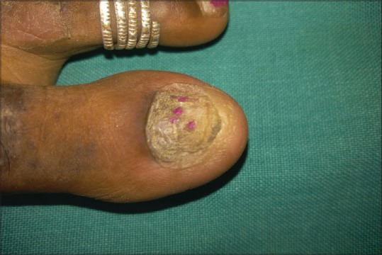
A 41-year-old female patient presented to our OPD with a history of dystrophy of nail of right great toe since last 1 year. She gave history of working in fisheries with continuous immersion of feet in water for longer periods. She had taken multiple oral and topical medications without any improvement. On examination, the nail appeared to be dystrophic with yellowish–brownish discoloration, nail plate thickening, and subungual hyperkeratosis [Figure 1]. There was no sign of inflammation. The nail clippings were taken and sent to microbiology for potassium hydroxide (KOH) mount and culture.
Figure 1.

Total dystrophic onychomycosis of right great toe
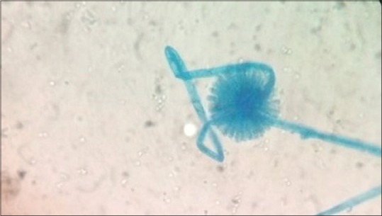
Direct wet mount of nail clippings with 40% of KOH showed thin, hyaline, branched, and septate hyphae. The nail sample was cultured on two different tubes containing Sabouraud Dextrose Agar with and without cycloheximide and incubated at 37°C for 48 h. The tube without cycloheximide showed colony growth but not the one with cycloheximide. The colonies were cotton to fluffy, white-to-gray in color. The lactophenol cotton blue mount showed aseptate hyphae branching sporangiospores with terminal ovoid vesicle, which bear finger-like microsporangia. Based on the above morphological characters, the isolate was identified as S. racemosum [Figure 2].
Figure 2.

Lactophenol cotton blue preparation of Syncephalastrumracemosum
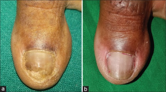
The patient was treated with oral pulse itraconazole therapy for 3 months combined with Q-switched Nd-YAG laser treatment with lasers settings adjusted to yield a fluency of 600 mJ/cm2 over a 5-mm spot at a 3 Hz frequency in a single session. Three sessions of laser treatment were performed at Day 0, 30, and 60. The patient was followed-up for 1 year. There was complete improvement of the nail both clinically and mycologically [Figure 3a and b].
Figure 3.

(a) Clearance of onychomycosis after 3 months of treatment. (b) Clearance of onychomycosis after 9 months of treatment
Discussion
Non-dermatophyte molds cause 2% of total cases of onychomycosis.[1] Only four cases of onychomycosis caused due to S. racemosum have been reported previously.[4,5,6,7] Mucorales have been classically described as having broad (10–50 μm), ribbon-like aseptate hyphae with right-angle branching, the hyphae are actually pauciseptate, and the angle of hyphal branching can vary from 45 to 90°.[8] The other published case of S. racemosum are systemic infections. In the previously reported cases, the patients were treated with debridement, topical nystatin, and oral fluconazole.[4,5,6,7] It is different from our case in which we treated our patient with oral pulse itraconazole combined with Q-switched Nd-YAG laser and the nail improved completely in the subsequent 1 year.
Trauma to nail is an important predisposing factor for onychomycosis. In our case the patient used to work in fisheries, from there she could have acquired the infection. Nowadays onychomycosis due to non-dermatophyte molds are increasing and infection due to S. racemosum is a rare condition. Appropriate diagnosis through culture and proper treatment is necessary because, though not life threatening, onychomycosis can cause significant discomfort to patient.
Declaration of patient consent
The authors certify that they have obtained all appropriate patient consent forms. In the form the patient(s) has/have given his/her/their consent for his/her/their images and other clinical information to be reported in the journal. The patients understand that their names and initials will not be published and due efforts will be made to conceal their identity, but anonymity cannot be guaranteed.
Financial support and sponsorship
Nil.
Conflicts of interest
There are no conflicts of interest.
References
- 1.Kalokasidis K, Onder M, Trakatelli M-G, Richert B, Fritz K, et al. The effect of Q-Switched Nd: YAG 1064nm/532nm laser in the treatment of onychomycosis in vivo. Dermatol Res Pract. 2013;2013:379725. doi: 10.1155/2013/379725. [DOI] [PMC free article] [PubMed] [Google Scholar]
- 2.Gupta AK, Humke S. The prevalence and management of onychomycosis in diabetic patients. Eur J Dermatol. 2000;10:379–84. [PubMed] [Google Scholar]
- 3.Roberts DT. Prevalence of dermatophyte onychomycosis in the United Kingdom: Results of an omnibus survey. Br J Dermatol. 1992;126:23–7. doi: 10.1111/j.1365-2133.1992.tb00005.x. [DOI] [PubMed] [Google Scholar]
- 4.Kumaran R, Rudramurthy KG. Total dystrophic onychomycosis caused by Syncephalastrumrecemosum: A case report. Int J Sci Stud. 2014;2:115–6. [Google Scholar]
- 5.Pavlovic MD, Bulajic N. Great toenail onychomycosis caused by Syncephalastrumracemosum. Dermatol Online J. 2006;12:7. [PubMed] [Google Scholar]
- 6.Baby S, Ramya TG, Geetha RK. Onychomycosis by Syncephalastrumracemosum: Case report from Kerala, India. Dermatol Reports. 2015;7:5527. doi: 10.4081/dr.2017.5527. [DOI] [PMC free article] [PubMed] [Google Scholar]
- 7.Jindal N, Kalra N, Arora S, Arora D, Bansal R. Onychomycosis of toenails caused by Syncephalastrumracemosum: A rare non-dermatophytemould. Indian J Med Microbiol. 2016;34:257–8. doi: 10.4103/0255-0857.176844. [DOI] [PubMed] [Google Scholar]
- 8.Prabhu RM, Patel R. Mucormycosis and entomophthoramycosis: A review of the clinical manifestations, diagnosis and treatment. Clin Microbiol Infect. 2004;10:31–47. doi: 10.1111/j.1470-9465.2004.00843.x. [DOI] [PubMed] [Google Scholar]


