Abstract
Background: Fractures of the radial shaft with disruption of the distal radial ulnar joint (DRUJ) or Galeazzi fractures are treated with reduction of the radius followed by stability assessment of the DRUJ. In rare instances, the reduction of the DRUJ is blocked by interposed structures requiring open reduction of this joint. The purpose of this study is to review all cases of irreducible Galeazzi fracture-dislocations reported in the literature to offer guidelines in the diagnosis and management of this rare injury. Methods: A search of the MEDLINE database, OVID database, and PubMed database was employed using the terms “Galeazzi” and “fracture.” Of the 124 articles the search produced, a total of 12 articles and 17 cases of irreducible Galeazzi fracture-dislocations were found. Results: The age range was 16 to 64 years (mean = 25 years). A high-energy mechanism of injury was the root cause in all cases. More than half of the irreducible DRUJ dislocations were not identified intraoperatively. In a dorsally dislocated DRUJ, a block to reduction in most cases (92.3%) was secondary to entrapment of one or more extensor tendons including the extensor carpi ulnaris, extensor digiti minimi, and extensor digitorum communis, with the remaining cases blocked by fracture fragments. Irreducible volar dislocations due to entrapment of the ulnar head occurred in 17.6% of cases with no tendon entrapment noted. Conclusions: In the presence of a Galeazzi fracture, a reduced/stable DRUJ needs to be critically assessed as more than half of irreducible DRUJs in a Galeazzi fracture-dislocation were missed either pre- or intraoperatively.
Keywords: Galeazzi, fracture, dislocation, ulnar styloid, distal radius
Introduction
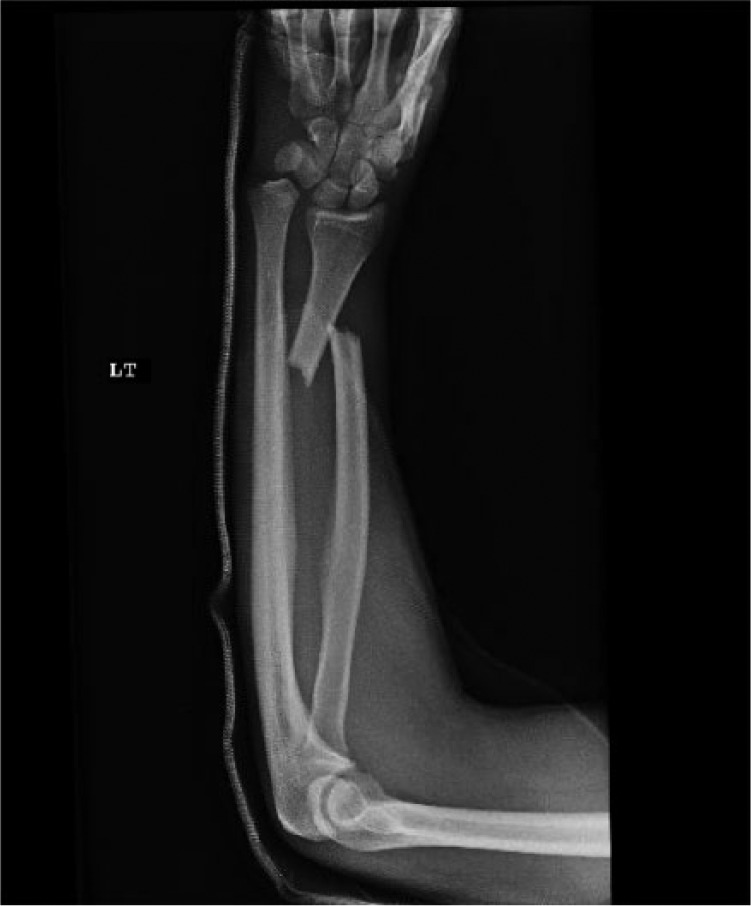
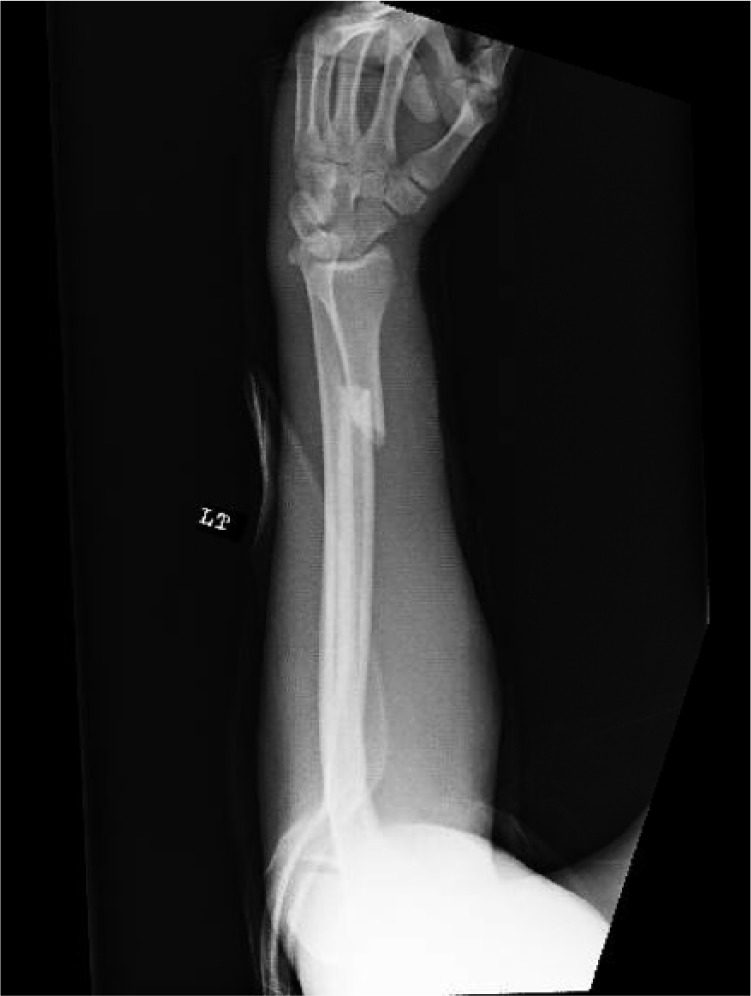
A Galeazzi fracture-dislocation is a fracture of the body of the radius with an associated dislocation or subluxation of the distal radial ulnar joint (DRUJ). The Galeazzi fracture-dislocation is a rare injury occurring in approximately 6.8% of forearm fractures (Figures 1 and 2).21,25 The general mechanism of injury is hyperpronation with axial loading. In 1822, Sir Astley Cooper first described the injury; however, the fracture pattern was named after Riccardo Galeazzi after his presentation of 18 cases in 1934.23
Figure 1.

Anteroposterior view of a left forearm demonstrating disruption of the distal radial ulnar joint.
Figure 2.

Lateral view of a left forearm demonstrating disruption of the distal radial ulnar joint.
There are several classifications for Galeazzi fractures. The AO classification lists the Galeazzi fracture-dislocation as 22-A2.3. Rettig and Raskin classified the Galeazzi fracture-dislocation according to the radius fracture’s proximity to the DRUJ: Type I < 7.5 cm and Type II > 7.5 cm.24 Radiographic signs suggesting rupture of the DRUJ include widening of the DRUJ on posteroanterior view, displacement of the ulna relative to the radius on the lateral view, fracture of the ulnar styloid base, and >5 mm of radial shortening. Moore et al determined that radial shortening of more than 5 mm occurs only with disruption of the triangular fibrocartilage complex (TFCC) or interosseous membrane. Shortening of more than 10 mm results in disruption of both the TFCC and interosseous membrane.21 Bruckner et al further classified the DRUJ dislocation as either simple (reduces spontaneously with reduction of the radius fracture) or complex (requires open reduction to remove any block to reduction to obtain satisfactory alignment of the DRUJ).4
After closed or open reduction, radiographic parameters of successful reduction mirror those used for diagnosis of DRUJ dislocations. Obtaining radiographs of the unaffected side is of critical importance in the diagnosis of continued DRUJ instability. Widening of the radioulnar space relative to the unaffected side on the anteroposterior view indicates continued instability. Widening on the lateral view more than 6 mm between the dorsal cortices indicates continued instability. Widening of 4 to 5 mm warrants further investigation with diagnostic studies such as computed tomography. Magnetic resonance imaging can be utilized but is less diagnostic for bony abnormalities.4,21,23,27
Hughston reported on the poor results of nonoperative management of Galeazzi fracture-dislocations, this was later reaffirmed by Mikic.9,18,26 Anatomic reduction with open reduction and internal fixation (ORIF) of the radius and indirect reduction of the DRUJ is the treatment; however, poor results are frequent secondary to persistent DRUJ instability.15,18,20,26 It is difficult to determine how many of these may be the result of a block to DRUJ reduction. Several cases have been reported describing irreducible Galeazzi fracture-dislocations, identified either pre or postoperatively, with tendon or fracture fragments causing a block to reduction. No study to date has compiled these results to ascertain the likelihood of encountering an irreducible Galeazzi fracture-dislocation either preoperatively or postoperative while possibly identifying the block to reduction requiring open reduction.
Materials and Methods
An electronic literature search was employed via the MEDLINE database, OVID database, and PubMed database (1950 through October 2016) using the search terms “Galeazzi” and “fracture.” The search resulted in 124 articles. Inclusion criteria for the study were a fracture of the radius and an irreducible DRUJ dislocation. Exclusion criteria included subjects with open distal radial physes, radius fracture involving the DRUJ, and radiocarpal dislocations. These cases were excluded as they could cause irreducible dislocations irrespective of the DRUJ dislocation and pediatric patients with open physes can also have causes of an irreducible dislocation that are inconsequential in the adult population.
The abstracts were evaluated for irreducible Galeazzi fracture-dislocations. Eleven articles were retrieved and reviewed. Cited references from these 11 retrieved articles provided an additional single article not identified in the primary search, yielding a total of 12 articles related to irreducible Galeazzi fracture-dislocations. Data were extracted from the information provided by the 12 articles, including from tables and photographs, and compiled into a tabulated form. Causes of irreducible dislocations were then recorded and analyzed.
Results
The search of MEDLINE for irreducible Galeazzi fracture-dislocations resulted in 11 articles. An additional article was identified via cited reference review. From these 12 articles, a total of 17 individuals meeting the inclusion criteria were identified to have sustained an irreducible Galeazzi fracture-dislocation.1-8,10-13
The age range was 16 to 64 years (mean = 25 years). Only 2 patients were older than 26 years; the others were in their late teens and early twenties. Of the articles where the mechanism of injury was specified (11 of 12), high-energy injury was the root cause. These injuries were the result of motorcycle accidents (47%), motor vehicle accidents (MVAs) (23.5%), falls (11.8%), and pedestrian struck (5.9%).
The location of the radius fracture was present in the distal third (41.2%), junction of the distal/middle third (41.2%), middle third (5.9%), and not specified in 2 cases (11.8%). An associated ulnar styloid fracture was identified in 16 of 17 (94.1%) cases. In general, the fractured ulnar styloid was displaced opposite from the ulnar head (76.5%). One ulnar styloid was blocking reduction of the DRUJ at the level of the dorsally displaced ulnar head, and another was missing due to an open DRUJ injury. Dorsal dislocation of the ulnar head accounted for 14 of 17 cases (82.4%).
More than half of irreducible DRUJ dislocations were not identified intraoperatively. The persistent DRUJ dislocations were discovered either on immediate postoperative radiographs or at the first follow-up office visit. One patient was found 2 months postoperatively to have a dislocated DRUJ. This patient eventually underwent a distal ulna excision. The timing to reduction or treatment of missed irreducible DRUJ dislocations ranged from postoperative day 1 to 5 months after the initial surgery.
An entrapped extensor tendon was implicated in 13 of 14 cases (92.3%) of dorsal dislocations. The entrapment of the extensor carpi ulnaris (ECU) alone or in combination with other tendons was present in 11 of 14 dorsal dislocations (78.5%), the extensor digiti minimi (EDM) in 4 of 14 dorsal dislocations (28.6%), and a combined ECU and EDM in 2 of 14 dorsal dislocations (14.3%). The ECU was displaced to the ulnar side of the ulnar head in 8 of 14 dorsal dislocations (57.1%) and to the radial side in 3 of 14 dorsal dislocations (21.4%). There were 3 cases of volar dislocation of the ulna with 2 reported blocks to reduction. One was caused by a volarly directed ulnar fracture fragment and the other was caused by the ulna dislocating through the volar capsule.
Discussion
A Galeazzi fracture-dislocation is a rare type of fracture pattern with an approximate incidence of 6.8% of all forearm fractures.21 The pattern of injury is usually a result of a high-energy injury with hyperpronation of the forearm and axial loading. The pattern of injury is a fracture of the body of the radius with an associated dislocation or subluxation of the DRUJ. The fracture of the radius occurs most often at the distal third or junction of the distal third of the radius, but may occur more proximally. When there is any fracture of the body of the radius, a Galeazzi fracture-dislocation must be suspected. Proper inspection and radiographs to evaluate for a DRUJ injury is a must as it is a commonly missed finding. The DRUJ dislocation is usually dorsal and often reduces well when anatomic alignment of the radius is restored; however, blocks to reduction do occur.2,18,24
The anatomy of the forearm is quite complex in regard to the articulations, osseous configurations, and muscular attachments. The proximal radioulnar joint and the DRUJ are pivot points for the radius to rotate on the ulna, providing the function of supination and pronation to the forearm. The stability of the DRUJ is dependent on multiple intrinsic and extrinsic stabilizers. Intrinsic factors include dorsal and volar radioulnar ligaments, the TFCC, the capsule, and the ulnar collateral ligament. Extrinsic factors include the pronator quadratus muscle, the intraosseous membrane, the ECU subsheath, and the compression of the flexors and extensors on the DRUJ. Instability of the DRUJ can be attributed to many clinical complaints including wrist pain and restricted or painful pronation/supination.27 While compromised pronation/supination may occur due to pathology anywhere along the forearm axis, missed dislocation of the DRUJ after a Galeazzi fracture can account for pain and loss of function14; therefore, restoration of the DRUJ is essential during ORIF of Galeazzi fractures.17,22,27
A fracture of the base of the ulnar styloid has been shown to increase the risk of DRUJ instability in distal radius fractures.16,19 One study reported 31% of Galeazzi fracture-dislocations had an associated fracture of the ulnar styloid.18 In our review of irreducible Galeazzi fracture-dislocations, a fracture of the ulnar styloid was present in 16 of 17 (94.1%) cases.1-8,10-13 Therefore, the presence of an ulnar styloid fracture should alert the treating surgeon to a possible irreducible Galeazzi fracture-dislocation and prompt strict evaluation of radiographs in addition to possible imaging of the contralateral arm.
Our review found extensor tendons to be implicated in the block to reduction in 13 of 14 dorsal dislocations (92.3%).1-6,10-12 The ECU was most frequently implicated, but the EDM must also be evaluated as a possible block to reduction. Volar dislocations were blocked by fracture fragments or the ulna dislocating through the volar capsule.Treating surgeons should be aware that dorsal dislocations are most likely blocked by extensor tendons which need to be extricated in order to properly reduce the DRUJ.
In our review, more than 50% of irreducible DRUJ dislocations were only identified postoperatively leading to persistent DRUJ instability and pain. Identification of irreducible/missed dislocations ranged from postoperative day 1 to 2 months after surgical fixation; therefore, it is imperative to thoroughly examine and perform close follow-up patients who sustain a Galeazzi fracture.1,2,4-8,10-13 Strict examination of radiographs must be performed and if persistent DRUJ dislocation is noted, revision ORIF with reduction of the DRUJ should be performed to prevent previously described sequela of chronic DRUJ dislocations.
One limitation of our study was the inconsistency of the data in the articles reviewed with respect to results and outcomes. We, therefore, were unable to utilize to the standard outcome testing measurement of the Mayo Wrist Score for a uniform presentation of results.
Conclusions
In summary, our review found the most common blocks to reduction in dorsal Galeazzi fracture-dislocations to be an extensor tendon. Blocks to reduction in volar dislocations are due to the ulna dislocating through the capsule, and a volar ulnar fracture fragment. The ECU was found to be the most frequently implicated extensor tendon in blocks to reduction of dorsal dislocations.1,2,4-8,10-13 This information can be helpful to treating surgeons when examining patients with irreducible Galeazzi fractures.
Footnotes
Ethical Approval: This study was approved by our institutional review board.
Statement of Human and Animal Rights: This article does not contain any studies with human or animal subjects.
Statement of Informed Consent: No patient data were included in this study.
Declaration of Conflicting Interests: The author(s) declared no potential conflicts of interest with respect to the research, authorship, and/or publication of this article.
Funding: The author(s) received no financial support for the research, authorship, and/or publication of this article.
References
- 1. Alexander AH, Lichtman DM. Irreducible distal radioulnar joint in a Galeazzi fracture—case report. J Hand Surg Am. 1981;6(3):258-261. [DOI] [PubMed] [Google Scholar]
- 2. Biyani A, Bhan S. Dual extensor tendon entrapment in Galeazzi fracture-dislocation: a case report. J Trauma. 1989;29(9):1295-1297. [DOI] [PubMed] [Google Scholar]
- 3. Borens O, Chehab EL, Roberts MM, et al. Bilateral Galeazzi fracture-dislocations. Am J Orthop. 2006;35(8):369-372. [PubMed] [Google Scholar]
- 4. Bruckner JD, Lichtman DM, Alexander AH. Complex dislocations of the distal radioulnar joint. Recognition and management. Clin Orthop Relat Res. 1992;275:90-103. [PubMed] [Google Scholar]
- 5. Budgen A, Lim P, Templeton P, et al. Irreducible Galeazzi injury. Arch Ortho Trauma Surg. 1998;118(3):176-178. [DOI] [PubMed] [Google Scholar]
- 6. Cetti NE. An unusual cause of blocked reduction of the Galeazzi injury. Injury. 1977;9(1):59-61. [DOI] [PubMed] [Google Scholar]
- 7. Giangarra CE, Chandler RW. Complex volar distal radioulnar joint dislocation occurring in a Galeazzi fracture. J Orthop Trauma. 1989;3(1):76-79. [DOI] [PubMed] [Google Scholar]
- 8. Gunes T, Erdem M, Sen C. Irreducible Galeazzi fracture-dislocation due to intra-articular fracture of the distal ulna. J Hand Surg Eur Vol. 2007;32(2):185-187. [DOI] [PubMed] [Google Scholar]
- 9. Hughston JC. Fracture of the distal radial shaft: mistakes in management. J Bone Joint Surg Am. 1957;39A(2):249-264. [PubMed] [Google Scholar]
- 10. Itoh Y, Horiuchi Y, Takahashi M, et al. Extensor tendon involvement in Smith’s and Galeazzi’s fractures. J Hand Surg Am. 1987;12(4):535-540. [DOI] [PubMed] [Google Scholar]
- 11. Jenkins NH, Mintowt-Czyz WJ, Fairclough JA. Irreducible dislocation of the distal radioulnar joint. Injury. 1987;18(1):40-43. [DOI] [PubMed] [Google Scholar]
- 12. Kikuchi Y, Nakamura T. Irreducible Galeazzi fracture-dislocation due to an avulsion fracture of the fovea of the ulna. J Hand Surg Br. 1999;24(3):379-381. [DOI] [PubMed] [Google Scholar]
- 13. Kim S, Ward JP, Rettig ME. Galeazzi fracture with volar dislocation of the distal radioulnar joint. Am J Orthop. 2012;41(11):E152-E154. [PubMed] [Google Scholar]
- 14. Kleinman WB, Graham TJ. The distal radioulnar joint capsule: clinical anatomy and role in posttraumatic limitation of forearm rotation. J Hand Surg Am. 1998;23(4):588-599. [DOI] [PubMed] [Google Scholar]
- 15. Kraus B, Horne G. Galeazzi fractures. J Trauma. 1985;25(11):1093-1095. [PubMed] [Google Scholar]
- 16. May MM, Lawton JN, Blazar PE. Ulnar styloid fractures associated with distal radius fractures: incidence and implications for distal radioulnar joint instability. J Hand Surg Am. 2002;27(6):965-971. [DOI] [PubMed] [Google Scholar]
- 17. McGinley JC, Kozin SH. Interosseous membrane anatomy and functional mechanics. Clin Orthop Relat Res. 2001;(383):108-122. [DOI] [PubMed] [Google Scholar]
- 18. Mikic ZD. Galeazzi fracture-dislocations. J Bone Joint Surg Am. 1975;57(8):1071-1080. [PubMed] [Google Scholar]
- 19. Mirarchi AJ, Hoyen HA, Knutson J, et al. Cadaveric biomechanical analysis of the distal radioulnar joint: influence of wrist isolation on accurate measurement and the effect of ulnar styloid fracture on stability. J Hand Surg Am. 2008;33(5):683-690. [DOI] [PubMed] [Google Scholar]
- 20. Moore TM, Klein JP, Patzakis MJ, et al. Results of compression-plating of closed Galeazzi fractures. J Bone Joint Surg Am. 1985;67(7):1015-1021. [PubMed] [Google Scholar]
- 21. Moore TM, Lester DK, Sarmiento A. The stabilizing effect of soft-tissue constraints in artificial Galeazzi fractures. Clin Orthop Relat Res. 1985;194:189-194. [PubMed] [Google Scholar]
- 22. Nakamura T, Yabe Y, Horiuchi Y. Functional anatomy of the triangular fibrocartilage complex. J Hand Surg Br. 1996;21(5):581-586. [DOI] [PubMed] [Google Scholar]
- 23. Reckling FW, Pletier LF. Riccardo Galeazzi and Galeazzi’s fracture. Surgery. 1965;58:453-459. [PubMed] [Google Scholar]
- 24. Rettig ME, Raskin KB. Galeazzi fracture-dislocation: a new treatment-oriented classification. J Hand Surg Am. 2001;26(2):228-235. [DOI] [PubMed] [Google Scholar]
- 25. Ring D, Rhim R, Carpenter C, et al. Isolated radial shaft fractures are more common than Galeazzi fractures. J Hand Surg Am. 2006;31(1):17-21. [DOI] [PubMed] [Google Scholar]
- 26. Strehle J, Gerber C. Distal radioulnar joint function after Galeazzi fracture-dislocations treated by open reduction and internal plate fixation. Clin Orthop Relat Res. 1993;293:240-245. [PubMed] [Google Scholar]
- 27. Wijffels M, Brink P, Schipper I. Clinical and non-clinical aspects of distal radioulnar joint instability. Open Orthop J. 2012;6:204-210. [DOI] [PMC free article] [PubMed] [Google Scholar]


