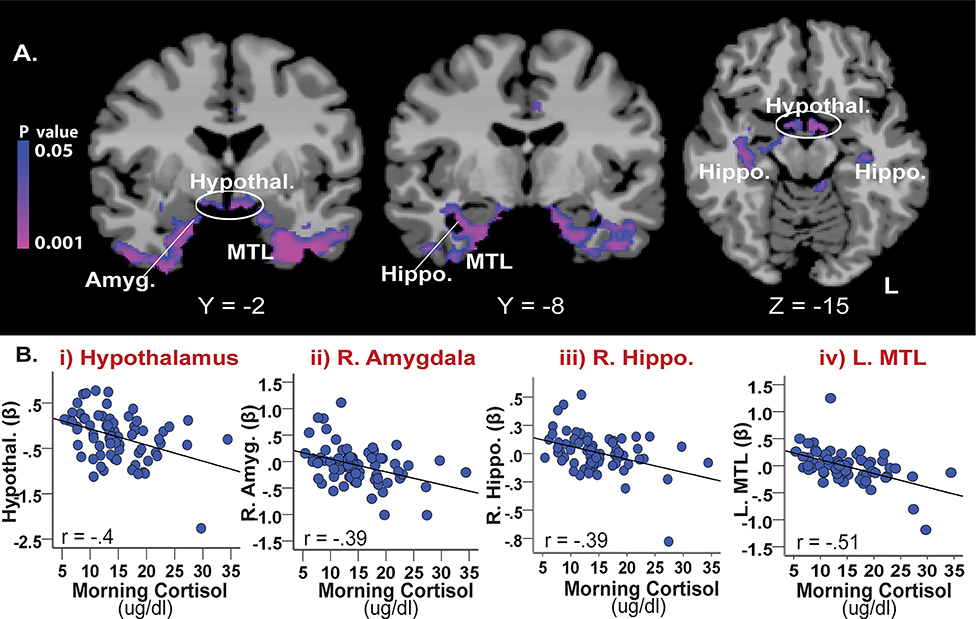
Figure 4.
The association between basal cortisol and stress-related neural activity (Stress-Baseline). (A) Results of a whole brain correlation analysis showed that basal cortisol levels were negatively correlated with stress-related activity in limbic-MTL regions including the amygdala, hippocampus, and medial temporal lobe as well as the hypothalamus (whole-brain FWE corrected, p <0.05). (B) The scatterplots show negative correlated patterns with no outliers between basal cortisol levels and stress-induced activity in the i) hypothalamus, ii) R. amygdala, iii) R. hippocampus, and iv) L. medial temporal lobe. Amyg. = amygdala; Hypothal. = hypothalamus; Hippo. = hippocampus, MTL = medial temporal lobe; R=right; L=left. Blue/purple colors = negative correlations.

