



Individuals with frontotemporal dementia (FTD) experience progressive behavioral changes accompanied by cognitive and functional decline. However, this process is not always one of inexorable decline, as occasionally loss in one area of function can lead to enhancement in others. This phenomenon, known as paradoxical functional facilitation, occurs when suppression or loss of activity in one part of the brain leads to a release of activity in another.1 Over the years, we have cared for many patients with FTD who experience a sudden onset increase in artistic creativity. Here we describe two patients who exemplify the phenomenon.
Patient 1
The patient, a right-handed manager, developed FTD and amyotrophic lateral sclerosis accompanied by an extraordinary surge of new artistic interest and output. His core FTD symptoms began insidiously his late 50s, although he did not come to our attention until several months before his death at the age of 64. Before his illness declared, he was an avid craftsman and builder who enjoyed woodworking and carrying out complicated home renovations. At age 59 he took up welding, first making solid, pragmatic furniture. Within months, he started exploring more creative possibilities, seeing “critters” within junk metal he scavenged: wild-looking birds with heads and beaks made from hammers or pickaxes, wings forged from saw blades, feet fashioned from rebar, and bodies painted royal blue or green, followed by amphibians, small mammals, and flying insect objects d’art (Figure 1).
Figure 1.
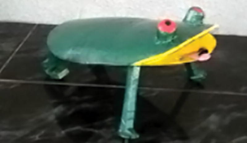


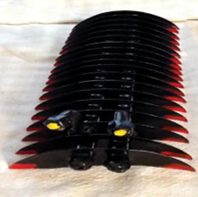

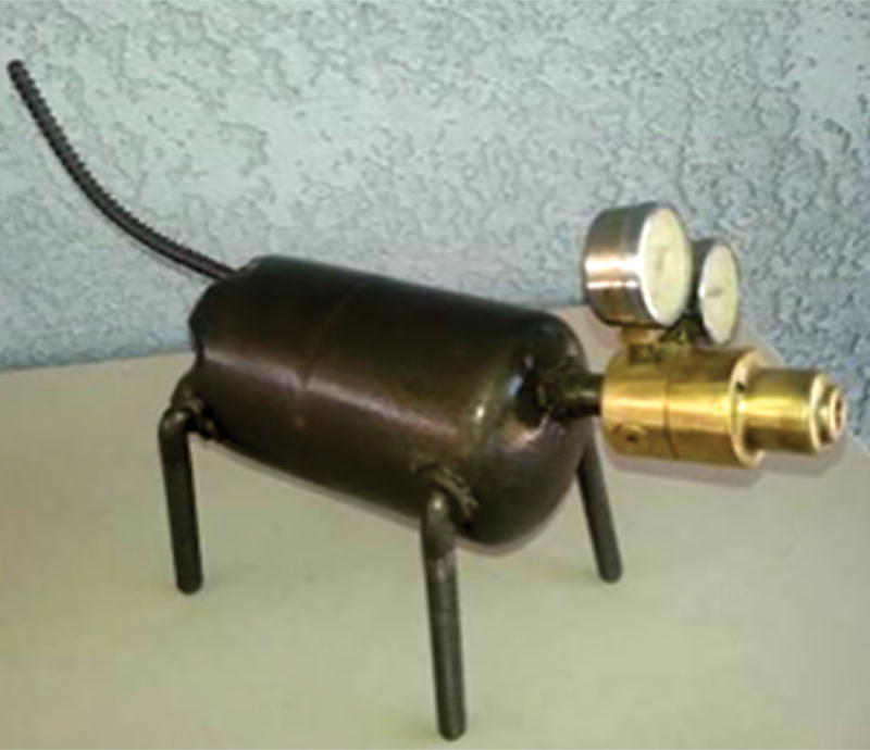

A sampling of artwork by Patient 1. All pieces were created between 2010 and 2013. 2010 (A), 2012 (E), and 2013 (B, C, D, F, G).
In retrospect, his artistic turn was perhaps the earliest manifestation of illness, accompanied by changes in temperament and reduced expressions of empathy. As his disease progressed the distinctive animal features of the sculptures began to disappear and took on more abstract and monochromatic forms. His brain MRI at age 64 was notable for cerebral atrophy with disproportionate involvement of the anterior temporal cortex (Figure 2), a common feature of individuals exhibiting paradoxical functional facilitation. 2 It was not until the final year of his life that he developed prominent changes in motor and language functioning. As for his passion for creating art, he was sculpting butterflies up until the week he died.
Figure 2.

T1-weighted brain MRI images of patient 1 at age 64. The image demonstrates anterior temporal atrophy that is more pronounced on the right.
Patient 2
The patient, a left-handed city employee, was initially admitted to the hospital at age 65 for progressive weakness from myasthenia gravis, during which time he composed 63 poems in 12 days during his stay. Neuropsychological testing, ordered to detect an underlying neurologic cause for the patient’s compulsive behavior, revealed mild impairments in executive function; an MRI showed frontal cortical atrophy affecting the right more than the left. He revealed that he was also a disillusioned painter because people did not like his latest work, which had shifted from peaceful landscapes or still lifes to erotic fantasy themes with mythical or religious undertones (Figure 3). Over the next five years, he demonstrated an increase in socially abnormal behaviors, impulsiveness, disinhibition, and hoarding. Despite his behavioral and cognitive deterioration, the patient’s creativity blossomed, and his artistic style evolved to mix painting and sculpture. At age 69 years old, he reached his artistic apex, embarking on his magnum opus: a 6-square-meter wooden relief painting of demons engaged in sexual acts (Figure 4). He was given the formal diagnosis with FTD at the age of 70, and continues to practice art now three years later.
Figure 3.





Progressive shift in aesthetic style by Patient 2. From left to right, paintings were produced in the 1980s (A & B), 2002 (C), 2008 (D), and 2017 (E).
Figure 4.

Patient 2’s “magnum opus”.
WHAT’S BEHIND THE RISE IN CREATIVITY IN FTD?
Prolific artistic production in the setting of neurodegenerative disease can be conceptualized as a type of compulsive behavior. Obsessive and compulsive tendencies often accompany FTD and are usually simplistic (e.g. pacing, hoarding) or hedonistic (e.g. overeating), but occasionally they are complex and idiosyncratic, as with the output of these two patients. Indeed, patient 2 stated that the themes of his art fill his mind, that he takes no pleasure from them, and that he needs to paint to extract his graphic visions from his mind as a form of relief. He spends most of the day painting, sometimes working through the night, and during a recent visit to the doctor’s office he drew seven separate sketches of perverse demons and mutant creatures. In contrast, patient 1’s greatest joy was gifting his sculptures to his friends, family, and even his neurologists.
The phenomenon of nascent artistic creativity in the setting of neurodegeneration was first described in a case report in The Lancet in 1996,2 then in a case series in 1998, and has been documented numerous times since.3,4 The primary neurologic symptom in most cases is loss of language due to left frontal or temporal lobe damage leading to diagnoses of primary progressive aphasia. When artistic behaviors co-occur in these cases, patients typically express themselves through visual art, but not exclusively so, as occasionally passions arise in music5 or poetry and verse. Overall the phenomenon is the result of a very specific set of changes in the brain where individuals maintain the capacity for visuospatial manipulation, display altered aesthetic preferences, and develop heightened interests in dedicated and focused effort.
In FTD, damage to the left frontal and temporal lobes can release prior and ongoing inhibition of the right parietal lobe, thereby increasing activity in a brain region critically involved in the visual creative process.3 With these changes in neural activity come changes in behavior, and in these cases heightened artistic expression. Right posterior regions of the brain are associated with visual perception, and their sparing in FTD enables the process of visual artistic ability. Increased activity in this region—even in the face of dysfunction in other parts of the brain—is linked to enhanced ability on simple visual search tasks, as well as a rise in artistic creativity.3 The neurological “teeter-totter” occurring between frontal and parietal structures – paradoxical functional facilitation – can be temporary induced through the use of psychedelic substances or transcranial magnetic stimulation or can be structural and long-lasting, as in the case with strokes or, as demonstrated here, neurodegenerative disease.
Within the group of patients with neurodegenerative disease who display this artistic behavior, aesthetic sensibilities appear to vary in relation to both the underlying pathology and anatomical site of their disorder6 and in many cases evolve over the course of illness.3 Art associated with frontal lobe dysfunction (e.g. patient 2) tends to be abstract and deconstructed, with provocative and haunting themes that convey a sense of frenetic energy. Conversely, the art of patients with primary temporal lobe damage (e.g. patient 1) is often representational depicting animals, people, or landscapes. Loss of conceptual knowledge, a hallmark of anterior temporal lobe disease, may have facilitated the remarkable “found art” creations of patient 1. In our clinical experience, patients with temporal-lobe-predominant FTD also often show a new predilection for bold, primary colors even in their daily life, dressing head to toe in Christmas red or deep purple, for example. Patients with this anatomy lose the ability to name, and possibly understand, muted, secondary colors,7 which may in part explain their changed color preferences. Meanwhile, patients with clinical presentations of Alzheimer’s disease (AD), a symptomatic presentation distinct from FTD, display opposite stylistic preferences, with color palates becoming more muted as the disease progresses.6 In AD, artistic expressions often become less complex and reductive, as famously documented in the cases of Willem de Kooning8 and William Utermohlen.9 The disrupted patterns of brain network connectivity that occur in patients with FTD have been demonstrated to be effectively the inverse to those seen in those with AD, suggesting that perhaps these differences in aesthetic style belie much deeper processes related to the functional anatomy of each illness.
FINDING MEANING THROUGH ART
Artistic expression reflects how one perceives, conceives of, and relates to the outer world, and does not require linguistic eloquence, memory, conceptual knowledge, or abstract reasoning. In this light, the artistic expressions of those with FTD and other forms of dementia, like AD, provide an otherwise hidden window into the rich and varied experiential inner world of patients who may otherwise struggle to communicate in more conventional ways. Burgeoning artistic behavior with the onset of neurodegeneration provides us with a rare opportunity to directly witness emerging artistic creativity and aesthetic sensibility. For the two cases described here, the visual arts provided a powerful avenue for self-expression, and their behavior, captured in its rawest form, informs our understanding of humanity as well as human biology. For this reason, we encourage our patients with FTD and other forms of dementia to pursue their artistic drive, for what they create has the ability to astound, educate, and inspire.
Acknowledgments
This work was supported by National Institutes of Health grants (K23 AG048291, P01 AG019724, P50 AG023501) and its contents are solely the responsibility of the authors and do not necessarily represent the official views of the National Institute on Aging or NIH, as well as additional funds from the Hellman foundation. The authors would like the acknowledge the contributions and efforts of Dana Smith, PhD and Samuel Lopez Alcalde, PhD for this work.
Bruce L. Miller, MD receives grant support from the NIH/NIA and the Centers for Medicare & Medicaid Services (CMS) as grants for the Memory and Aging Center. As an additional disclosure, Dr. Miller serves as Medical Director for the John Douglas French Foundation; Scientific Director for the Tau Consortium; Director/Medical Advisory Board of the Larry L. Hillblom Foundation; Scientific Advisory Board Member for the National Institute for Health Research Cambridge Biomedical Research Centre and its subunit, the Biomedical Research Unit in Dementia (UK); and Board Member for the American Brain Foundation (ABF). For additional information call: 415-476-5591
Dr. Zachary Miller, MD, is funded by an NIH grant, K23 AG048291.
Footnotes
Author Disclosures:
Michael Erkkinen, MD, Raquel Gutiérrez Zúñiga, MD, and Cristóbal Carnero Pardo, MD, PhD have no conflicts to disclose.
References
- 1.Kapur N. Paradoxical functional facilitation in brain-behaviour research. A critical review. Brain. 1996;119(Pt 5):1775–1790. doi: 10.1093/brain/119.5.1775. [DOI] [PubMed] [Google Scholar]
- 2.Miller BL, Ponton M, Benson DF, Cummings JL, Mena I. Enhanced artistic creativity with temporal lobe degeneration. Lancet. 1996;348(9043):1744–1745. doi: 10.1016/s0140-6736(05)65881-3. [DOI] [PubMed] [Google Scholar]
- 3.Seeley WW, Matthews BR, Crawford RK, et al. Unravelling bolero: Progressive aphasia, transmodal creativity and the right posterior neocortex. Brain. 2008;131(Pt 1):39–49. doi: 10.1093/brain/awm270. [DOI] [PubMed] [Google Scholar]
- 4.Mell JC, Howard SM, Miller BL. Art and the brain: The influence of frontotemporal dementia on an accomplished artist. Neurology. 2003;60(10):1707–1710. doi: 10.1212/01.wnl.0000064164.02891.12. [DOI] [PubMed] [Google Scholar]
- 5.Boeve BF, Geda YE. Polka music and semantic dementia. Neurology. 2001;57(8):1485. doi: 10.1212/wnl.57.8.1485. [DOI] [PubMed] [Google Scholar]
- 6.Rankin KP, Liu AA, Howard S, et al. A case-controlled study of altered visual art production in alzheimer’s and FTLD. Cogn Behav Neurol. 2007;20(1):48–61. doi: 10.1097/WNN.0b013e31803141dd. [DOI] [PMC free article] [PubMed] [Google Scholar]
- 7.Rogers TT, Graham KS, Patterson K. Reprint of: Semantic impairment disrupts perception, memory, and naming of secondary but not primary colours. Neuropsychologia. 2015;76:276–288. doi: 10.1016/j.neuropsychologia.2015.05.022. [DOI] [PubMed] [Google Scholar]
- 8.Espinel CH. De kooning’s late colours and forms: Dementia, creativity, and the healing power of art. Lancet. 1996;347(9008):1096–1098. doi: 10.1016/s0140-6736(96)90285-8. [DOI] [PubMed] [Google Scholar]
- 9.van Buren B, Bromberger B, Potts D, Miller B, Chatterjee A. Changes in painting styles of two artists with alzheimer’s disease. Psychology of Aesthetics, Creativity, and the Arts. 2013;7(1):89. [Google Scholar]