

Extracellular vesicles (EVs) play important roles in cellular communication and pathogenesis. The RNA molecules in EVs have been implicated in a variety of processes. EV-associated RNA classes have recently been described in pathogenic fungi; however, only a few reports of studies describing the RNAs in fungal EVs are available. Improved knowledge of EV-associated RNA will contribute to the understanding of their role during infection. In this study, we described the RNA content in EVs produced by two isolates of Histoplasma capsulatum. Our results add this important pathogen to the current short list of fungal species with the ability to use EVs for the extracellular release of RNA.
KEYWORDS: Histoplasma capsulatum, RNA, extracellular vesicles
ABSTRACT
Eukaryotic cells, including fungi, release extracellular vesicles (EVs). These lipid bilayered compartments play essential roles in cellular communication and pathogenesis. EV composition is complex and includes proteins, glycans, pigments, and RNA. RNAs with putative roles in pathogenesis have been described in EVs produced by fungi. Here we describe the RNA content in EVs produced by the G186AR and G217B strains of Histoplasma capsulatum, an important human-pathogenic fungal pathogen. A total of 124 mRNAs were identified in both strains. In this set of RNA classes, 93 transcripts were enriched in EVs from the G217B strain, whereas 31 were enriched in EVs produced by the G186AR strain. This result suggests that there are important strain-specific properties in the mRNA composition of fungal EVs. We also identified short fragments (25 to 40 nucleotides in length) that were strain specific, with a greater number identified in EVs produced by the G217B strain. Remarkably, the most highly enriched processes were stress responses and translation. Half of these fragments aligned to the reverse strand of the transcript, suggesting the occurrence of microRNA (miRNA)-like molecules in fungal EVs. We also compared the transcriptome profiles of H. capsulatum with the RNA composition of EVs, and no correlation was observed. Taking the results together, our study provided information about the RNA molecules present in H. capsulatum EVs and about the differences in composition between the strains. In addition, we found no correlation between the most highly expressed transcripts in the cell and their presence in the EVs, reinforcing the idea that the RNAs were directed to the EVs by a regulated mechanism.
IMPORTANCE Extracellular vesicles (EVs) play important roles in cellular communication and pathogenesis. The RNA molecules in EVs have been implicated in a variety of processes. EV-associated RNA classes have recently been described in pathogenic fungi; however, only a few reports of studies describing the RNAs in fungal EVs are available. Improved knowledge of EV-associated RNA will contribute to the understanding of their role during infection. In this study, we described the RNA content in EVs produced by two isolates of Histoplasma capsulatum. Our results add this important pathogen to the current short list of fungal species with the ability to use EVs for the extracellular release of RNA.
INTRODUCTION
Histoplasma capsulatum is a major human fungal pathogen on the global stage that causes disease in both immunocompetent and immunocompromised individuals, albeit the risk for severe disease increases with compromised immunity (e.g., in patients with HIV infection or cancer as well as in individuals receiving steroids or tumor necrosis factor alpha [TNF-α] blockers). In the United States, it is the most common cause of fungal pneumonia (1). H. capsulatum is of particular concern in certain developing regions (2), especially in Latin American countries, including Brazil (3, 4), Guatemala (5), and French Guiana, where it is considered the “first cause of AIDS-related death” (6). Despite its clear importance, enormous gaps exist in our understanding of the pathogenesis of histoplasmosis, the disease caused by H. capsulatum. An interesting facet of the biology of H. capsulatum is its ability to release extracellular vesicles (EVs) (7, 8).
EVs are bilayered lipid structures released by remarkably diverse cells across all kingdoms (9). We have demonstrated that EVs are present in both ascomycetes and basidiomycetes (7, 10–14). This observation implies that mechanisms for EV production and release are truly ancient, as they appear to predate the divergence of these branches 0.5–1.0 billion years ago. Fungal EVs can carry biologically active proteins, carbohydrates, lipids, pigments and nucleic acids (15, 16), many of which are constituents of the fungal cell wall and diverse others are associated with stress response and pathogenesis.
EV-mediated transport of fungal RNA was recently shown in both commensal and opportunistic fungi. EV RNA molecules, mostly smaller than 250 nucleotides (nt), were identified in Cryptococcus neoformans, Paracoccidioides brasiliensis, Candida albicans, Saccharomyces cerevisiae, and Malassezia sympodialis (17, 18). Since H. capsulatum packages diverse compounds within EVs, we postulated that it too would use these compartments to export RNA. In this study, the EV-associated RNA components were characterized in two different isolates of H. capsulatum. As described in other fungi, H. capsulatum EVs carry both mRNAs and noncoding RNAs (ncRNAs). In addition, proteomic data allowed the identification of 139 RNA-binding proteins (RBPs) in the EVs, suggesting that proteins involved in RNA metabolism might play an important role in cell communication through the EVs. Our results add this important pathogen to the list of fungal species with the ability to use EVs for the extracellular release of RNA.
RESULTS
Histoplasma capsulatum EVs contain RNA.
We characterized the RNA molecules contained in EVs isolated from culture supernatant samples of H. capsulatum strains G186AR and G217B. These strains belong to distinct clades, and G217B has been shown to be more virulent than G186AR in experimental models (19, 20). The best-known difference between these two strains is that G217B lacks alpha-1,3-glucan on the yeast form cell wall (19, 20).
The reads obtained from the mRNA libraries (reads of >200 nt) were aligned with each strain-specific genome available at the NCBI (G186AR ABBS02 and G217B ABBT01). For data validation, we considered only sequences with expression values of transcripts per million (TPM) of ≥100 in all biological replicates and transcripts with reads covering at least 50% of the coding DNA sequence (CDS). The small RNA (sRNA) fraction was analyzed for the presence of different species of noncoding RNAs (ncRNAs) by aligning the sRNA fraction (reads of <200 nt) with the H. capsulatum G186AR strain. These RNA molecules were compared between the strains in order to gain insights into the role of the EV RNA in this fungus and also to determine if there were differences with respect to composition between the two strains with distinct phenotypes.
Strain-specific content of EV RNA in H. capsulatum.
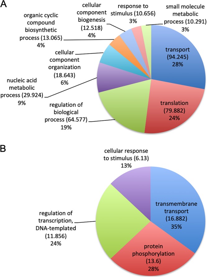
We identified a total of 124 mRNA sequences in EV samples from the two strains and carried out paired comparisons between the G186AR and G217B samples. We applied the statistical negative binomial test with filters corresponding to TPM values of ≥100, log2 values of ≥2, and false-discovery-rate (FDR) values of ≤0.05. We observed 93 transcripts enriched in EVs derived from the G217B strain, while 31 transcripts were enriched in the G186AR strain (see Table S1 in the supplemental material). In the G217B-associated transcripts, we observed enrichment in biological processes for vesicle-mediated transport (18%), oxidation-reduction mechanisms (12%), transmembrane transport (11%), and translation (8%) (Fig. 1). In the G186AR strain, the mRNA sequences were enriched only in general cellular and metabolic processes (59%). These results suggest that there are important differences with respect to the mRNA composition of EVs derived from these two strains of H. capsulatum.
FIG 1.

Gene ontology analysis. The pie charts present the gene ontology of mRNA sequences enriched in EVs isolated from (A) H. capsulatum G217B (n = 93) and (B) H. capsulatum G186AR (n = 31).
List of transcripts differentially enriched in H. capsulatum G217B and G186AR strains. Download Table S1, XLSX file, 1.4 MB (1.4MB, xlsx) .
Copyright © 2019 Alves et al.
This content is distributed under the terms of the Creative Commons Attribution 4.0 International license.
H. capsulatum EVs contain mRNA fragments and microRNA (miRNA)-like molecules.
In addition to the identification of full-length transcripts in EVs, we also detected short reads of averages of 25 to 40 nt in length that aligned consistently in the CDS but at specific positions of the mRNAs (3′ end, 5′ end, or middle sequence); about 50% of these short fragments aligned to the reverse strand, including 172 (G217B) and 80 (G186AR) sequences of this type (Table 1). A total of 172 fragments were represented in the G217B sample compared to only 80 in the G186AR EVs (Table 1). About 47% of the reference mRNA translate proteins of unknown biological processes; this could be explained by the fact that around 33% of the genes annotated in H. capsulatum genome code hypothetical proteins and/or do not present a conserved domain, which impedes our current ability to determine specific biological activities. Those associated with DNA metabolism/biogenesis were the second most abundant for both EV samples (22 for G217B versus 16 for G186AR), followed by transport for G217B and by protein modification for both strain EVs. Other processes related to short RNAs identified in both strain EVs were oxidation-reduction, signaling, and carbohydrate and lipid metabolism (Table 1). RNA fragments associated with translation were highly enriched in G217B (n = 11) but not in G186AR (n = 2) EVs, while those related to response to stress were found exclusively in the G217B sample. The corresponding proteins are stress response protein whi2, DNA repair protein rad5, and a thermotolerance protein (Table 1). Analysis of translation-related sequences allowed identification of mRNA fragments associated with distinct steps of the translation process, such as ribosome biogenesis and processing. Other metabolic pathways identified in both strains were protein modification, carbohydrate, and lipid metabolism, signaling, oxidation-reduction, and transmembrane transport, among others (Table 1).
TABLE 1.
Fragments of mRNAs identified in the EVs isolated from the G217B and G186AR strainsa
| Feature ID | G217B alignment |
G186AR alignment |
Sequence description | GO |
|---|---|---|---|---|
| Protein modification | ||||
| HCBG_03026 | 5′R | 5′R | Tetratricopeptide-like helical | Amino acid metabolic process |
| HCBG_05660 | MR | CMGC SRPK protein kinase | Protein modification process | |
| HCBG_05782 | MF | Dihydrofolate synthetase fol3 | Cofactor metabolic process | |
| HCBG_06582 | 5′F | Aspartyl aminopeptidase | Peptidase activity | |
| HCBG_07777 | MF | Mitochondrial processing peptidase alpha | Peptidase activity | |
| HCBG_08965 | MF | MF | Tyrosine phosphatase | Protein modification process |
| HCBG_09127 | 3′R / 3′F | Proteasome component C5 | Peptidase activity | |
| HCBG_09175 | 5′F | 5′F | Aspartic-type endopeptidase | Peptidase activity |
| HCBG_09182 | MR | Protein kinase | Protein modification process | |
| HCBG_01228 | 5′F | Oxidative stress-induced growth inhibitor 2 | Peptidase activity | |
| HCBG_01665 | MF | MF | pH domain-containing protein | Protein modification process |
| HCBG_03811 | MR | 3′R | Heat shock protein Hsp98 Hsp104 | ATPase activity, peptidase activity |
| HCBG_00544 | MF | Ubiquitin conjugating enzyme | Ligase activity | |
| HCBG_02715 | 3′F | 3′F | Ubiquitin family protein | |
| HCBG_05116 | 3′F | Protein | Protein modification process | |
| HCBG_07497 | 3′F | Protein | Peptidase activity | |
| Carbohydrate metabolism | ||||
| HCBG_00058 | 5′R | Mannosyl-oligosaccharide alpha-mannosidase | Catabolic process | |
| HCBG_00633 | 3′R / 3′NS | Class V chitinase | Catabolic process | |
| HCBG_06620 | 3′R | 3′R | Transaldolase | Carbohydrate metabolic process |
| Lipid metabolism | ||||
| HCBG_02433 | MF | 5′F | Acyl carrier protein | Biosynthetic process |
| HCBG_01540 | MF | MF | Predicted protein | Lipid metabolic process |
| HCBG_04372 | 3′R | GPI anchor biosynthesis protein (Pig-f) | Lipid metabolic process | |
| Response to stress | ||||
| HCBG_02224 | 3′F | General stress response protein Whi2 | ||
| HCBG_01643 | 3′R | DNA repair protein Rad5 | Response to stress | |
| HCBG_06196 | 3′R | Thermotolerance protein | ||
| Translation | ||||
| HCBG_00808 | MF | MF | 60S ribosomal protein L15 | |
| HCBG_00853 | 3′F | Small nucleolar ribonucleoprotein complex | ||
| HCBG_01544 | 5′R / F | 5′R | Ribosome biogenesis protein | |
| HCBG_02168 | 5′F / MF | 60S ribosomal protein l25 | Translation | |
| HCBG_02499 | 5′R | rRNA processing protein Utp6 | Oxidoreductase activity | |
| HCBG_02762 | 3′F | 60S ribosomal protein L31 | Translation | |
| HCBG_04580 | MR | Prenyl cysteine carboxyl methyltransferase Ste14 | mRNA processing | |
| HCBG_08644 | 5′R | Leucyl-tRNA synthetase | Translation | |
| HCBG_03984 | 5′R | Transcription initiation protein Spt5 | Translation | |
| HCBG_04793 | 5′R | U5 small nuclear ribonucleoprotein component | Chromosome organization | |
| HCBG_06802 | 5′R | Ribosome biogenesis protein Ssf2 | ||
| Signaling process | ||||
| HCBG_00598 | 5′F / 5′NS | MinD kinetochore complex component Nnf1 | Signal transduction | |
| HCBG_03086* | 5′R / F | Ste Ste20 paka protein kinase | Reproduction | |
| HCBG_04646* | 3′R | Protein Ras-2 | Signal transduction | |
| Oxidation-reduction | ||||
| HCBG_00763 | 3′R | 3′R / 3′NS | Benzoate 4-monooxygenase cytochrome p450 | Oxidoreductase activity |
| HCBG_03251 | 3′R / 3 F | Tim-barrel enzyme family protein | Oxidoreductase activity | |
| HCBG_04436 | 5′R / 3′R | Flavin-containing monooxygenase | Oxidoreductase activity | |
| HCBG_05481 | 3′F | 3′F | Like subfamily b member 4 | Protein folding |
| HCBG_05591 | 3′F | 3′F | Fmn-binding split-barrel-like protein | Oxidoreductase activity |
| HCBG_06890 | 5′F | Glutaredoxin | Homeostatic process | |
| HCBG_08366 | 3′F | Conserved hypothetical protein | Oxidoreductase activity | |
| HCBG_01233 | 5′R / 5′F | Galactose oxidase beta-propeller | ||
| HCBG_00232 | 5′F | Tyrosinase | Oxidoreductase activity | |
| HCBG_03159 | MR | Ste Ste7 Mek1 protein kinase | Reproduction | |
| Transport | ||||
| HCBG_00485 | 3′R | Vacuolar ABC heavy-metal transporter | Transmembrane transport | |
| HCBG_00680 | 3′F | Arsenine resistance protein | Transmembrane transport | |
| HCBG_00850 | MR | MFS monocarboxylate | Transmembrane transport | |
| HCBG_01089 | 5′F / 5′NS | 5′R / 5′NS | Mitochondrial carrier | Transport |
| HCBG_02374 | 5′R | Endosomal cargo receptor | Vesicle-mediated transport | |
| HCBG_02985 | 5′R | 5′R | V-type proton ATPase proteolipid subunit | Vesicle-mediated transport |
| HCBG_03067 | 5′R | 5′R | Mitochondrial dicarboxylate carrier | Transmembrane transport |
| HCBG_03738 | MF | Exocyst complex component Sec10 | Vesicle-mediated transport | |
| HCBG_04312 | 3′F | 5′R / 3′F | Nonrepetitive nucleoporin | Nucleocytoplasmic transport |
| HCBG_04317 | 5′F | mRNA transport regulator | Transport | |
| HCBG_04719 | 5′F | Nucleoporin | ||
| HCBG_04608 | 3′R | MFS transporter | Transmembrane transport | |
| HCBG_05671 | MR | Actin-associated protein | Vesicle-mediated transport | |
| HCBG_05941 | 5′F | 5′R | Potassium uptake protein | Transmembrane transport |
| HCBG_05942 | MR | Potassium uptake protein | Transmembrane transport | |
| HCBG_06437 | MF | MF | Oligopeptide transporter | Transport |
| HCBG_06658 | MR | PX domain-containing protein | Transmembrane transport | |
| HCBG_07112 | MF | Ap-2 adaptor complex subunit | Vesicle-mediated transport | |
| HCBG_07566 | 3′R | 3′R / MR | Actin cytoskeleton-regulatory complex protein Pan1 | Vesicle-mediated transport |
| HCBG_08252* | 5′F | MFS multidrug transporter | Transmembrane transport | |
| HCBG_09093 | 5′R | Kinetoplast-associated protein Kap | Transmembrane transport | |
| HCBG_09150 | 5′R / 3′R | Cap binding protein | Transport | |
| HCBG_04513 | 5′F | 3-Oxoacyl-acyl-carrier-protein synthase | ||
| DNA metabolism or biogenesis | ||||
| HCBG_00397 | MF | PHD finger domain | Chromosome organization | |
| HCBG_00799 | 5′F | 5′F | Transcriptional regulator Ngg1 | Peptidase activity |
| HCBG_01145 | 5′R | 5′R / 3′F | C6 zinc finger domain-containing protein | Biosynthetic process |
| HCBG_02996 | 3′F | Recombination hot spot-binding protein | DNA metabolic process | |
| HCBG_01721 | 3′F | Nitrogen assimilation transcription factor nira | Chromosome organization | |
| HCBG_03125 | MF | White collar | Signal transduction | |
| HCBG_03879 | MR | MR | DNA-directed RNA polymerase I subunit | Biosynthetic process |
| HCBG_04485 | 3′F | Centromere protein Cenp-o | Chromosome organization | |
| HCBG_04625 | MR | C6 finger domain | Biosynthetic process | |
| HCBG_04221 | 3′R | Chromatin remodeling complex subunit | Helicase activity | |
| HCBG_05411 | 3′R | 3′R | Transcription factor SteA | Reproduction |
| HCBG_05417 | MF | Elongator complex protein 3 | Biosynthetic process | |
| HCBG_05986 | 5′F | G1/S regulator | DNA metabolic process | |
| HCBG_05814 | 3′R | 3′R | Histone H2a | Chromosome organization |
| HCBG_06244 | MF | double-strand-break repair protein | DNA metabolic process, reproduction | |
| HCBG_07395 | MR | CP2 transcription factor | Biosynthetic process | |
| HCBG_07428 | 3′F | Caf1 family ribonuclease | ||
| HCBG_09164 | MF | MF | C2H2 finger domain transcription factor | Biosynthetic process |
| HCBG_00846 | 5′F | Transcription factor Tau55-like protein | ||
| HCBG_04340 | 3′R | 3′R | Formamidopyrimidine-DNA glycosylase | DNA metabolic process |
| HCBG_01534 | MF | MF | Telomere length regulation protein Elg1 | Ion binding, lipid binding |
| HCBG_06146 | 5′R | 5′R | Telomerase-binding protein Est1a | |
| HCBG_07560 | 5′R / 5′F | 5′R / 5′F | DNA repair protein protein | |
| HCBG_05625 | 3′R | 3′R | p60-like cell wall | |
| HCBG_09024 | MR | Hlh transcription factor | ||
| HCBG_06915 | 5′F | 5′F | Proline-rich protein-15 | Chromosome segregation |
| Other/unknown function | ||||
| HCBG_00048 | 5′R | 5′R | Hypothetical protein HCBG_00048 | |
| HCBG_00453 | 5′R | MIZ zinc finger protein | Ion binding | |
| HCBG_00947 | 3′F | Predicted protein | ||
| HCBG_00975 | 5′R | 5′R | ATPase AAA-5 protein | Ion binding |
| HCBG_01015 | MF | MF | Predicted protein | |
| HCBG_01082 | 3′R / 3′F | 3′R | Zinc knuckle domain protein | |
| HCBG_01086 | 5′R | Predicted protein | ||
| HCBG_01127 | 5′R / 3′R | Predicted protein | ||
| HCBG_01146 | MF | Predicted protein | ||
| HCBG_01161 | MF | Predicted protein | ||
| HCBG_01256 | 3′R | Conserved hypothetical protein | ||
| HCBG_01258 | MR | Predicted protein | ||
| HCBG_01500 | MR | Predicted protein | ||
| HCBG_01656 | MF | Predicted protein | ||
| HCBG_01888 | 3′R | 3′R | Conserved hypothetical protein | |
| HCBG_01952 | 3′F | Conserved hypothetical protein | ||
| HCBG_02098 | 5′R | Protein | ||
| HCBG_02107 | 5′F | Predicted protein | ||
| HCBG_02158 | 3′F | Conserved hypothetical protein | ||
| HCBG_02464 | 3′R / 3′F | 3′F / 3′R / 3′NS | Carbohydrate-binding module family 48 protein | |
| HCBG_02569 | MR / MF | MF | Predicted protein | |
| HCBG_02659 | MR / MF | MR | Predicted protein | |
| HCBG_02697 | 3′R | 3′R | Predicted protein | |
| HCBG_02981 | MF | Phosphotransferase enzyme family protein | ||
| HCBG_02986 | MF | 5′F | Predicted protein | |
| HCBG_03093 | MR | PH domain protein | ||
| HCBG_03374 | MF | MF | Glutathione transferase | |
| HCBG_03658 | 3′R / 3F | Conserved hypothetical protein | Helicase activity | |
| HCBG_03692 | 3′R / 3F | Predicted protein | ||
| HCBG_03693 | MR / MF | MR / MF | Predicted protein | |
| HCBG_03805 | MF | MF | mtDNA inheritance protein | |
| HCBG_03899 | MR | MR / 3′R | WD repeat protein | |
| HCBG_03911 | 3′R | 3′R | Protein | |
| HCBG_03913 | MR | Hypothetical protein HCBG_03913 | ||
| HCBG_03980 | MR | Phosphatidylserine decarboxylase | ||
| HCBG_04009 | MR | Hypothetical protein HCBG_04009 | ||
| HCBG_04186 | MR | Conserved hypothetical protein | ||
| HCBG_04193 | 3′R | 3′R | Conserved hypothetical protein | |
| HCBG_04201 | 3′F | Hypothetical protein HCBG_04201 | ||
| HCBG_04208 | 3′F | 3′F | Conserved hypothetical protein | |
| HCBG_04365 | MF | Hypothetical protein HCBG_04365 | ||
| HCBG_04371 | 5′R / 5′F | Bifunctional uridylyltransferase uridylyl-removing enzyme | ||
| HCBG_04380 | 3′R | 3′R | Predicted protein | |
| HCBG_04393 | 3′R | Protein | ||
| HCBG_04452 | 3′R | 3′R | Predicted protein | |
| HCBG_04780 | 5′R | 5′R | Bromodomain-containing protein | |
| HCBG_04887 | MR | Predicted protein | ||
| HCBG_05336 | 5′R | UPF0160 domain protein | ||
| HCBG_05404 | 3′R / 3′F | Predicted protein | ||
| HCBG_05580 | 3′R | Methyltransferase domain-containing protein | ||
| HCBG_05638 | 5′R | Predicted protein | ||
| HCBG_05703 | 5′R | Conserved hypothetical protein | ||
| HCBG_05744 | 5′F | T-complex protein 1 subunit beta | ||
| HCBG_05763 | 3′R | 3′F | Conserved hypothetical protein | |
| HCBG_05878 | 3′F | Hypothetical protein HCBG_05878 | ||
| HCBG_06018 | 5′F | Cytomegalovirus GH-receptor family | ||
| HCBG_06054 | MR | Phosphotransferase family protein | Ion binding, kinase activity | |
| HCBG_06071 | MF | MF | Protein | |
| HCBG_06082 | MR | Conserved hypothetical protein | ||
| HCBG_06114 | 3′F | Protein | ||
| HCBG_06176 | 3′F | KH domain protein | RNA binding | |
| HCBG_06239 | 5′R | Nonsense-mediated mRNA decay protein | ||
| HCBG_06270 | MR | Predicted protein | ||
| HCBG_06364 | MR | F-box domain-containing protein | ||
| HCBG_06436 | MF | Predicted protein | ||
| HCBG_06661 | 5′NS | Predicted protein | ||
| HCBG_06677 | 3′F | Predicted protein | ||
| HCBG_06927 | 3′R / 3′F | Predicted protein | ||
| HCBG_07002 | 5′R / 5′F | 5′R / 5′F | Ketoreductase | |
| HCBG_07065 | 5′F | Predicted protein | ||
| HCBG_07214 | 5′R | 5′R | Predicted protein | |
| HCBG_07247 | MR | Acyltransferase 3 | Transferring acyl groups | |
| HCBG_07296 | MR | MR | Hypothetical protein HCBG_07296 | |
| HCBG_07377 | MF | MR | Predicted protein | |
| HCBG_07484 | 3′F | Rhomboid family membrane protein | Peptidase activity | |
| HCBG_07611 | MR / MF | MR / MF / MNS | Protein | |
| HCBG_07676 | 3′R / 3′F | Lyr family protein | ||
| HCBG_07802 | 3′R / 3′F | 3′R / 3′F | Predicted protein | |
| HCBG_07811 | 3′F | 3′F | Predicted protein | |
| HCBG_08059 | MR | MF | DUF833 domain protein | Protein complex assembly |
| HCBG_08505 | 3′F | Sucrase ferredoxin domain-containing protein | ||
| HCBG_08661 | MF | MF | Predicted protein | |
| HCBG_08693 | 3′R | Set domain protein | ||
| HCBG_08838 | 5′R | WW domain | ||
| HCBG_08850 | 5′R | Integral membrane protein | ||
| HCBG_09013 | 5′F | 5′F | Predicted protein | |
| HCBG_09099 | 5′R | 5′R | Conserved hypothetical protein | |
| HCBG_09144 | MF | Predicted protein | ||
For some transcripts, there was an alignment in specific positions of the mRNA, not covering the entire sequence. 5′, 3′, or M (middle of the mRNA) followed by an “F” or an “R” represents forward (F) or reverse (R) orientation. GO, gene ontology; GPI, glycosylphosphatidylinositol; ID, identifier; mtDNA, mitochondrial DNA.
To gain further insight into the role of EV RNAs, to determine if they could be derived from a miRNA-like pathway, and to assess if they could play a biological role in the recipient cell, we searched for RNA secondary structures, since they are fundamental for gene expression regulation (21). A broad study of RNA structures in distinct cells revealed regulatory effects of the RNA structure throughout mRNA life cycle such as polyadenylation, splicing, translation, and turnover (22, 23). Using the entire range of EV RNA sequencing (RNA-seq) data, a total of 33 RNAs with putative structures were generated by a probability distribution, using a free energy (ΔG) value of less than or equal to −7.0 (Table S2). On the basis of this parameter, we identified transcripts for U3 small nucleolar RNA-associated protein, l-isoaspartate O-methyltransferase, serine/threonine-protein kinase, proteasome component C5, pre-rRNA processing protein Utp22, C-x8-C-x5-C-x3-H zinc finger protein, fungus-specific transcription factor domain-containing protein, and DNA damage-responsive transcriptional repressor RPH1 (Fig. 2; see also Table S2).
FIG 2.

RNA secondary structure. We used ppFold software to predict the secondary structure from the putative miRNAs extracted from the obtained reads. The numbers in parentheses represent the alignment E values. The colors indicated for the nucleotides represent the reliability percentage for each position of the RNA molecule (bottom panel). The stability value corresponding to each structure is given in kilocalories/mole.
Comparison of the RNAs with predicted secondary structure with the H. capsulatum genome. Download Table S2, XLSX file, 0.01 MB (14.1KB, xlsx) .
Copyright © 2019 Alves et al.
This content is distributed under the terms of the Creative Commons Attribution 4.0 International license.
Comparison of EV ncRNA classes in H. capsulatum EVs.
We used the ncRNA database from H. capsulatum to identify the classes of ncRNA present in EV RNAs. The data analysis revealed 73 different sequences of ncRNA in H. capsulatum EVs from the G186AR strain and 38 from the G217B isolate. A total of 33 molecular species were common to both strains, 40 were exclusively identified in the G186AR strain, and the most abundant class of ncRNA found in H. capsulatum EVs consisted of tRNAs (Table 2).
TABLE 2.
Classes of ncRNA sequences identified in EV preparations from H. capsulatum strains G186AR and G217Ba
| RNA category and ncRNA | G186AR | G217B |
|---|---|---|
| rRNA | ||
| 15S_rRNA | — | X |
| NTS1-2 | X | — |
| RDN18-1 | X | X |
| RDN18-2 | X | X |
| RDN25-1 | X | — |
| RDN25-2 | X | X |
| RDN37-1 | X | — |
| RDN37-2 | X | — |
| RDN5-1 | X | X |
| RDN5-2 | X | X |
| RDN5-3 | X | X |
| RDN5-4 | X | X |
| RDN5-5 | X | X |
| RDN5-6 | X | X |
| RDN58-1 | X | X |
| RDN58-2 | X | X |
| ncRNA | ||
| RUF21 | X | X |
| snoRNA | ||
| snR54 | X | X |
| tRNA | ||
| tRNA-Ser | — | X |
| tRNA-Met | — | X |
| tRNA-Gln | — | X |
| tRNA-Cys | — | X |
| tRNA-Ser | X | X |
| tRNA-Pro | X | X |
| tRNA-Ala | X | X |
| tRNA-Thr | X | X |
| tRNA-Ala | X | X |
| tRNA-Phe | X | X |
| tRNA-Ala | X | X |
| tRNA-Asn | X | X |
| tRNA-Met | X | X |
| tRNA-Arg | X | X |
| tRNA-Trp | X | X |
| tRNA-Gly | X | X |
| tRNA-Asp | X | X |
| tRNA-Pro | X | X |
| tRNA-Thr | X | X |
| tRNA-His | X | X |
| tRNA-Glu | X | X |
| tRNA-Gln | X | X |
| tRNA-Tyr | X | X |
| tRNA-Gln | X | X |
| tRNA-Gly | X | — |
| tRNA-Lys | X | — |
| tRNA-Ile | X | — |
| tRNA-Leu | X | — |
| tRNA-Met | X | — |
| tRNA-Gly | X | — |
| tRNA-Ile | X | — |
| tRNA-Thr | X | — |
| tRNA-Lys | X | — |
| tRNA-Met | X | — |
| tRNA-Val | X | — |
| tRNA-Phe | X | — |
| tRNA-Ile | X | — |
| tRNA-Sec | X | — |
| tRNA-Asp | X | — |
| tRNA-Thr | X | — |
| tRNA-Ile | X | — |
| tRNA-Ser | X | — |
| tRNA-Ser | X | — |
| tRNA-Arg | X | — |
| tRNA-Lys | X | — |
| tRNA-Leu | X | — |
| tRNA-Ser | X | — |
| tRNA-Leu | X | — |
| tRNA-Ala | X | — |
| tRNA-Cys | X | — |
| tRNA-Thr | X | — |
| tRNA-His | X | — |
| tRNA-Tyr | X | — |
| tRNA-Ser | X | — |
| tRNA-Leu | X | — |
| tRNA-Lys | X | — |
| tRNA-Ala | X | — |
| tRNA-Pro | X | — |
| tRNA-Arg | X | — |
| tRNA-Glu | X | — |
X, present; —, absent.
Analysis of proteins putatively associated with RNA metabolism in the EVs.
As a rule, cellular RNAs are covered with proteins and exist as ribonucleoprotein (RNP) complexes. The proteins associated with RNAs are named RNA-binding proteins (RBPs). These proteins participate in several biological processes, ranging from transcription to RNA decay (24). In this context, we investigated the presence of RBPs in the H. capsulatum EVs. We analyzed the proteomic EV data available for the G217B strain (25), and we identified 139 proteins related to RNA metabolism (8) (Table 3; see also Table S3). We found many RBPs, such as poly(A) binding protein (PABP), Nrd1, Prp24, and Snd1; splicing factors, exosome complex components, and ribosomal proteins (Table 3; see also Table S3) were identified. In addition, we also found quelling-deficient protein 2 (QDE2), an Argonaute protein important in the RNA machinery in fungi. Because we identified the QDE2 in EVs, we searched for the components of the RNA interference (RNAi) machinery in H. capsulatum and compared them with the proteins from Neurospora crassa and Schizosaccharomyces pombe, which are the fungal species for which the RNAi machinery was best described previously (26, 27). H. capsulatum EVs contained one Argonaute protein (QDE2), two Dicer-like proteins, the QIP (quelling interaction protein), and the RNA-dependent RNA polymerase (QDE1) (Table 4).
TABLE 3.
Proteins related to RNA metabolism identified in EV preparations from H. capsulatum strain G217B
| Majority protein ID | Protein name | Gene name |
|---|---|---|
| C0NMG7 | QDE2 protein | HCBG_03944 |
| C0P170 | Cap binding protein | HCBG_09150 |
| C0NJ23 | Exosome complex exonuclease RRP4 | HCBG_03153 |
| C0NM03 | Exosome complex exonuclease RRP45 | HCBG_04533 |
| C0NCT3 | KH domain RNA-binding protein | HCBG_00929 |
| C0NUH0 | KH domain RNA-binding protein | HCBG_07001 |
| C0NIU5 | KH domain-containing protein | HCBG_02352 |
| C0NUS5 | mRNA 3′-end-processing protein RNA14 | HCBG_06689 |
| C0NNW0 | mRNA cleavage and polyadenylation factor CLP1 | CLP1 HCBG_04840 |
| C0NP91 | mRNA decapping enzyme | HCBG_04971 |
| C0NC87 | mRNA export factor Mex67 | HCBG_00733 |
| C0NJ33 | Nuclear and cytoplasmic polyadenylated RNA-binding protein Pub1 | HCBG_03163 |
| C0NQQ9 | Poly(A)+ RNA export protein | HCBG_05339 |
| C0NSS5 | Polyadenylate-binding protein (PABP) | HCBG_06205 |
| C0NKR4 | Ribonucleoprotein | HCBG_03744 |
| C0NSY4 | RNA binding domain-containing protein | HCBG_06264 |
| C0NWH9 | RNA-binding protein | HCBG_07509 |
| C0NB22 | RNA-binding protein | HCBG_00318 |
| C0NPA1 | RNA-binding protein Nrd1 | HCBG_04981 |
| C0NZI9 | RNA-binding protein Prp24 | HCBG_08569 |
| C0NTZ5 | RNA-binding protein Snd1 | HCBG_06625 |
| C0NMQ0 | RNP domain-containing protein | HCBG_04027 |
| C0NLQ4 | RRM domain-containing protein | HCBG_04434 |
| C0NJ27 | Transcription elongation factor Spt6 | HCBG_03157 |
| C0NTQ1 | Transcription initiation factor TFIID complex 60-kDa subunit | HCBG_06531 |
| C0NRU6 | U1 snRNP-associated protein Usp106 | HCBG_05876 |
| C0NZZ2 | U1 snRNP-associated protein Usp107 | HCBG_08722 |
| C0NBS3 | U2 snRNP auxiliary factor large subunit | HCBG_00569 |
| C0NAD4 | U3 small nucleolar RNA-associated protein | HCBG_00080 |
| C0NZA3 | U3 small nucleolar RNA-associated protein 22 | HCBG_08483 |
| C0NLW4 | U3 snoRNP-associated protein Rrp5 | HCBG_04494 |
| C0P0R0 | U6 snRNA-associated Sm-like protein LSm2 | HCBG_08990 |
| C0P041 | 30S ribosomal protein S10 | HCBG_08883 |
| C0NFV8 | 40S ribosomal protein S15 | HCBG_01774 |
| C0NX47 | 40S ribosomal protein S18 | HCBG_08039 |
| C0NZD2 | 40S ribosomal protein S20 | HCBG_08512 |
| C0NBD0 | 40S ribosomal protein S21 | HCBG_00426 |
| C0NUD0 | 40S ribosomal protein S3 | HCBG_06961 |
| C0NLP3 | 40S ribosomal protein S4 | HCBG_04423 |
| C0NF40 | 40S ribosomal protein S5A | HCBG_01506 |
| C0NLR5 | 40S ribosomal protein S9 | HCBG_04445 |
| C0NTH6 | 5′–3′ exoribonuclease 1 (EC 3.1.13.-) | HCBG_06456 |
| C0NKI2 | 60S ribosomal protein L1 | HCBG_03662 |
| C0NNL2 | 60S ribosomal protein L3 | HCBG_04742 |
| C0NCP3 | 60S ribosomal protein L30 | HCBG_00889 |
| C0NRD6 | 60S ribosomal protein L5 | HCBG_05566 |
| C0NQR6 | 60S ribosomal protein L9B | HCBG_05346 |
| C0NPC0 | Acyl-RNA-complex subunit | HCBG_05000 |
| C0NKL8 | Alanine-tRNA ligase (EC 6.1.1.7) (alanyl-tRNA synthetase) (AlaRS) | ALA1 HCBG_03698 |
| C0NCS0 | Alternative oxidase (EC 1.-.-.-) | HCBG_00916 |
| C0ND66 | Arginyl-tRNA synthetase | HCBG_01062 |
| C0NT82 | Asparagine-rich protein | HCBG_06362 |
| C0NP94 | Asparaginyl-tRNA synthetase | HCBG_04974 |
| C0NGY7 | Aspartyl-tRNA synthetase | HCBG_02609 |
| C0NNJ3 | ATP-dependent helicase NAM7 | HCBG_04723 |
| C0NIT7 | ATP-dependent RNA helicase DOB1 | HCBG_02344 |
| C0NAN2 | ATP-dependent RNA helicase EIF4A | HCBG_00178 |
| C0NFC7 | Cell cycle control protein | HCBG_01593 |
| C0NT49 | Cleavage and polyadenylation specific factor 5 | HCBG_06329 |
| C0NW18 | Clustered mitochondria protein homolog (protein TIF31 homolog) | CLU1 TIF31 HCBG_07348 |
| C0NTW5 | Cysteinyl-tRNA synthetase | HCBG_06595 |
| C0NZE4 | d-Aminoacyl-tRNA deacylase (EC 3.1.1.-) (EC 3.1.1.96) | HCBG_08524 |
| C0NSH0 | DNA-directed RNA polymerase II polypeptide | HCBG_06100 |
| C0NB61 | DNA-directed RNA polymerase subunit beta (EC 2.7.7.6) | HCBG_00357 |
| C0NKS3 | Elicitor protein | HCBG_03753 |
| C0NRY6 | Eukaryotic peptide chain release factor GTP-binding subunit | HCBG_05916 |
| C0P0 × 7 | Eukaryotic translation initiation factor 3 subunit D (EIF3D) | HCBG_09057 |
| C0NEV9 | Fibrillarin | HCBG_01425 |
| C0NZT8 | Glutaminyl-tRNA synthetase | HCBG_08668 |
| C0NKS5 | Glutamyl-tRNA synthetase | HCBG_03755 |
| C0NE28 | Glycyl-tRNA synthetase | HCBG_02121 |
| C0NN35 | Histidyl-tRNA synthetase | HCBG_04162 |
| C0NL66 | Isoleucyl-tRNA synthetase, cytoplasmic | HCBG_03896 |
| C0NZR4 | Leucyl-tRNA synthetase | HCBG_08644 |
| C0NH95 | Leucyl-tRNA synthetase | HCBG_02717 |
| C0NI62 | Lysine-tRNA ligase (EC 6.1.1.6) (lysyl-tRNA synthetase) | HCBG_03034 |
| C0NMS8 | Mitotic control protein dis3 | HCBG_04055 |
| C0NBJ8 | mRNA splicing protein PRP8 | HCBG_00494 |
| C0NY83 | NAM9+ protein | HCBG_07877 |
| C0NG69 | Nucleic acid-binding protein | HCBG_01885 |
| C0NUD1 | Phenylalanyl-tRNA synthetase subunit beta | HCBG_06962 |
| C0NBD1 | Phenylalanyl-tRNA synthetase subunit beta cytoplasmic | HCBG_00427 |
| C0NUP1 | Polymerase II polypeptide D | HCBG_06655 |
| C0NNC4 | Pre-mRNA-processing factor 39 | HCBG_04251 |
| C0NJB4 | Pre-mRNA-processing protein prp40 | HCBG_03244 |
| C0NXM8 | Pre-mRNA-splicing factor | HCBG_08220 |
| C0NLW7 | Prolyl-tRNA synthetase | HCBG_04497 |
| C0NW72 | Ribonuclease T2-like protein | HCBG_07402 |
| C0NEF9 | Ribonuclease Z | HCBG_01275 |
| C0NIJ3 | Ribosomal biogenesis protein Gar2 | HCBG_02250 |
| C0NHN4 | Ribosomal protein L14 | HCBG_02856 |
| C0NI43 | Ribosomal protein L6 | HCBG_03015 |
| C0NVX9 | Ribosomal protein S5 | HCBG_07309 |
| C0NN82 | RNA helicase (EC 3.6.4.13) | HCBG_04209 |
| C0NEY2 | RNA polymerase II largest subunit | HCBG_01448 |
| C0NL28 | RNA polymerase subunit | HCBG_03858 |
| C0NYA7 | RNase H domain-containing protein | HCBG_07901 |
| C0NH14 | RNP domain-containing protein | HCBG_02636 |
| C0NDP9 | RNP domain-containing protein | HCBG_01992 |
| C0NC99 | SAM domain-containing protein | HCBG_00745 |
| C0NE91 | Seryl-tRNA synthetase | HCBG_02184 |
| C0NSR2 | Signal recognition particle subunit SRP68 (SRP68) | HCBG_06192 |
| C0NDB1 | Small nuclear ribonucleoprotein | HCBG_01107 |
| C0NTA0 | Splicing factor 3A subunit 3 | HCBG_06380 |
| C0NUB9 | Splicing factor 3B | HCBG_06950 |
| C0NBR2 | Splicing factor 3B subunit 1 | HCBG_00558 |
| C0NGZ9 | Threonyl-tRNA synthetase | HCBG_02621 |
| C0NSB0 | Transfer RNA-Trp synthetase | HCBG_06040 |
| C0NL23 | tRNA (cytosine-5-)-methyltransferase NCL1 | HCBG_03853 |
| C0NUP2 | tRNA [guanine(37)-N1]-methyltransferase (EC 2.1.1.228) | TRM5 HCBG_06656 |
| C0NEY0 | tRNA guanylyltransferase | HCBG_01446 |
| C0NJJ2 | tRNA ligase (EC 6.5.1.3) | HCBG_03322 |
| C0NM44 | tRNA pseudouridine synthase | HCBG_04574 |
| C0NSG9 | Tyrosine-tRNA ligase (EC 6.1.1.1) (Tyrosyl-tRNA synthetase) | HCBG_06099 |
| C0NP46 | Uncharacterized protein | HCBG_04926 |
| C0NZF6 | Uncharacterized protein | HCBG_08536 |
| C0NIA9 | Uncharacterized protein | HCBG_03081 |
| C0NMF3 | Uncharacterized protein | HCBG_04683 |
| C0NPI9 | Uncharacterized protein | HCBG_05069 |
| C0NKI6 | Uncharacterized protein | HCBG_03666 |
| C0NF97 | Uncharacterized protein | HCBG_01563 |
| C0NEJ1 | Uncharacterized protein | HCBG_01307 |
| C0NEC3 | Uncharacterized protein | HCBG_01239 |
| C0NJN9 | Uncharacterized protein | HCBG_03369 |
| C0NYC3 | Uncharacterized protein | HCBG_07917 |
| C0NIB5 | Uncharacterized protein | HCBG_03087 |
| C0NYN4 | Uncharacterized protein | HCBG_08264 |
| C0NBT4 | Uncharacterized protein | HCBG_00580 |
| C0NKE4 | Uncharacterized protein | HCBG_03624 |
| C0NGB7 | Uncharacterized protein | HCBG_02389 |
| C0NM01 | Uncharacterized protein | HCBG_04531 |
| C0NG47 | Uncharacterized protein | HCBG_01863 |
| C0NEU7 | Uncharacterized protein | HCBG_01413 |
| C0NG27 | Valyl-tRNA synthetase | HCBG_01843 |
| C0P019 | Vip1 protein | HCBG_08749 |
| C0NG23 | Ribosome biogenesis protein RPF2 | HCBG_01839 |
| C0NGE8 | Ribosome biogenesis protein TSR3 | TSR3 HCBG_02420 |
| C0NAE4 | Ribosome biogenesis protein YTM1 | YTM1 HCBG_00090 |
TABLE 4.
Proteins associated with the RNAi machinery in H. capsulatum G186AR EVs compared to S. pombe and N. crassa
| Protein |
H. capsulatum
product |
G186AR ID |
E value | % identity |
% positives |
|---|---|---|---|---|---|
| NP_587782.1, argonaute (Schizosaccharomyces pombe) | QDE2 protein | HCBG_03944 | 1.00E−85 | 28 | 45 |
|
ESA42122.1, posttranscriptional silencing protein QDE-2 (Neurospora crassa OR74A) |
QDE2 protein | HCBG_03944 | 1.00E−178 | 37 | 53 |
| NP_588215.2, dicer (Schizosaccharomyces pombe) | Dicer-like protein | HCBG_01751 | 1.00E−113 | 28 | 44 |
| EAA34302.3, dicer-like protein 2 (Neurospora crassa OR74A) | Dicer-like protein 2 | HCBG_01136 | 3.00E−97 | 31 | 49 |
|
XP_959047.1, RNA-dependent RNA polymerase (Neurospora crassa OR74A) |
RNA-dependent RNA polimerase |
HCBG_06604 | 3.00E−92 | 31 | 46 |
| XP_964030.3, RecQ family helicase (Neurospora crassa OR74A) | Dicer-like protein | HCBG_01751 | 0.00E + 00 | 45 | 60 |
| ABQ45366.1, QDE-2-interacting protein (Neurospora crassa) | QDE-2-interacting protein (QIP) |
HCBG_07373 | 2.00E−50 | 27 | 43 |
Proteins related to RNA metabolism identified in EVs derived from the H. capsulatum G217B strain (25). Download Table S3, XLSX file, 0.06 MB (63.6KB, xlsx) .
Copyright © 2019 Alves et al.
This content is distributed under the terms of the Creative Commons Attribution 4.0 International license.
Comparisons of cellular RNA versus EV RNA showed a distinct enrichment of molecules in the vesicles.
We next assessed the composition of cellular RNA from H. capsulatum yeast cells (28) and compared this information to that obtained from analyses of EV-associated RNA composition under the same conditions. There was no correlation between the transcripts with highest expression levels and their presence in the EVs (Table S4). Examples of highly expressed cellular transcripts included histones 4, 2B, and 2A, allergen Aspf4, chaperones, and translation factors, among others (Table S4). In contrast, zinc knuckle domain-containing protein, vacuolar ATP synthase subunit C, G1/S regulator, thermotolerance protein, histone variant H2A.Z, and proteasome component C5 had an enrichment value of greater than 7,000 in the EVs, while they showed low expression values in the cell (Table S4). The differences in composition between cells and EVs were also evaluated by grouping the transcripts into biological processes (Fig. 3). For the yeast cells, the main pathways were associated with transport, translation, and general metabolic processes (Fig. 3). For the EVs, the enriched pathways were transmembrane transport, protein phosphorylation, and transcription regulation (Fig. 3). This result demonstrates the low levels of correlation between the most highly expressed cellular mRNAs and EV cargo, providing evidence that there might be a mechanism directing the RNA molecules to the EVs.
FIG 3.
Gene ontology analysis. The pie charts present the gene ontology of mRNA sequences enriched in H. capsulatum cells (A) and in EVs isolated from H. capsulatum (B).
Comparison the H. capsulatum transcriptome (G186AR and G217B strains) (28) with the vesicular RNA sequences. Download Table S4, XLSX file, 2.2 MB (2.2MB, xlsx) .
Copyright © 2019 Alves et al.
This content is distributed under the terms of the Creative Commons Attribution 4.0 International license.
DISCUSSION
As previously described (17, 18), RNA molecules associated with fungal EVs are remarkably diverse. For instance, mRNAs, tRNA fragments, snoRNAs, small nucleolar RNAs (snRNAs), and miRNA-like molecules were characterized in EVs from C. albicans, C. neoformans, P. brasiliensis, and S. cerevisiae (17). We observed similar distributions of RNA molecules in H. capsulatum EVs. The comparison between the G186AR and G217B EVs revealed important differences in the variety of mRNAs identified. When the mRNA composition was compared to what was described for other fungi, important similarities were observed. For example, the most abundant biological process identified in G217B EVs was vesicle-mediated transport, which was also the most abundant process in C. albicans EVs (17). Molecules required for ribosome biogenesis, which were observed in G217B EVs, belonged to the most highly enriched process in S. cerevisiae EVs (17). However, in the comparisons of the ncRNA molecules, different profiles were observed. Most of the ncRNAs in H. capsulatum strains derived from tRNAs; a similar profile was obtained with C. albicans (17). In addition, almost no snoRNAs were identified in H. capsulatum, but this class of ncRNAs was one of the most abundant in the EVs of other fungi (17). Differences in EV composition were observed previously in C. neoformans; the EV-associated RNA produced by mutant cells with defective unconventional secretion differed considerably from similar samples produced by wild-type cells (29).
In our study, we identified short reads that aligned specifically to exons; however, these sequences did not correspond to complete mRNAs in the EVs. They instead corresponded to 25-nt-long fragments that were enriched in specific exons of the transcript. These fragments of mRNAs were previously described in human cells (30), where most of the transcripts identified in the EVs corresponded to a fraction of the mRNA with an enrichment of the 3′ UTR of the transcript (30). The results of that human study led to the hypothesis that the mRNA fragments had a role in gene expression regulation in the recipient cells as the secreted mRNA could act as competitors to regulate stability, localization, and translation of mRNAs in target cells (30). In Mucor circinelloides cells, the presence of the RNA silencing pathway (sRNA) resulted in the production of both sense and antisense sRNAs (31–33). Sequencing analysis of the sRNA content of this fungus showed the existence of exonic small interfering RNAs (exo-siRNAs) as a new type of sRNA. They were produced from exons of the same genes that are later regulated through the repression of the corresponding mRNA (34). This result agrees with our observation of short reads in the exonic regions of the transcripts. We therefore hypothesize that, similarly to what was described for M. circinelloides cells, H. capsulatum EV fragments can regulate expression of their own mRNAs. Of note, we also found a highly represented population of putative exonic RNA in Paracoccidioides strains (R. Peres da Silva, L. V. G. Longo, J. P. C. da Cunha, T. J. P. Sobreira, H. Faoro, M. L. Rodrigues, S. Goldenberg, L. R. Alves, and R. Puccia, unpublished data).
As H. capsulatum EVs contain different RNA molecules, it is reasonable to hypothesize that proteins that regulate RNA metabolism are also present in the EVs, probably associated with RNA. If validated, this hypothesis could indicate how the RNAs in a specific subset are directed to the vesicles and exported. RNA-binding proteins (RBPs) participate in several biological processes, from RNA transcription to decay (24). We detected a number of RNA-binding proteins in H. capsulatum EVs (25). These proteins were also identified in association with EVs in other systems. For example, in the EVs produced by human epithelial cells, 30 RBPs were identified (35), including heterogeneous nuclear ribonucleoproteins (hnRNPs). These proteins are responsible for directing pre-mRNAs in the maturation processes that culminate in transcriptional regulation, alternative splicing, transport, and localization (35). In addition, RBPs in EVs were identified in distinct models as hepatocytes, human embryonic kidney (HEK) cells, and mouse myoblast cells (35–37). Interestingly, one of the RBPs identified in EVs was SND1 (staphylococcal nuclease domain-containing protein 1), which is a main component of the RNA-induced silencing complex (RISC) that plays an important role in miRNA function (37).
Another example of a protein identified in the EVs of H. capsulatum and distinct organisms is an endonuclease of the Ago2 family. An infection model with Plasmodium falciparum demonstrated that infected red blood cells released EVs containing functional miRNA-Argonaute 2 complexes (38). Moreover, endothelial cells internalized the P. falciparum EVs, and the miRNA-Argonaute 2 complexes were transferred to the cells and acted in regulation of gene expression and in the barrier properties of the recipient cells (38). The Argonaute protein named QDE2 in H. capsulatum was identified as enriched in the EVs of the G217B strain.
The small silencing RNAs include a variety of molecules, such as microRNAs (miRNAs) and various small interfering RNAs (siRNAs), including exo-siRNAs, endogenous siRNAs (endo-siRNAs), and Piwi-interacting RNAs (piRNAs) (39). Previous studies of small RNAs in fungi identified the RNAi machinery in the fission yeast species Schizosaccharomyces pombe, in the budding yeast species Saccharomyces castellii and C. albicans, and in filamentous fungi (26, 27, 40). One of the best-characterized models is represented by the filamentous fungus N. crassa (27, 41–45). The RNAi machinery in that organism functions in defense against transposons (46). A similar process has been described in C. neoformans, where RNAi is involved in the regulation of transposon activity and genome integrity during vegetative growth (47). In N. crassa, the QDE2 gene encodes an Argonaute protein that is homologous to the rde-1 gene in C. elegans, encoding a protein required for double-stranded RNA (dsRNA)-induced silencing (27). The characterization of RNAs associated with QDE2 in N. crassa led to the identification of miRNA-like RNAs (milRNAs) in this organism (48). The identification of QDE2 in H. capsulatum EVs in association with the small RNAs indicated that the QDE2-milRNA complex might be directed to the EVs and possibly delivered to recipient cells, with the potential to interfere with gene expression regulation and/or cell-cell communication.
Fungal EVs have been implicated in a number of communication processes, including transfer of virulence (49) and antifungal resistance (50). In Cryptococcus gattii, pathogen-to-pathogen communication via EVs resulted in reversion of an avirulent phenotype through mechanisms that required vesicular RNA (49). The sequences required for this process, however, remained unknown. This is an efficient illustration of the potential derived from the characterization of EV-associated RNA in fungi. In this context, our study results provide information from the H. capsulatum model that will allow the design of pathogenic experimental models aiming at characterizing the role of extracellular RNAs in fungal pathogenesis.
MATERIALS AND METHODS
Fungal strains and growth conditions.
The H. capsulatum strains were subjected to long-term storage at −80°C. Aliquots were inoculated into Ham’s F-12 media (Gibco; catalog no. 21700-075) supplemented with glucose (18.2 g/liter), l-cysteine (8.4 mg/liter), HEPES (6 g/liter), and glutamic acid (1 g/liter) and cultivated at 37°C with constant shaking at 150 rpm. Viability assessments were performed using Janus green 0.02%, and all aliquots used had >99% live yeast cells. EVs were then isolated from fungal culture supernatants as previously described (12).
sRNA isolation.
Small RNA-enriched fractions were isolated using a miRNeasy minikit (Qiagen) and were then treated with an RNeasy MinElute cleanup kit (Qiagen), according to the manufacturer’s protocol, to obtain small RNA-enriched fractions. The sRNA profile was assessed in an Agilent 2100 Bioanalyzer (Agilent Technologies).
RNA sequencing.
Purified sRNA (100 ng) was used for RNA-seq analysis with two independent biological replicates. The RNA-seq analysis was performed using a SOLiD 3 Plus platform and an RNA-Seq kit (Life Sciences) according to the manufacturer's recommendations.
In silico data analysis.
The sequencing data were analyzed using version 10.1 of CLC Genomics Workbench. The reads were trimmed on the basis of quality, with a threshold Phred score of 25. The reference genomes used for mapping were obtained from the NCBI database (H. capsulatum G186AR strain ABBS02 and G217B strain ABBT01). The alignment was performed using the following parameters: additional number of bases of upstream and downstream sequences, 100; minimum number of reads, 10; maximum number of mismatches, 2; nonspecific match limit, −2, minimum fraction length, 0.7 for the genome mapping or 0.8 for the RNA mapping. The minimum proportion of read similarity mapped on the reference genome was 80%. Only uniquely mapped reads were considered in the analysis. The libraries were normalized per million, and the expression values for the transcripts were recorded in RPKM (reads per kilobase per million). We also analyzed other expression values, including TPM (transcripts per million) and CPM (counts per million). The statistical test applied was the DGE (differential gene expression) test. For the ncRNA analysis, the database used was the ncRNA database from Histoplasma capsulatum (EnsemblFungi G186AR GCA_000150115 assembly ASM15011v1). The secondary structure analysis was performed using the PPFold plugin in CLC Genomics Workbench v. 10.1 and the default parameters. The entire RNA-seq database was subjected to PPFold analysis, and the putative structures were determined. Analysis of the relationship between the profile of RNA sequences detected in this study and the protein composition of H. capsulatum EVs was based on results recently obtained with strain G217B using a proteomic approach (25). The cellular RNA used in this analysis was assessed using the Sequence Read Archive (SRA) database (accession numbers SRR2015219 and SRR2015223) (28).
Data availability.
The data were deposited into the SRA database under study accession number PRJNA514312.
ACKNOWLEDGMENTS
J.D.N. was supported in part by NIH R01AI052733 and R21AI124797. M.L.R. is currently on leave from the position of Associate Professor at the Microbiology Institute of the Federal University of Rio de Janeiro, Rio de Janeiro, Brazil. He was supported by grants from the Brazilian agency Conselho Nacional de Desenvolvimento Científico e Tecnológico (CNPq; grants 405520/2018-2, 440015/2018-9, and 301304/2017-3) and Fiocruz (grants VPPCB-007-FIO-18 and VPPIS-001-FIO18). R.P. was supported by FAPESP (grant 13/25950-10). We also acknowledge support from Coordenação de Aperfeiçoamento de Pessoal de Nível Superior (CAPES; Finance Code 001) and the Instituto Nacional de Ciência e Tecnologia de Inovação em Doenças de Populações Negligenciadas (INCT-IDPN).
We declare that we have no conflicts of interest.
REFERENCES
- 1.Heydon K, Zaoutis TE, Chu JH, Feudtner C, Walsh TJ. 2006. Hospitalizations for endemic mycoses: a population-based national study. Clin Infect Dis 42:822–825. doi: 10.1086/500405. [DOI] [PubMed] [Google Scholar]
- 2.Adenis AA, Aznar C, Couppié P. 2014. Histoplasmosis in HIV-infected patients: a review of new developments and remaining gaps. Curr Trop Med Rep 1:119–128. doi: 10.1007/s40475-014-0017-8. [DOI] [PMC free article] [PubMed] [Google Scholar]
- 3.Prado M, da Silva MB, Laurenti R, Travassos LR, Taborda CP. 2009. Mortality due to systemic mycoses as a primary cause of death or in association with AIDS in Brazil: a review from 1996 to 2006. Mem Inst Oswaldo Cruz 104:513–521. doi: 10.1590/S0074-02762009000300019. [DOI] [PubMed] [Google Scholar]
- 4.Cordeiro RA, Pereira JF, Caetano ÉP, Castelo-Branco D, Rocha MFG, Sidrim JJC, Camargo ZP, Fechine MAB, Monteiro AJ, Lima RAC, Mesquita JRL, Brilhante R. 2012. Histoplasmosis in HIV-positive patients in Ceará, Brazil: clinical-laboratory aspects and in vitro antifungal susceptibility of Histoplasma capsulatum isolates. Trans R Soc Trop Med Hyg 106:484–488. doi: 10.1016/j.trstmh.2012.05.003. [DOI] [PubMed] [Google Scholar]
- 5.Lopes-Bezerra LM, Schubach A, Costa RO. 11 May 2006. Sporothrix schenckii and sporotrichosis. An Acad Bras Cienc doi: 10.1590/S0001-37652006000200009. [DOI] [PubMed] [Google Scholar]
- 6.Carme B, Bessieres M-H, Adenis A, Fillaux J, Couppie P, Demar M, Menard S, Iriart X, Chauvin P, Nacher M, Aznar C, Lavergne R-A, Blanchet D, Magnaval J-F, Berry A, Cassaing S. 2014. A complementary tool for management of disseminated Histoplasma capsulatum var. capsulatum infections in AIDS patients. Int J Med Microbiol 304:1062–1065. doi: 10.1016/j.ijmm.2014.07.016. [DOI] [PubMed] [Google Scholar]
- 7.Albuquerque PC, Nakayasu ES, Rodrigues ML, Frases S, Casadevall A, Zancope-Oliveira RM, Almeida IC, Nosanchuk JD. 2008. Vesicular transport in Histoplasma capsulatum: an effective mechanism for trans-cell wall transfer of proteins and lipids in ascomycetes. Cell Microbiol 10:1695–1710. doi: 10.1111/j.1462-5822.2008.01160.x. [DOI] [PMC free article] [PubMed] [Google Scholar]
- 8.Matos Baltazar L, Sobreira TJP, Choi H, Nakayasu ES, Casadevall A, Nimrichter L, Nosanchuk JD. 2016. Antibody binding alters the characteristics and contents of extracellular vesicles released by Histoplasma capsulatum. mSphere 1:e00085-15. doi: 10.1128/mSphere.00085-15. [DOI] [PMC free article] [PubMed] [Google Scholar]
- 9.Rodrigues ML, Godinho RMC, Zamith-Miranda D, Nimrichter L. 3 December 2015. Traveling into outer space: unanswered questions about fungal extracellular vesicles. PLoS Pathog doi: 10.1371/journal.ppat.1005240. [DOI] [PMC free article] [PubMed] [Google Scholar]
- 10.Rodrigues ML, Nakayasu ES, Oliveira DL, Nimrichter L, Nosanchuk JD, Almeida IC, Casadevall A. 2008. Extracellular vesicles produced by Cryptococcus neoformans contain protein components associated with virulence. Eukaryot Cell 7:58–67. doi: 10.1128/EC.00370-07. [DOI] [PMC free article] [PubMed] [Google Scholar]
- 11.Da Silva TA, Roque-Barreira MC, Casadevall A, Almeida F. 24 October 2016. Extracellular vesicles from Paracoccidioides brasiliensis induced M1 polarization in vitro. Sci Rep doi: 10.1038/srep35867. [DOI] [PMC free article] [PubMed] [Google Scholar]
- 12.Rodrigues ML, Nimrichter L, Oliveira DL, Frases S, Miranda K, Zaragoza O, Alvarez M, Nakouzi A, Feldmesser M, Casadevall A. 2007. Vesicular polysaccharide export in Cryptococcus neoformans is a eukaryotic solution to the problem of fungal trans-cell wall transport. Eukaryot Cell 6:48–59. doi: 10.1128/EC.00318-06. [DOI] [PMC free article] [PubMed] [Google Scholar]
- 13.Vallejo MC, Matsuo AL, Ganiko L, Medeiros LCS, Miranda K, Silva LS, Freymüller-Haapalainen E, Sinigaglia-Coimbra R, Almeida IC, Puccia R. 2011. The pathogenic fungus Paracoccidioides brasiliensis exports extracellular vesicles containing highly immunogenic α-galactosyl epitopes. Eukaryot Cell 10:343–351. doi: 10.1128/EC.00227-10. [DOI] [PMC free article] [PubMed] [Google Scholar]
- 14.Gehrmann U, Qazi KR, Johansson C, Hultenby K, Karlsson M, Lundeberg L, Gabrielsson S, Scheynius A. 22 July 2011. Nanovesicles from malassezia sympodialis and host exosomes induce cytokine responses—novel mechanisms for host-microbe interactions in atopic eczema. PLoS One doi: 10.1371/journal.pone.0021480. [DOI] [PMC free article] [PubMed] [Google Scholar]
- 15.Brown L, Wolf JM, Prados-Rosales R, Casadevall A. 1 September 2015. Through the wall: extracellular vesicles in Gram-positive bacteria, mycobacteria and fungi. Nat Rev Microbiol doi: 10.1038/nrmicro3480. [DOI] [PMC free article] [PubMed] [Google Scholar]
- 16.Nimrichter L, De Souza MM, Del Poeta M, Nosanchuk JD, Joffe L, Tavares PDM, Rodrigues ML. 8 July 2016. Extracellular vesicle-associated transitory cell wall components and their impact on the interaction of fungi with host cells. Front Microbiol doi: 10.3389/fmicb.2016.01034. [DOI] [PMC free article] [PubMed] [Google Scholar]
- 17.Peres da Silva R, Puccia R, Rodrigues ML, Oliveira DL, Joffe LS, César GV, Nimrichter L, Goldenberg S, Alves LR. 14 January 2015. Extracellular vesicle-mediated export of fungal RNA. Sci Rep doi: 10.1038/srep07763. [DOI] [PMC free article] [PubMed] [Google Scholar]
- 18.Rayner S, Bruhn S, Vallhov H, Andersson A, Billmyre RB, Scheynius A. 4 January 2017. Identification of small RNAs in extracellular vesicles from the commensal yeast Malassezia sympodialis. Sci Rep doi: 10.1038/srep39742. [DOI] [PMC free article] [PubMed] [Google Scholar]
- 19.Tewarl RP, Berkhout FJ. 1972. Comparative pathogenicity of albino and brown types of Histoplasma capsulatum for mice. J Infect Dis 125:504–508. doi: 10.1093/infdis/125.5.504. [DOI] [PubMed] [Google Scholar]
- 20.Mayfield JA, Rine J. 2007. The genetic basis of variation in susceptibility to infection with Histoplasma capsulatum in the mouse. Genes Immun 8:468–474. doi: 10.1038/sj.gene.6364411. [DOI] [PubMed] [Google Scholar]
- 21.Bevilacqua PC, Ritchey LE, Su Z, Assmann SM. 14 September 2016. Genome-wide analysis of RNA secondary structure. Annu Rev Genet doi: 10.1146/annurev-genet-120215-035034. [DOI] [PubMed] [Google Scholar]
- 22.Kertesz M, Iovino N, Unnerstall U, Gaul U, Segal E. 23 September 2007. The role of site accessibility in microRNA target recognition. Nat Genet 39:1278–1284. doi: 10.1038/ng2135. [DOI] [PubMed] [Google Scholar]
- 23.Kertesz M, Wan Y, Mazor E, Rinn JL, Nutter RC, Chang HY, Segal E. 2 September 2010. Genome-wide measurement of RNA secondary structure in yeast. Nature doi: 10.1038/nature09322. [DOI] [PMC free article] [PubMed] [Google Scholar]
- 24.Glisovic T, Bachorik JL, Yong J, Dreyfuss G. 13 March 2008. RNA-binding proteins and post-transcriptional gene regulation. FEBS Lett doi: 10.1016/j.febslet.2008.03.004. [DOI] [PMC free article] [PubMed] [Google Scholar]
- 25.Baltazar LM, Zamith-Miranda D, Burnet MC, Choi H, Nimrichter L, Nakayasu ES, Nosanchuk JD. 23 May 2018. Concentration-dependent protein loading of extracellular vesicles released by Histoplasma capsulatum after antibody treatment and its modulatory action upon macrophages. Sci Rep doi: 10.1038/s41598-018-25665-5. [DOI] [PMC free article] [PubMed] [Google Scholar]
- 26.Colmenares SU, Buker SM, Buhler M, Dlakić M, Moazed D. 2007. Coupling of double-stranded RNA synthesis and siRNA generation in fission yeast RNAi. Mol Cell 27:449–461. doi: 10.1016/j.molcel.2007.07.007. [DOI] [PubMed] [Google Scholar]
- 27.Catalanotto C, Azzalin G, Macino G, Cogoni C. 2000. Gene silencing in worms and fungi. Nature 404:245. doi: 10.1038/35005169. [DOI] [PubMed] [Google Scholar]
- 28.Gilmore SA, Voorhies M, Gebhart D, Sil A. 15 July 2015. Genome-wide reprogramming of transcript architecture by temperature specifies the developmental states of the human pathogen Histoplasma. PLoS Genet doi: 10.1371/journal.pgen.1005395. [DOI] [PMC free article] [PubMed] [Google Scholar]
- 29.da Silva RP, Martins S. d T, Rizzo J, Dos Reis FCG, Joffe LS, Vainstein M, Kmetzsch L, Oliveira DL, Puccia R, Goldenberg S, Rodrigues ML, Alves LR. 8 August 2018. Golgi reassembly and stacking protein (GRASP) participates in vesicle-mediated RNA export in Cryptococcus neoformans. Genes (Basel) doi: 10.3390/genes9080400. [DOI] [PMC free article] [PubMed] [Google Scholar]
- 30.Batagov AO, Kurochkin IV. 7 July 2013. Exosomes secreted by human cells transport largely mRNA fragments that are enriched in the 3′-untranslated regions. Biol Direct doi: 10.1186/1745-6150-8-12. [DOI] [PMC free article] [PubMed] [Google Scholar]
- 31.Nicolás FE, de Haro JP, Torres-Martínez S, Ruiz-Vázquez RM. 2007. Mutants defective in a Mucor circinelloides Dicer-like gene are not compromised in siRNA silencing but display developmental defects. Fungal Genet Biol 44:504–516. doi: 10.1016/j.fgb.2006.09.003. [DOI] [PubMed] [Google Scholar]
- 32.Cervantes M, Calo S, Ruiz-Vázquez RM, Torres-Martínez S, Nicolás FE, de Haro JP. 2009. A single Dicer gene is required for efficient gene silencing associated with two classes of small antisense RNAs in Mucor circinelloides. Eukaryot Cell 8:1486–1497. doi: 10.1128/EC.00191-09. [DOI] [PMC free article] [PubMed] [Google Scholar]
- 33.Cervantes M, Vila A, Nicolás FE, Moxon S, de Haro JP, Dalmay T, Torres-Martínez S, Ruiz-Vázquez RM. 2013. A single Argonaute gene participates in exogenous and endogenous RNAi and controls cellular functions in the basal fungus Mucor circinelloides. PLoS One doi: 10.1371/journal.pone.0069283. [DOI] [PMC free article] [PubMed] [Google Scholar]
- 34.Nicolas FE, Moxon S, de Haro JP, Calo S, Grigoriev IV, Torres-MartÍnez S, Moulton V, Ruiz-Vázquez RM, Dalmay T. 2010. Endogenous short RNAs generated by Dicer 2 and RNA-dependent RNA polymerase 1 regulate mRNAs in the basal fungus Mucor circinelloides. Nucleic Acids Res 38:5535–5541. doi: 10.1093/nar/gkq301. [DOI] [PMC free article] [PubMed] [Google Scholar]
- 35.Statello L, Maugeri M, Garre E, Nawaz M, Wahlgren J, Papadimitriou A, Lundqvist C, Lindfors L, Collén A, Sunnerhagen P, Ragusa M, Purrello M, Pietro CD, Tigue N, Valadi H. 2018. Identification of RNA-binding proteins in exosomes capable of interacting with different types of RNA: RBP-facilitated transport of RNAs into exosomes. PLoS One doi: 10.1371/journal.pone.0195969. [DOI] [PMC free article] [PubMed] [Google Scholar]
- 36.Alonzi T, Weisz A, Battistelli C, Tarallo R, Tripodi M, Giurato G, Montaldo C, Santangelo L, Cicchini C, Mancone C. 2016. The RNA-binding protein SYNCRIP is a component of the hepatocyte exosomal machinery controlling MicroRNA sorting. Cell Rep 17:799–808. doi: 10.1016/j.celrep.2016.09.031. [DOI] [PubMed] [Google Scholar]
- 37.Sork H, Corso G, Krjutskov K, Johansson HJ, Nordin JZ, Wiklander OPB, Lee YXF, Westholm JO, Lehtiö J, Wood MJA, Mäger I, El Andaloussi S. 17 July 2018. Heterogeneity and interplay of the extracellular vesicle small RNA transcriptome and proteome. Sci Rep doi: 10.1038/s41598-018-28485-9. [DOI] [PMC free article] [PubMed] [Google Scholar]
- 38.Marti M, Padmanabhan P, Ghiran I, Walch M, Kuo WP, Nilsson S, Brancucci NM, Ravel D, Trachtenberg A, Ma S, Mantel P-Y, Filgueira L, Huttenhower C, Hjelmqvist D, Ankarklev J, Ribeiro M, Grüring C, Duraisingh MT, Kharoubi-Hess S, Martinelli R. 10 October 2016. Infected erythrocyte-derived extracellular vesicles alter vascular function via regulatory Ago2-miRNA complexes in malaria. Nat Commun doi: 10.1038/ncomms12727. [DOI] [PMC free article] [PubMed] [Google Scholar]
- 39.Ghildiyal M, Zamore PD. 1 February 009. Small silencing RNAs: an expanding universe. Nat Rev Genet doi: 10.1038/nrg2504. [DOI] [PMC free article] [PubMed] [Google Scholar]
- 40.Drinnenberg IA, Weinberg DE, Xie KT, Mower JP, Wolfe KH, Fink GR, Bartel DP. 2009. RNAi in budding yeast. Science 326:544–550. doi: 10.1126/science.1176945. [DOI] [PMC free article] [PubMed] [Google Scholar]
- 41.Pallotta M, Cogoni C, Sachs MS, Catalanotto C, Macino G, Vayssie L, ReFalo P. 2004. Redundancy of the two Dicer genes in transgene-induced posttranscriptional gene silencing in Neurospora crassa. Mol Cell Biol 24:2536–2545. [DOI] [PMC free article] [PubMed] [Google Scholar]
- 42.Choudhary S, Liu Y, Cheng P, Maiti M, Lee H-C, Liu Q, He Q. 2007. A double-stranded-RNA response program important for RNA interference efficiency. Mol Cell Biol 27:3995–4005. doi: 10.1128/MCB.00186-07. [DOI] [PMC free article] [PubMed] [Google Scholar]
- 43.Cogoni C, Macino G. 1999. Gene silencing in Neurospora crassa requires a protein homologous to RNA-dependent RNA polymerase. Nature 399:166–169. doi: 10.1038/20215. [DOI] [PubMed] [Google Scholar]
- 44.Cogoni C, Macino G. 1999. Posttranscriptional gene silencing in Neurospora by a RecQ DNA helicase. Science 286:2342–2344. doi: 10.1126/science.286.5448.2342. [DOI] [PubMed] [Google Scholar]
- 45.Chicas A, Cogoni C, Macino G. 2004. RNAi-dependent and RNAi-independent mechanisms contribute to the silencing of RIPed sequences in Neurospora crassa. Nucleic Acids Res 32:4237–4243. doi: 10.1093/nar/gkh764. [DOI] [PMC free article] [PubMed] [Google Scholar]
- 46.Nolan T, Braccini L, Azzalin G, De Toni A, Macino G, Cogoni C. 2005. The post-transcriptional gene silencing machinery functions independently of DNA methylation to repress a LINE1-like retrotransposon in Neurospora crassa. Nucleic Acids Res 33:1564–1573. doi: 10.1093/nar/gki300. [DOI] [PMC free article] [PubMed] [Google Scholar]
- 47.Jiang N, Yang Y, Janbon G, Pan J, Zhu X. 2012. Identification and functional demonstration of miRNAs in the fungus Cryptococcus neoformans. PLoS One 7:e52734. doi: 10.1371/journal.pone.0052734. [DOI] [PMC free article] [PubMed] [Google Scholar]
- 48.Selker EU, Lewis ZA, Gu W, Mello CC, Li L, Xue Z, Crosthwaite SK, Pertsemlidis A, Freitag M, Liu Y, Lee H-C. 2010. Diverse pathways generate microRNA-like RNAs and Dicer-independent small interfering RNAs in fungi. Mol Cell 38:803–814. doi: 10.1016/j.molcel.2010.04.005. [DOI] [PMC free article] [PubMed] [Google Scholar]
- 49.Bielska E, Sisquella MA, Aldeieg M, Birch C, O’Donoghue EJ, May RC. 19 April 2018. Pathogen-derived extracellular vesicles mediate virulence in the fatal human pathogen Cryptococcus gattii. Nat Commun doi: 10.1038/s41467-018-03991-6. [DOI] [PMC free article] [PubMed] [Google Scholar]
- 50.Mitchell KF, Azadi P, Jaromin A, Sanchez H, Dominguez E, Andes DR, Mitchell A, Zarnowski R, Bernhardt J, Heiss C, Covelli AS. 2018. Candida albicans biofilm–induced vesicles confer drug resistance through matrix biogenesis. PLoS Biol 16:e2006872. doi: 10.1371/journal.pbio.2006872. [DOI] [PMC free article] [PubMed] [Google Scholar]
Associated Data
This section collects any data citations, data availability statements, or supplementary materials included in this article.
Supplementary Materials
List of transcripts differentially enriched in H. capsulatum G217B and G186AR strains. Download Table S1, XLSX file, 1.4 MB (1.4MB, xlsx) .
Copyright © 2019 Alves et al.
This content is distributed under the terms of the Creative Commons Attribution 4.0 International license.
Comparison of the RNAs with predicted secondary structure with the H. capsulatum genome. Download Table S2, XLSX file, 0.01 MB (14.1KB, xlsx) .
Copyright © 2019 Alves et al.
This content is distributed under the terms of the Creative Commons Attribution 4.0 International license.
Proteins related to RNA metabolism identified in EVs derived from the H. capsulatum G217B strain (25). Download Table S3, XLSX file, 0.06 MB (63.6KB, xlsx) .
Copyright © 2019 Alves et al.
This content is distributed under the terms of the Creative Commons Attribution 4.0 International license.
Comparison the H. capsulatum transcriptome (G186AR and G217B strains) (28) with the vesicular RNA sequences. Download Table S4, XLSX file, 2.2 MB (2.2MB, xlsx) .
Copyright © 2019 Alves et al.
This content is distributed under the terms of the Creative Commons Attribution 4.0 International license.
Data Availability Statement
The data were deposited into the SRA database under study accession number PRJNA514312.