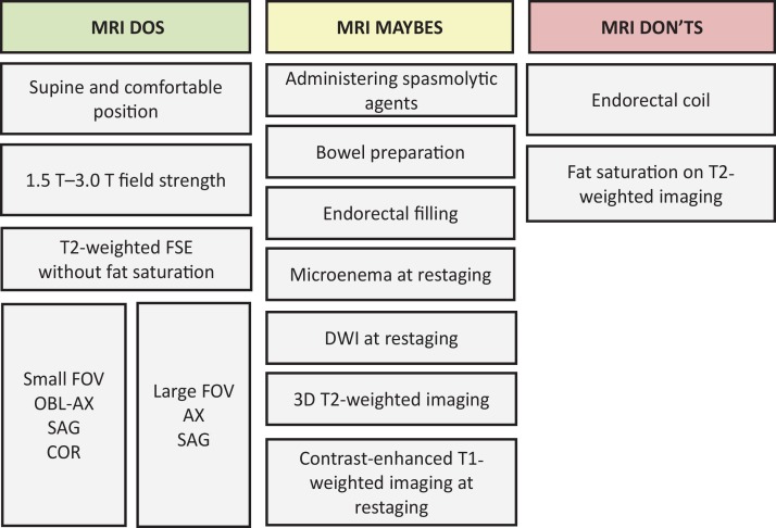
Figure 3.
Chart categorizes rectal MRI protocol according to techniques that are frequently recommended (“Dos”), those that are not recommended (“Don’ts”), and some that are controversial practices (“Maybes.”) T2-weighted imaging with fat saturation is rarely necessary but may be useful for imaging mucinous tumors. AX = axial, COR = coronal, DWI = diffusion-weighted imaging, FOV = field of view, FSE = fast spin-echo, OBL-AX = oblique axial, SAG = sagittal, 3D = three dimensional.

