Dear editor,
Bulla is defined as a fluid-filled blister on skin measuring five millimeters or more. Common causes are infection (Vibrio sp, Streptococcal sp), trauma, drugs, burns, porphyria, allergy (contact dermatitis), bullous pemphigoid and malignancy [1, 2]. The color of the bulla depends on the etiology, e.g., infection-related bullae can be white, yellow or red, while traumatic bullae are usually red (hemorrhagic).
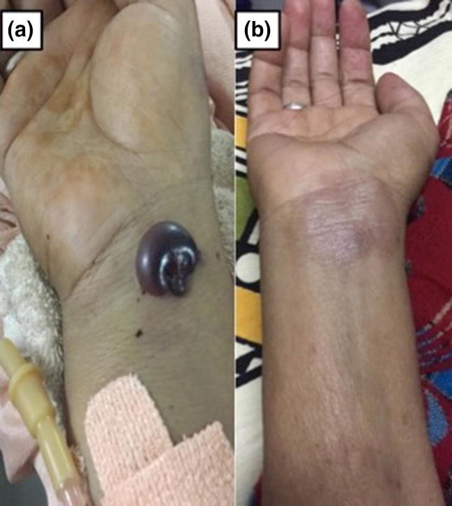
A 50-year-old previously healthy lady presented with a generalized weakness for 2 months and red colored blisters over left forearm for 1 week. There was no history of fever, trauma, itching, and exposure to the extreme of climate or drug intake. She had no history of such lesions in the past. Clinical examination showed a sick looking patient with multiple, well defined, non-tender, cherry-red colored, tense, bullous eruptions over both upper limbs and trunk on the otherwise healthy skin (Fig. 1a). Oral and conjunctival mucosa was normal. She also had pallor and splenomegaly. Investigation showed; hemoglobin 66 g/L, Platelet 17 × 109/L and WBC 40 × 109/L with 88% immature cells on the peripheral blood smear. Her bone marrow showed 90% blasts, which were myeloperoxidase negative and CD19, CD20, CD22, and TdT positive, confirming the diagnosis of pre-B acute lymphoblastic leukemia. A biopsy from the edge of the bulla did not show any infiltration by malignant cells. Blood culture and wound swab (including Tzanck smear) were sterile. She was put on the pre-phase steroid (prednisolone @60 mg/m2) along with supportive care (allopurinol, hydration, PRBC, and SDAP). On day-7 of steroid, the bullous lesions subsided (Fig. 1b). Her chemotherapy was continued as per modified-BFM protocol.
Fig. 1.

a Clinical photograph showing well defined, cherry-red colored, tense, bullous eruptions (hemorrhagic bullae) over forearm at presentation. b Receding hemorrhagic bullae after 01 week of oral prednisolone
Hemorrhagic bulla has been reported at the presentation in cases of hematological malignancy. Among patients with hematological malignancies, who are usually neutropenic, it usually results from skin infection like impetigo and Ecthyma [3–5]. However, rarely it can be due to leukemic infiltration (leukemia cutis) [6, 7]. In the index case, there was no evidence of infection or leukemic infiltration and the patient improved with steroid and platelet transfusion. We propose thrombocytopenia as the underlying mechanism, which probably caused hemorrhagic bullae in the index case. Hemorrhagic bullae can be considered as the counterpart of wet purpura (often seen in patients with life-threatening thrombocytopenia) over mucosa [8]. We conclude that any case of hemorrhagic bullae of skin should be thoroughly evaluated if no other apparent cause is found on initial evaluation because the treatment, as well as the outcome, depends on the underlying etiology.
Conflict of interest
There is no conflict of interest between the authors.
Ethical Approval
All procedures performed in studies involving human participants were in accordance with the ethical standards of the institutional and/or national research committee and with the 1964 Helsinki declaration and its later amendments or comparable ethical standards.
Human and Animals Rights
No animals were involved in the study.
Informed Consent
Informed signed written consent was taken from the patient involved.
Footnotes
Publisher's Note
Springer Nature remains neutral with regard to jurisdictional claims in published maps and institutional affiliations.
References
- 1.Hsiao CT, Lin LJ, Shiao CJ, et al. Hemorrhagic bullae are not only skin deep. Am J Emerg Med. 2008;26(3):316–319. doi: 10.1016/j.ajem.2007.07.014. [DOI] [PubMed] [Google Scholar]
- 2.Miguel-Gomez L, Fonda-Pascual P, Carrillo-Gijon R, et al. Bullous hemorrhagic dermatosis probably associated with enoxaparin. Indian J Dermatol Venereol Leprol. 2016;82:319–320. doi: 10.4103/0378-6323.175915. [DOI] [PubMed] [Google Scholar]
- 3.Ochonisky S, Aractingi S, Dombret H, et al. Acute undifferentiated myeloblastic leukemia revealed by specific hemorrhagic bullous lesions. Arch Dermatol. 1993;129(4):512–513. doi: 10.1001/archderm.1993.01680250128025. [DOI] [PubMed] [Google Scholar]
- 4.Mishra K, Yanamandra U, Prakash G, et al. Ecthyma gangrenosum in a case of acute lymphoblastic lymphoma. BMJ Case Rep. 2017;4:2017. doi: 10.1136/bcr-2016-218501. [DOI] [PMC free article] [PubMed] [Google Scholar]
- 5.Mishra K, Jandial A, Bal A, et al. Verrucous skin lesions in a case of acute lymphoblastic leukemia: a rare manifestation of cytomegalovirus infection. Indian J Hematol Blood Transfus. 2018;34(2):378–380. doi: 10.1007/s12288-017-0855-3. [DOI] [PMC free article] [PubMed] [Google Scholar]
- 6.Piette WW. An approach to cutaneous changes caused by hematologic malignancies. Dermatol Clin. 1989;7(3):467–479. doi: 10.1016/S0733-8635(18)30578-3. [DOI] [PubMed] [Google Scholar]
- 7.Jandial A, Mishra K, Rajpal S, et al. “Blueberry muffin spots” in an adult male successfully treated with imatinib. Indian J Hematol Blood Transfus. 2018;1:11. doi: 10.1007/s12288-018-1024-z. [DOI] [PMC free article] [PubMed] [Google Scholar]
- 8.Mishra K, Jandial A, Malhotra P, et al. Wet purpura: a sinister sign in thrombocytopenia. BMJ Case Rep. 2017;1:2017. doi: 10.1136/bcr-2017-222008. [DOI] [PMC free article] [PubMed] [Google Scholar]


