Dear Editor,
A 25-year-old, unmarried girl presented to emergency services with severe lower abdominal pain for 12 h. There was no history of trauma. She was diagnosed with von Willebrand disease (vWD) type 3 at 2 years of age when she had presented with mucocutaneous bleeding episodes following a trivial fall, and she was managed with tranexamic acid and fresh frozen plasma with complete recovery. She did not give a history of any other episode of significant bleeding in the past. On examination, she had pallor, pulse rate was 126 per minute, and blood pressure was 90/60 mm of Hg. She had generalized abdominal distension with rigidity and diffuse tenderness but no rebound tenderness. An ill-defined lump was palpable in the hypogastrium and both iliac fossae. Investigations revealed, hemoglobin of 57 g/L, WBC 64 × 10^9/L, and platelet 246 × 10^9/L. Prothrombin time (12 s) and INR (1.2) were normal, but activated partial thromboplastin time (aPTT) was significantly prolonged (51 s, control 31 s).
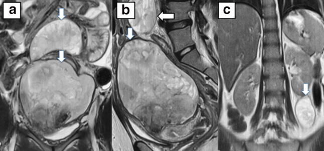
Ultrasound examination for evaluation of the acute abdomen revealed multiple intra-abdominal non-drainable heteroechoic mass like collections. Magnetic resonance imaging (MRI) was done to discern further the nature of these heteroechoic lesions, which revealed multiple large abdominopelvic and retroperitoneal heterogeneously hyperintense hematomas (Fig. 1).A diagnosis of vWD with intra-abdominal hematoma was made. She was managed with packed red blood cell (04 units), fresh frozen plasma (FFP) (12 units), and cryoprecipitate (24 units) transfusions and tramadol (for pain relief) over next 3 days, following which she had significant symptomatic improvement with no further drop in hemoglobin. Follow-up ultrasonography after 10 days of treatment showed a 20–30% decrease in the size of abdominal mass lesions. One year later, on follow up, she was asymptomatic, and a repeat MRI abdomen showed near complete resolution of the hematoma.
Fig. 1.

a Coronal T2 weighted MRI reveals a 10 cm large abdominopelvic mass (pseudotumor) which was heterogeneously hyperintense and showed a T2 hypointense rim, consistent with a hematoma. Another similar localized intraperitoneal lesion was also seen measuring 7 × 4 cm. b Saggital MR image shows that the larger mass is compressing and distorting the uterus. c Coronal MRI at the level of kidneys reveals another infrarenal retroperitoneal hematoma of similar MR morphology
Von Willebrand disease is the most frequent inherited bleeding disorder with a worldwide prevalence of approximately 0.6–1.3%. The disease is classified into three types; type 1, 2 and 3. Type 3 vWD, an autosomal recessive disease, is the most severe and the rarest of all three types [1]. Patients of vWD type 3 are well known to have a recurrent hematoma, which can either present as painful swellings in anatomical locations frequently exposed to external trauma (more common) or less commonly as slow-growing organized swellings (pseudotumor) [2, 3]. However, there are only a few of case reports of massive hematoma and pseudotumor in vWD involving muscle or abdominal cavity [4–6]. The treatment for acute hematoma typically requires replacement of von Willebrand factors (vWF). The commercially available vWF concentrates have limited availability in developing countries, and it is usually beyond the financial capability of the patient. The next best option includes cryoprecipitate (80U vWF) and fresh frozen plasma (1U factor VIII/mL). The treatment for slow-growing hematoma (pseudotumor) requires a trial of factor replacement followed by assessment for need of surgical excision.Our case represented a unique aspect of vWD in which the patient had multiple large intrabdominal pseudotumors and also depicts the typical and rarely illustrated MR images of these pseudotumors. The prompt evaluation and detection, and aggressive management resulted in a favorable outcome. Though the factor VIII and vWF concentrates were not available, FFP and cryoprecipitate could tide over the acute crisis. MRI was instrumental in the diagnosis of this case as MRI showed characteristic imaging findings precluding any need of urgent surgical exploration for acute abdomen. We suggest that a patient of VWD with acute abdominal pain/abdominal distension should be suspected for pseudotumor which may be confirmed by MR imaging for appropriate and timely management.
Conflict of interest
The authors declare that they have no conflict of interest.
Informed Consent
Informed signed written consent was taken from the patient involved.
Ethical Approval
All procedures performed in studies involving human participants were in accordance with the ethical standards of the institutional and/or national research committee and with the 1964 Helsinki declaration and its later amendments or comparable ethical standards.
Human and Animals Rights
No animals were involved in the study.
References
- 1.Leebeek FW, Eikenboom JC. Von Willebrand’s disease. N Engl J Med. 2016;375(21):2067–2080. doi: 10.1056/NEJMra1601561. [DOI] [PubMed] [Google Scholar]
- 2.Argyris PP, Anim SO, Koutlas IG. Maxillary pseudotumor as initial manifestation of von Willebrand disease, type 2: report of a rare case and literature review. Oral Surg Oral Med Oral Pathol Oral Radiol. 2016;121(2):e27–e31. doi: 10.1016/j.oooo.2015.05.018. [DOI] [PubMed] [Google Scholar]
- 3.Biri A, Kurdoglu M, Kurdoglu Z, Gultekin S, Gursel T. Acute abdominal pain caused by a spontaneous intramyometrial hematoma in type III von Willebrand disease. Pediatr Emerg Care. 2006;22(9):650–652. doi: 10.1097/01.pec.0000235838.06331.c3. [DOI] [PubMed] [Google Scholar]
- 4.Keikhaei B, Soltani Shirazi A. Spontaneous iliopsoas muscle hematoma in a patient with von Willebrand disease: a case report. J Med Case Rep. 2011;Jul2; 5:274. doi: 10.1186/1752-1947-5-274. [DOI] [PMC free article] [PubMed] [Google Scholar]
- 5.Eby CS, Caracioni AA, Badar S, Joist JH. Massive retroperitoneal pseudotumour in a patient with type 3 von Willebrand disease. Haemophilia. 2002;8:136–141. doi: 10.1046/j.1365-2516.2002.00588.x. [DOI] [PubMed] [Google Scholar]
- 6.Wexler S, Edgar M, Thomas A, Learmonth I, Scott G. Pseudotumour in von Willebrand disease. Haemophilia. 2001;7(6):592–594. doi: 10.1046/j.1365-2516.2001.00552.x. [DOI] [PubMed] [Google Scholar]


