Abstract
The progression of Alzheimer's dementia is associated with neurovasculature impairment, which includes inflammation, microthromboses, and reduced cerebral blood flow. Here, we investigate the effects of β amyloid peptides on the function of platelets, the cells driving haemostasis. Amyloid peptide β1-42 (Aβ1-42), Aβ1-40, and Aβ25-35 were tested in static adhesion experiments, and it was found that platelets preferentially adhere to Aβ1-42 compared to other Aβ peptides. In addition, significant platelet spreading was observed over Aβ1-42, while Aβ1-40, Aβ25-35, and the scAβ1-42 control did not seem to induce any platelet spreading, which suggested that only Aβ1-42 activates platelet signalling in our experimental conditions. Aβ1-42 also induced significant platelet adhesion and thrombus formation in whole blood under venous flow condition, while other Aβ peptides did not. The molecular mechanism of Aβ1-42 was investigated by flow cytometry, which revealed that this peptide induces a significant activation of integrin αIIbβ3, but does not induce platelet degranulation (as measured by P-selectin membrane translocation). Finally, Aβ1-42 treatment of human platelets led to detectable levels of protein kinase C (PKC) activation and tyrosine phosphorylation, which are hallmarks of platelet signalling. Interestingly, the NADPH oxidase (NOX) inhibitor VAS2870 completely abolished Aβ1-42-dependent platelet adhesion in static conditions, thrombus formation in physiological flow conditions, integrin αIIbβ3 activation, and tyrosine- and PKC-dependent platelet signalling. In summary, this study highlights the importance of NOXs in the activation of platelets in response to amyloid peptide β1-42. The molecular mechanisms described in this manuscript may play an important role in the neurovascular impairment observed in Alzheimer's patients.
1. Introduction
Alzheimer's disease (AD) is a multifactorial age-related neurodegenerative disorder representing 60-80% of dementia cases [1]. Prominent morphological hallmarks of the disease include pathological accumulation of insoluble aggregates of polymeric protein fragments known as β amyloid peptides deposited in the brain parenchyma (amyloid plaques) and within the cerebral vessel walls (cerebral amyloid angiopathy (CAA)), formation of neurofibrillary tangles within neurons (tau pathology), oxidative stress and chronic neurovascular inflammation resulting in blood hypoperfusion, and damages to the blood brain barrier (BBB) [2]. The manifestation of these pathological conditions eventually lead to neurovascular dysfunction, neuronecrosis, cognitive decline, and ultimately death [3].
Epidemiological data, postmortem pathological examination, and experimental studies on both human and animal AD brains have revealed significant correlations and shared pathophysiological mechanisms between Alzheimer's and vascular diseases [4–9]. Common contributing causes include conditions such as hypertension, diabetes mellitus, hypercholesterolemia, apolipoprotein E (APOE) 4 polymorphism, and traumatic brain injury [10].
The potential role of platelets in Alzheimer's disease has been investigated in a number of studies. The initial work of Rosenberg et al. in 1997 highlighted possible platelet activation in AD patients due to altered APP processing [11]. His work was followed up by Sevush et al. in 1998 and by other groups later on, and it was confirmed that there is an aberrant and chronic preactivation of platelets that can eventually contribute towards atherothrombosis, CAA, and progression of AD [12]. Several studies showed a correlation between AD and platelet abnormalities, including abnormal membrane fluidity, increased β-secretase activity, and altered APP metabolism [13]; α-degranulation, P-selectin surface expression, and integrin αIIbβ3 activation [14]; platelet adhesion [15, 16]; formation of leukocyte-platelet complexes [12]; coagulation abnormalities [17–19]; and platelet adhesion and accumulation at vascular β amyloid deposition sites, where they were shown to modulate β amyloid complexation into aggregates [20].
Several authors utilised both soluble and fibril forms of β amyloid peptides as agonists and demonstrated that Aβ peptides are able to promote platelet activation, adhesion, and aggregation. For example, fibrillar Aβ1-40 was shown to induce platelet aggregation by binding to scavenger receptors CD36 and GP1bα and activating p38 MAPK/COX1 pathways. This induces the release of the potent aggregation agonist thromboxane A2 (TxA2) [21]. Donner et al. more recently showed that Aβ1-40 can bind to integrin αIIbβ3 and trigger the release of ADP and clusterin (a chaperone protein), which promoted the formation of Aβ1-40 fibrils [22]. In addition, the use of synthetic Aβ25-35, which retains the biological and toxic properties of the full length Aβ1-40 and Aβ1-42, has been shown to activate the PAR1 thrombin receptor and stimulate an intracellular signalling cascade involving Ras/Raf, PI3K, P38MAPK, and cPLA2 and TxA2 formation and release [23].
NADPH oxidases (NOXs) are the only enzyme family recognized for their sole primary function of generating reactive oxygen species (ROS), and they have been proposed as the main source of ROS in platelets during haemostasis [24]. Recently, two types of NOXs have been identified in human and mouse platelets (NOX1 and NOX2) [25], but a comprehensive understanding of their activation signalling pathways in response to β amyloid peptides remains elusive. An interesting paper published by Walsh et al. demonstrated that oligomeric and fibrillar forms of Aβ1-42 can act as a ligand for the GPVI receptor and activate platelets [26]. Since NOX1 has been shown to play a key role in signalling for the GPVI receptor [25, 27], this may suggest that Aβ1-42 acts through a NOX1-dependent activation of platelets.
Recently, we demonstrated that upon stimulation of platelets by both monomeric or fibril forms of Aβ1-42, significant intracellular superoxide anion formation can be detected using a novel flow cytometry method using the molecular probe dihydroethidium (DHE) [28]. Here, we further investigate the effects of different Aβ (Aβ1-42, Aβ1-40, Aβ25-35, and scrambled Aβ1-42 control) on platelet activation and adhesion in static and physiological flow conditions. The use of pan-NOX inhibitor VAS2870 [28] allows the assessment of the role of NOXs on platelet adhesion and activation. Our primary objective is to understand the mechanism underlining β amyloid peptide-dependent regulation of platelets, which can potentially improve our understanding of AD and facilitate the development of pharmacological tools to combat the progression of this disease.
2. Materials and Methods
2.1. Reagents
Dimethylsulfoxide (DMSO), indomethacin, prostaglandin E1 (PGE1), bovine serum albumin (BSA), sodium citrate solution (4% w/v), fibrinogen, thrombin from human plasma, 4% w/v paraformaldehyde, TRITC-conjugated phalloidin, 3,3′-dihexyloxacarbocyanine iodide (DiOC6), VAS2870, D-(+)-glucose monohydrate, and 4-(2-hydroxyethyl) piperazine-1-ethanesulfonic acid (HEPES) were from Sigma-Aldrich (Poole, UK). Fibrillar collagen was from Chrono-Log Corporation (Havertown, PA US). The anti-phosphotyrosine antibody (4G10) was from Upstate Biotechnology Inc. (Lake Placid, US). Anti-PKC phosphor-substrate antibody was from Cell Signaling Technology (Danvers, US). Anti-pleckstrin antibody was from Abcam (Cambridge, UK). FITC-PAC1 and PE-Cy5-CD62P (P-selectin) antibodies were from Becton Dickinson, (Wokingham, UK). Peroxidase-conjugated anti-IgG antibodies were from Bio-Rad (Hercules, US). The chemiluminescent substrate kit was from Merck Millipore (Burlington, US). Amyloid peptides were synthesized by LifeTein (New Jersey, US). The sequences of the peptides are as follows:
Aβ1-40 (4.3 kDa): DAEFRHDSGYEVHHQKLVFFAEDVGSNKGAIIGLMVGGVV
Aβ1-42 (4.5 kDa): DAEFRHDSGYEVHHQKLVFFAEDVGSNKGAII GLMVGGVVIA
Scrambled Aβ1-42: (4.5 kDa): DEFAKNIGHHDGVAVHMYKGRQVEFIGSIALVFEDVGSAGLV
Aβ23-35 (1.0 kDa): GSNKGAIIGLM
2.2. Preparation of Washed Platelets
Human whole blood was obtained from healthy volunteers following Royal Devon and Exeter NHS Foundation Trust Code of Ethics and Research Conduct and under NRES South West – Central Bristol committee approval (Rec n. 14/SW/1089). 20–30 ml of blood were drawn in the presence of the anticoagulant sodium citrate (0.5% w/v). Platelet rich plasma (PRP) was then isolated from whole blood by centrifugation at 200 × g for 20 mins. PRP was then subjected to a second centrifugation at 500 × g for 10 mins in the presence of indomethacin (10 μM) and PGE1 (40 ng/ml). Platelets were resuspended in modified Tyrode's HEPES buffer (145 mM NaCl, 2.9 mM KCl, 10 mM HEPES, 1 mM MgCl2, and pH 7.3; 5 mM D-glucose was added before use).
2.3. Adhesion Assay
Round coverslips (22 mm) were placed in a clear flat-bottom 6-well plate and then coated overnight at 4°C with 10 μM Aβ1-42, Aβ1-40, Aβ25-35, scrambled Aβ1-42, or 5 mg/ml BSA, all diluted in PBS. Excess solution was then gently removed, and the coverslips were blocked with 0.5% w/v BSA in PBS for 1 h at room temperature. Coated dishes were then washed gently with PBS. Washed platelets were resuspended at a density of 2 × 10⁷ platelets/ml. 0.5 ml of washed platelets were incubated for 30 mins at 37°C. Nonadherent platelets were discarded, and the adherent ones were gently washed with PBS and then fixed with 4% w/v paraformaldehyde for 10 mins at RT. 0.1% v/v Triton X-100/PBS was added to permeabilize the platelets. After 5 mins, 0.1% Triton X-100/PBS was removed, and the coated coverslips were washed with PBS and then blocked with 5 mg/ml BSA in PBS for 30 mins. Fixed platelets were then stained with 10 μM TRITC-conjugated phalloidin for 1 hour at RT and then washed with PBS. Coverslips were mounted onto microscope slides using Vectashield. Evaluation of platelet adhesion and spreading was performed using a Leica LED fluorescence microscope, and digital images were acquired at 10x and 100x magnification objectives. Platelet coverage and surface area were measured using ImageJ (version 1.52e, Wayne Rasband, NIH).
2.4. Thrombus Formation Assay under Physiological Flow
Human peripheral blood was anticoagulated with sodium citrate 0.25% w/v. Platelets were fluorescently labelled by incubation with 1 μM 3,3′-dihexyloxacarbocyanine iodide (DiOC6) for 10 minutes. ibidi Vena8 Fluoro+ microchips (ibidi GmbH, Martinsried, Germany) were coated with 10 μM Aβ peptides or 0.1 mg/ml fibrillar collagen. Nonspecific binding sites were saturated with 0.1% w/v BSA. Physiological flow conditions (200–1000 sec−1) were applied using an ExiGo pump (Cellix Ltd. Microfluidics Solutions, Dublin, Ireland). Images of the thrombi formed after 10 minutes of flow were obtained with an EVOS Fl microscope (Thermo Fisher Scientific, Waltham, MA, US). Platelet coverage was measured using ImageJ (version 1.52e, Wayne Rasband, NIH).
2.5. Flow Cytometry
Platelets isolated as described above were resuspended at 2 × 10⁷ cells/ml density. After stimulation in suspension as described (5–20 μM Aβ1-42, Aβ1-40, Aβ25-35, scrambled Aβ1-42, or 0.5 unit/ml thrombin) for 10 minutes at 37°C, platelets were incubated for a further 10 minutes with PAC1 and anti-P-selectin antibodies conjugated to FITC and PE-Cy5, respectively. 1 in 10 dilution in Tyrode's buffer was used to stop the immunolabelling of the platelets. Surface fluorescence was assessed using a FACSAria III flow cytometer (BD Biosciences, San Jose, USA).
2.6. Immunoblotting
Platelet samples (0.2 ml, 1 × 10⁹ platelets/ml) were stimulated at 37°C under stirring conditions (1,200 rpm) with Aβ1-42, Aβ1-40, and Aβ25-35 (including 1 mM CaCl2) in a 490D aggregometer (Chrono-Log Corporation, Havertown, PA, US). Where indicated, the NOX inhibitor VAS2870 (10 μM) was preincubated for 10 minutes before treatments with Aβ peptides. The reaction was stopped after 3 minutes by the addition of a half volume of 3x SDS sample buffer (37.5 mM Tris, pH 8.3, 288 mM glycine, 6% SDS, 1.5% DTT, 30% glycerol, and 0.03% bromophenol blue) followed by heating the samples at 95°C for 5 minutes. Platelet proteins were separated on SDS-PAGE gels, transferred to a PVDF membrane, and analysed in immunoblotting using anti-phosphotyrosine antibody (4G10), anti-phospho-PKC substrate antibody, and anti-pleckstrin antibodies. Reactive proteins were visualized by ECL.
2.7. Statistical Analysis
Data were analysed by one-way ANOVA with Bonferroni posttest using the statistical software GraphPad Prism. Results were expressed as the mean ± standard error (SEM). Differences were considered significant at P value < 0.05.
3. Results
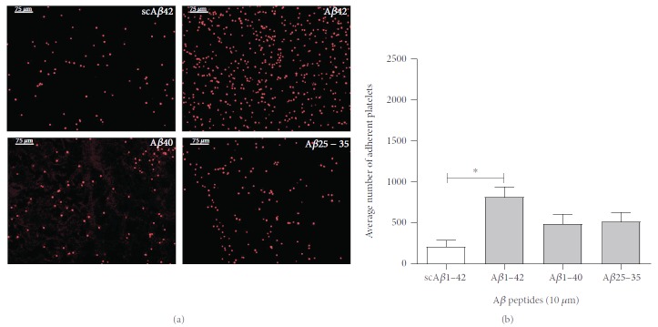
Initial experiments were carried out to examine the effect of amyloidogenic peptides on human platelet adhesion in static conditions by allowing platelet suspensions to rest on coverslips coated with scrambled Aβ1-42, Aβ1-42, Aβ1-40, and Aβ25-35 for 30 minutes. Adhering platelets were then fixed, permeabilized, and stained with TRITC-phalloidin. Phalloidin is a poisonous molecule extracted from the mushroom Amanita phalloides that binds strongly to filaments of actin (F-actin), which is a major component of the cell cytoskeleton [29]. In these experiments, phalloidin allows effective visualization of adhered platelets. Figure 1(a) shows the high number of adhered platelets to Aβ1-42. Adhesion to Aβ1-40, or Aβ25-35 is higher than control scrambled Aβ1-42 peptide, but this difference is not statistically significant. The quantification of adhered platelets from 4 independent experiments is shown in Figure 1(b). The statistical analysis (one-way ANOVA with Bonferroni posttest) shows significance (P < 0.05) for the increase in platelet adhesion to Aβ1-42 compared to scrambled Aβ1-42.
Figure 1.
β amyloid peptides in static conditions support platelet adhesion. Human platelet suspensions were plated on glass coverslips coated with 10 μM scrambled Aβ1-42 (scAβ42), Aβ1-42, Aβ1-40, and Aβ25-35. The adhered platelets after 30 minutes shown in (a) were fixed, permeabilized, and stained with TRITC-conjugated phalloidin and are representative images at 10x magnification. The quantification of the adhered platelets evaluated as the mean number of platelets per optical field is shown in (b). Statistical significance for 4 independent experiments was analysed by one-way ANOVA with Bonferroni posttest (∗P < 0.05), with bars representing standard error of the mean (SEM).
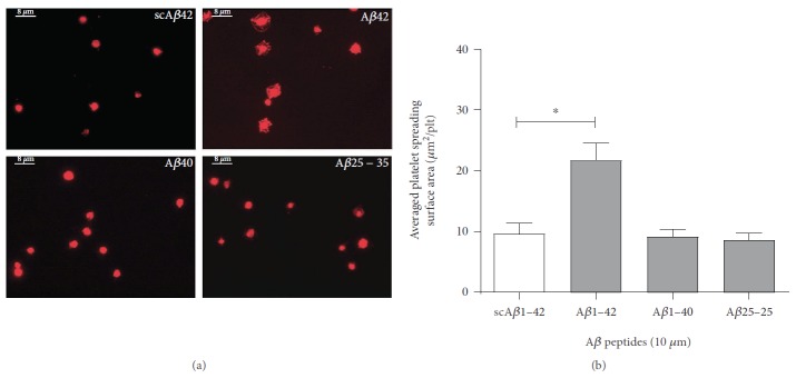
In order to assess platelet spreading on Aβ peptides, Figure 2(a) displays the morphological changes in adhering platelets at a higher magnification (100x). Initial adhesion is characterised by shape change from discoid into irregular or round shape and filopodia formation, while complete adhesion and signalling activation is associated with extensive platelet spreading and formation of lamellipodia. Aβ1-40 and Aβ25-35 induced only the morphological changes of the early phase of platelet adhesion, with few fine processes extending into different directions (filopodia). On the other hand, platelets adhering to Aβ1-42 show full spreading, with the formation of extensive lamellipodia and a significant increase in surface area coverage. Figure 2(b) quantifies the average surface area of the platelets confirming the effectiveness of Aβ1-42 as a substrate for signalling activation and platelet spreading, while no platelet spreading is observed for Aβ1-40 and Aβ25-35.
Figure 2.
β amyloid peptides in static conditions support platelet spreading. Human platelet suspensions were plated on glass coverslips coated with 10 μM scrambled Aβ1-42 (scAβ42), Aβ1-42, Aβ1-40, and Aβ25-35. The adhered platelets shown in (a) were fixed, permeabilized, and stained with TRITC-conjugated phalloidin and are representative images at 100x magnification. The quantification of the mean surface area of the adhered platelets (μm2/plt) is shown in (b). Statistical significance for 4 independent experiments was analysed by one-way ANOVA with Bonferroni posttest (∗P < 0.05), with bars representing standard error of the mean (SEM).
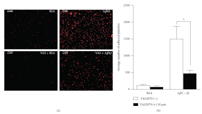
Since platelets demonstrated significant adhesion and spreading on Aβ1-42, we next investigated the effects of the NOX inhibitor VAS2870 on this substrate. Both platelet adhesion and spreading were strongly impaired. In Figure 3(a), we show the quantification of platelets adhering to Aβ1-42 in the presence or absence of VAS2870. Although the number of adhering platelets is not completely abated by VAS2870, there was a statistically significant decrease in the number of adhering platelets in the presence of this inhibitor (Figure 3(b)).
Figure 3.
Effect of NOX inhibitor VAS2870 on platelet adhesion to Aβ1-42. Human platelet suspension was preincubated with NOX inhibitor VAS2870 (10 μM) for 30 mins then plated on glass coverslips coated with 10 μM Aβ1-42 and 5 mg/ml BSA in PBS. The numbers of the adhered platelets fixed, permeabilized, and stained with TRITC-conjugated phalloidin are shown in (a), and representative images at 10x magnification are displayed. The quantification of the adhered platelets evaluated as the mean number of the adhered platelets per optical field is shown (b). Statistical significance for 4 independent experiments was analysed by one-way ANOVA with Bonferroni posttest (∗P < 0.05), with bars representing standard error of the mean (SEM).
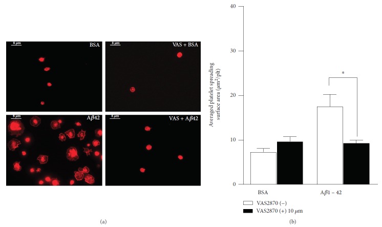
When observing adhering platelets at a higher magnification (100x), VAS2870 appeared to significantly reduce the platelet spreading and the resulting surface area per platelet was lower than in the absence of the NOX inhibitor (Figure 4(a)). Statistical analysis revealed statistical significance of the effect of VAS2870 on platelet spreading (Figure 4(b)).
Figure 4.
Effect of NOX inhibitor VAS2870 on platelet spreading on Aβ1-42. Human platelet suspension was preincubated with NOX inhibitor VAS2870 (10 μM) for 30 mins then plated on glass coverslips coated with 10 μM Aβ1-42 and 5 mg/ml BSA in PBS. The surface area of the adhered platelets fixed, permeabilized, and stained with TRITC-conjugated phalloidin is shown in (a), and representative images at 100x magnification are displayed. The quantification of the surface area of the adhered platelets (μm2/plt) is shown in (b). Statistical significance for 4 independent experiments was analysed by one-way ANOVA with Bonferroni posttest (∗P < 0.05), with bars representing standard error of the mean (SEM).
Next, we tested whether Aβ peptides can stimulate platelet adhesion under physiological shear. We tested both arterial (1,000 sec−1) and venous shear (200 sec−1) [30]. At 1,000 sec−1, the tensile strength of the binding of platelets to Aβ peptides is not sufficient to guarantee effective adhesion and thrombus formation (Figures 5(a) and 5(b)). At lower shear rate corresponding to venous circulation (200 sec−1), Aβ1-42 but not Aβ1-40, Aβ25-35, or scrambled Aβ1-42 induced convincing platelet adhesion and thrombus formation (Figures 5(c), 5(e) and 5(f)). Similarly to what was observed in static conditions, the inhibition of NOX with VAS2870 (10 μM) inhibited platelet adhesion (and thrombus formation) in response to Aβ1-42 (Figures 5(d) and 5(g)).
Figure 5.
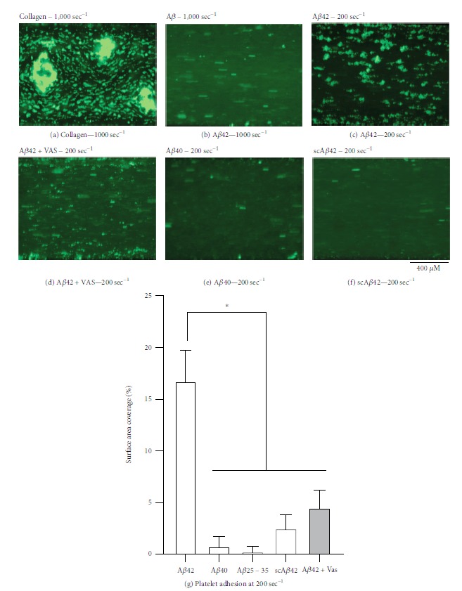
Adhesion of platelets to amyloid peptides under physiological shear stress. Flow biochips (ibidi Vena8 Fluoro+) were coated with 0.1 mg/ml fibrillary collagen or 10 μM scrambled Aβ1-42 (scAβ42), Aβ1-42, Aβ1-40, and Aβ25-35. Platelet adhesion was tested in human whole blood at shear rates of 1,000 sec−1 and 200 sec−1. Where indicated, 10 μM VAS2870 was added to the blood to inhibit NOXs. Pictures shown here are representative of 3 independent experiments. Surface coverage analysis was performed using ImageJ and statistically analysed by one-way ANOVA with Bonferroni posttest (∗P < 0.05).
As the mechanisms leading to platelet adhesion to Aβ1-42 remain unclear, but previous work suggest a role for integrin αIIbβ3 on the adhesion to Aβ1-40 [22, 31], we tested the activation of this integrin using the antibody PAC1 by flow cytometry. These experiments suggested that only Aβ1-42 (but not Aβ1-40, Aβ2535, or scrambled Aβ1-42) induced a convincing activation of integrin αIIbβ3 (Figures 6(a)–6(g)). NOX inhibition with 10 μM VAS2870 abolished Aβ1-42-induced αIIbβ3 activation (Figure 6(d)). Interestingly, no significant translocation of P-selectin to the surface of the platelets was observed, suggesting that Aβ peptides (including Aβ1-42) cannot stimulate effective platelet degranulation on their own.
Figure 6.
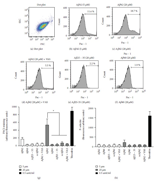
Activation of integrin αIIbβ3 by Aβ1-42. Washed platelets were stimulated as indicated with Aβ1-42, Aβ1-40, Aβ25-35, or scrambled Aβ1-42 or 0.5 units/ml thrombin for 10 minutes and then labelled with FITC-PAC1 (b-g) and PE-Cy5-P-selectin (h) for a further 10 minutes. A side-scattering (SSC)/forward-scattering (FSC) dot plot is shown in (a) and suggests the high purity of the platelet preparation. The histograms for the intensity of PAC1 staining in the different conditions are shown in (b-f) (representative of 3 independent experiments). Data analyses are shown in (g) and (h). Statistical analysis by one-way ANOVA with Bonferroni posttest is shown in (g) and (h) (n = 3, ∗P < 0.05).
We next investigated the effect of Aβ1-42 on intracellular platelet signalling using phosphospecific immunoblotting. Both unstimulated and Aβ1-42-stimulated human platelets were treated with either DMSO (control) or VAS2870. They were then lysed, and the resulting protein extracts were separated by SDS-PAGE. Immunoblotting for phosphotyrosine and phosphorylated PKC substrates is shown in Figure 7. These antibodies are used to determine whether tyrosine phosphorylation cascades and PKC are activated by Aβ1-42 treatment, but they do not allow the identification of the targets of the phosphorylation events and function as a qualitative evidence of activation of the abovementioned signalling pathways. In these experiments, pleckstrin is used simply as a loading control. Tyrosine phosphorylation is one of the key events that occur upon platelet activation, therefore detecting tyrosine phosphorylation of platelet proteins provides a proof that Aβ1-42 induces platelet signalling activation. Figure 7(a) shows the phosphotyrosine profile of resting and Aβ1-42-treated platelets in the absence or presence of VAS2870. Several bands are observed upon stimulation with Aβ1-42 (compared to DMSO-treated controls), which suggests activation of tyrosine kinase-dependent pathways and generation of tyrosine-phosphorylated protein substrates. Very significantly, the pretreatment with VAS2870 leads to the abolishment of tyrosine phosphorylation in response to Aβ1-42, which suggests that the activity of NADPH oxidases is necessary for the signalling induced by this peptide.
Figure 7.
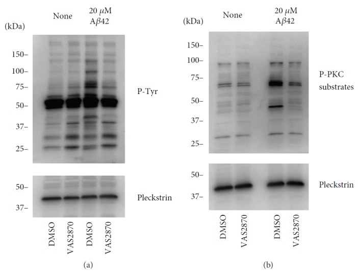
Aβ42-induced signalling in platelets. (a, b) Unstimulated and Aβ42-stimulated (20 μM) human platelets were treated with 10 μM DMSO or VAS2870. Total proteins were separated by SDS-PAGE as described in the Materials and Methods, and protein phosphorylation was analysed by immunoblotting with the indicated antibodies: (a) anti-p-Tyr and anti-pleckstrin antibody and (b) anti-PKC phosphosubstrate and anti-pleckstrin antibody. The figure represents blots from three independent experiments.
PKC is an essential protein kinase enzyme that is activated by diacylglycerol (DAG) and Ca2+ released from internal stores. The activation of PKC is a well-known intracellular event induced by platelet activation, which is usually accompanied by phosphorylation of regulatory serine/threonine residues in PKC substrate proteins [32–34]. In Figure 7(b), several bands corresponding to different substrate proteins for PKC appear more intensely upon Aβ1-42 stimulation, suggesting that PKC is activated by this peptide. PKC activation is strongly associated to platelet activation and induction of thrombus formation [34–36]. The abolishment of phospho-PKC immunostaining by VAS2870 suggests that NADPH oxidase activity is required for the Aβ1-42-dependent platelet signalling leading to PKC activation.
4. Discussion
The differential ability of the tested β amyloid peptides to induce platelet adhesive responses in static and flow conditions is extremely interesting, but it remains difficult to explain. Nonetheless, our observations are not isolated, as Aβ1-42 has been identified as the most biologically active of the amyloid peptides [37–39]. The biological activity of Aβ1-42 is associated to a marked toxicity of this peptide in several experimental systems [37–39]. One possible explanation is the marked propensity of Aβ1-42 to form fibrils compared to Aβ1-40 [40], which is due to the promotion of intermolecular interactions between amyloid monomers induced by the hydrophobic properties of the extra amino acids of Aβ1-42 compared to Aβ1-40. Recent studies have confirmed the enhanced propensity of Aβ1-42 to form fibrils compared to other Aβ peptides [41, 42]. This draws an interesting parallel with the effect of collagen on platelets. The activation of GPVI on platelets and induction of thrombus formation depends on the fibrillary structure of collagen (i.e., monomeric collagen does not induce platelet activation) [43]. Therefore, similarly to collagen, the β amyloid peptides may also need to be in a fibrillar form to bind adjacent receptors and induce effective intracellular signalling in platelets. This seems to favour the hypothesis that GPVI is the receptor for β amyloid peptides on platelets, as this receptor preferentially binds substrates in a fibrillary form, which allows the contemporaneous interaction of the same fibril to different receptors (GPVI and integrin α2β1 in the case of collagen) [44, 45].
Our static adhesion results are in partial disagreement with older studies describing the ability of Aβ25-35 and Aβ1-40 to promote platelet adhesion [16, 22, 31, 46]. In reality, in our experiments, Aβ1-40 and Aβ25-35 coating led to a noticeable increase in platelet adhesion (from less than 200 platelets per optical field on scrambled peptides to around 400). Possibly because of the extent of the effect of Aβ1-42 (over 800 platelets per optical field), the effect of these two peptides did not have statistically significant results. Analysis of adhering platelets at a higher magnification revealed that Aβ1-40 and Aβ25-35 did not induce extensive platelet spreading, with most platelets adhering to these substrates displaying a spherical morphology and modest filopodia formation, which is indicative of partial activation. These data are therefore suggesting some ability of Aβ25-35 and Aβ1-40 to induce platelet adhesion, but a significantly higher platelet adhesion to Aβ1-42, which is likely to induce more extensive platelet intracellular signalling and full spreading (as suggested by spreading data on Figure 3). The conditions utilised for the resuspension of the peptides and the coating of the surfaces in this and Canobbio et al.'s study of 2013 [16] are different, which is likely to affect the level of peptide fibrillation and ability to bind platelets. Further investigation of this discrepancy is necessary to fully understand how platelet binding of β amyloid peptides is regulated. As platelet adhesion under static conditions recapitulates platelet adhesion in the bloodstream, these data suggest the possibility that microthrombosis observed in the neurovasculature of AD patients is due to platelet adhesion to Aβ1-42 accumulating in the perivascular space and migrating into the bloodstream via endothelial cell transport [47].
We also tested Aβ peptides for their ability to induce thrombus formation in whole blood under physiological shear. In previous studies, it was shown that Aβ25-35 was not able to induce thrombus formation on its own [35]. This was confirmed in the present study. The ability of amyloid peptides to potentiate platelet adhesion on collagen that we showed in previous studies was not investigated in this manuscript because the Aβ1-42 peptide showed a remarkable ability to induce platelet adhesion on its own. Although Aβ1-42 has been shown to potentiate platelet adhesion to collagen and other substrates previously [14, 48], here we present the first evidence that this peptide alone is sufficient to induce thrombus formation under flow.
Integrin αIIbβ3 has been suggested as the receptor on platelets for Aβ1-40 [22, 31]. Therefore, we analysed whether this integrin is activated in the presence of Aβ peptides. Integrins are adhesion receptors characterised by two activation states (active and inactive), with only the active state able to interact and bind its substrates. The signalling leading to integrin activation is known as inside-out signalling, while the signalling triggered by the engagement of the integrin with its substrate is known as outside-in signalling [49]. With the PAC1 antibody, we were able to assess the activation of αIIbβ3, which is significant for Aβ1-42 (but not for Aβ1-40, Aβ25-35, or scrambled Aβ1-42). This is a significant finding suggesting profound differences in the biological effect of Aβ peptides, with only Aβ1-42 inducing signalling activation in platelets. This is in contrast with previous studies showing the signalling response induced by Aβ1-40 [22, 31] or Aβ25-35 [35]. This discrepancy remains difficult to explain, but the differences in the experimental conditions and the preparation of the peptide are a likely explanation. In addition, our current study cannot categorically exclude some level of platelet activation by other Aβ peptides (as shown, e.g., in the static adhesion experiments where Aβ1-40 and Aβ25-35 induce a moderate increase compared to controls). Certainly, Aβ1-42 represents by far the most active Aβ peptide in our hands.
One important question that remains unanswered relates to the receptor responsible for the initial engagement of Aβ1-42. Integrin αIIbβ3 is the most expressed and functionally crucial adhesion receptor in platelets [50]. Its activation is the consequence of a signalling cascade known as inside-out signalling, which requires receptor-dependent activation. Therefore, although integrin αIIbβ3 is likely to participate in platelet adhesion to Aβ peptides, an alternative receptor is likely to exist. Different receptors have been suggested, including protease-activated receptor 1 [23], GPVI [27], and CD36 [21]. Our current data do not help to resolve this impasse.
The intracellular signalling involved in platelet adhesion and activation by β amyloid peptides has been studied by several groups. For example, Sonkar et al. showed that exposure to Aβ25-35 resulted in increased myosin light chain (MLC) phosphorylation and RhoA-GTP levels. This led to the conclusion that Aβ25-35 induces cellular activation via RhoA-dependent modulation of actin and cytoskeletal reorganisation [51]. Our previous investigations also showed that Aβ25-35 promoted intracellular calcium increase by entry from the extracellular environment, which led to dense granule and ADP release, and in turn to the activation of the P2Y12 receptor, the small GTPase Rap1b, and both PI3K and MAP kinase pathways [35]. In this study, we utilised tyrosine phosphorylation and PKC-dependent phosphorylation tested by immunoblotting (Figures 7(a) and 7(b)) as markers of platelet activation and to confirm that NOX activity is crucial to trigger Aβ-dependent signalling in platelets. No further detail on the signalling cascades triggered by Aβ peptides can be drawn from this study. The activation of tyrosine phosphorylation and PKC-dependent protein phosphorylation cascades are central to platelet activation and common to most platelet agonists [52, 53]. We have shown previously the modulation of PKC activity by NOX inhibitors, possibly via dampening of GPVI receptor signalling [25]. Other investigators highlighted the link between NOX activation and PKC activity. In fact, this appears to be a bidirectional interaction, not only with different PKC isozymes showing the ability to activate NOXs (e.g., [54]) but also with NOX-dependent ROS leading to oxidation and activation of PKC enzymes [55]. The data from our current study could be explained by either direct PKC stimulation or triggering of cell signalling leading to PKC activation.
Interestingly, although the effect on αIIbβ3 by Aβ1-42 was very evident (i.e., similar to thrombin for 20 μM Aβ1-42), there was no apparent platelet degranulation, as measured by P-selectin immunostaining. This implies that differently to canonical agonists such as thrombin, collagen, or thromboxane A2, Aβ1-42 induces integrin activation without full platelet activation (i.e., partial stimulation). This may explain the poor activity of Aβ1-42 as a platelet agonist in some traditional assays, such as platelet aggregation [28].
In general, the variability in the peptides utilised (e.g., Aβ25-35, Aβ1-40, or Aβ1-42) and the focus of different studies on different receptors and signalling pathways led to apparently contradicting results. For example, an intriguing study reported the reduction of Aβ peptide-dependent platelet activation by fibrinogen [56]. Although the underlying mechanisms remain difficult to explain, this observation may be correlated to our data on Aβ peptide-dependent activation of integrin αIIbβ3 (which is the main fibrinogen receptor on platelets). The use by authors of the above study of a different Aβ peptide for their stimulations (i.e., Aβ25-35) makes any comparison of our and their studies difficult. Further studies are required to resolve these contradictions.
This study highlights the importance of NADPH oxidase activation and platelet oxidative responses in the prothrombotic responses induced by Aβ1-42, which is the β amyloid peptide accumulating in the brain of Alzheimer's and cerebral amyloid angiopathy (CAA) patients. In addition to giving us some direction in the elucidation of the molecular mechanisms underlying platelet activation by β amyloid peptides, these data suggest a potential therapeutic opportunity aiming at limiting the vascular component of Alzheimer's disease by targeting NADPH oxidase activity.
Acknowledgments
The authors would like to thank Dr. Pia Leete at the University of Exeter Medical School for her support with cell imaging techniques. This work was funded by Alzheimer's Research UK (ARUK-PG2017A-3) and the British Heart Foundation (PG/15/40/31522). This article presents independent research funded by the above funders and supported by the National Institute for Health Research (NIHR) Exeter Clinical Research Facility.
Data Availability
The manuscript does not contain data-intensive results and did not require the use of online repositories. Raw data are available on request by contacting the corresponding author.
Disclosure
The views expressed are those of the author and not necessarily those of the NHS, the NIHR, or the Department of Health.
Conflicts of Interest
The authors have no conflicts of interest.
References
- 1.Kljajevic V. Overestimating the effects of healthy aging. Frontiers in Aging Neuroscience. 2015;7:p. 164. doi: 10.3389/fnagi.2015.00164. [DOI] [PMC free article] [PubMed] [Google Scholar]
- 2.Smith E. E., Greenberg S. M. β-Amyloid, blood vessels, and brain function. Stroke. 2009;40(7):2601–2606. doi: 10.1161/STROKEAHA.108.536839. [DOI] [PMC free article] [PubMed] [Google Scholar]
- 3.Nelson A. R., Sweeney M. D., Sagare A. P., Zlokovic B. V. Neurovascular dysfunction and neurodegeneration in dementia and Alzheimer’s disease. Biochimica et Biophysica Acta (BBA) - Molecular Basis of Disease. 2016;1862(5):887–900. doi: 10.1016/j.bbadis.2015.12.016. [DOI] [PMC free article] [PubMed] [Google Scholar]
- 4.Werring D. J., Gregoire S. M., Cipolotti L. Cerebral microbleeds and vascular cognitive impairment. Journal of the Neurological Sciences. 2010;299(1-2):131–135. doi: 10.1016/j.jns.2010.08.034. [DOI] [PubMed] [Google Scholar]
- 5.Nakata-Kudo Y., Mizuno T., Yamada K., et al. Microbleeds in Alzheimer disease are more related to cerebral amyloid angiopathy than cerebrovascular disease. Dementia and Geriatric Cognitive Disorders. 2006;22(1):8–14. doi: 10.1159/000092958. [DOI] [PubMed] [Google Scholar]
- 6.Haglund M., Sjobeck M., Englund E. Severe cerebral amyloid angiopathy characterizes an underestimated variant of vascular dementia. Dementia and Geriatric Cognitive Disorders. 2004;18(2):132–137. doi: 10.1159/000079192. [DOI] [PubMed] [Google Scholar]
- 7.Humpel C. Chronic mild cerebrovascular dysfunction as a cause for Alzheimer’s disease? Experimental Gerontology. 2011;46(4):225–232. doi: 10.1016/j.exger.2010.11.032. [DOI] [PMC free article] [PubMed] [Google Scholar]
- 8.van Rooden S., Goos J. D. C., van Opstal A. M., et al. Increased number of microinfarcts in Alzheimer disease at 7-T MR imaging. Radiology. 2014;270(1):205–211. doi: 10.1148/radiol.13130743. [DOI] [PubMed] [Google Scholar]
- 9.Chi N. F., Chien L. N., Ku H. L., Hu C. J., Chiou H. Y. Alzheimer disease and risk of stroke: a population-based cohort study. Neurology. 2013;80(8):705–711. doi: 10.1212/WNL.0b013e31828250af. [DOI] [PubMed] [Google Scholar]
- 10.O’Brien J. T., Markus H. S. Vascular risk factors and Alzheimer’s disease. BMC Medicine. 2014;12(1):p. 218. doi: 10.1186/s12916-014-0218-y. [DOI] [PMC free article] [PubMed] [Google Scholar]
- 11.Rosenberg R. N., Baskin F., Fosmire J. A., et al. Altered amyloid protein processing in platelets of patients with Alzheimer disease. Archives of Neurology. 1997;54(2):139–144. doi: 10.1001/archneur.1997.00550140019007. [DOI] [PubMed] [Google Scholar]
- 12.Sevush S., Jy W., Horstman L. L., Mao W. W., Kolodny L., Ahn Y. S. Platelet activation in Alzheimer disease. Archives of Neurology. 1998;55(4):530–536. doi: 10.1001/archneur.55.4.530. [DOI] [PubMed] [Google Scholar]
- 13.Johnston J. A., Liu W. W., Coulson D. T. R., et al. Platelet beta-secretase activity is increased in Alzheimer’s disease. Neurobiology of Aging. 2008;29(5):661–668. doi: 10.1016/j.neurobiolaging.2006.11.003. [DOI] [PubMed] [Google Scholar]
- 14.Gowert N. S., Donner L., Chatterjee M., et al. Blood platelets in the progression of Alzheimer’s disease. PLoS One. 2014;9(2, article e90523) doi: 10.1371/journal.pone.0090523. [DOI] [PMC free article] [PubMed] [Google Scholar]
- 15.Prodan C. I., Ross E. D., Stoner J. A., Cowan L. D., Vincent A. S., Dale G. L. Coated-platelet levels and progression from mild cognitive impairment to Alzheimer disease. Neurology. 2011;76(3):247–252. doi: 10.1212/WNL.0b013e3182074bd2. [DOI] [PMC free article] [PubMed] [Google Scholar]
- 16.Canobbio I., Catricala S., Di Pasqua L. G., et al. Immobilized amyloid Abeta peptides support platelet adhesion and activation. FEBS Letters. 2013;587(16):2606–2611. doi: 10.1016/j.febslet.2013.06.041. [DOI] [PubMed] [Google Scholar]
- 17.Suidan G. L., Singh P. K., Patel-Hett S., et al. Abnormal clotting of the intrinsic/contact pathway in Alzheimer disease patients is related to cognitive ability. Blood Advances. 2018;2(9):954–963. doi: 10.1182/bloodadvances.2018017798. [DOI] [PMC free article] [PubMed] [Google Scholar]
- 18.Zamolodchikov D., Renne T., Strickland S. The Alzheimer’s disease peptide β-amyloid promotes thrombin generation through activation of coagulation factor XII. Journal of Thrombosis and Haemostasis. 2016;14(5):995–1007. doi: 10.1111/jth.13209. [DOI] [PMC free article] [PubMed] [Google Scholar]
- 19.Cortes-Canteli M., Zamolodchikov D., Ahn H. J., Strickland S., Norris E. H. Fibrinogen and altered hemostasis in Alzheimer’s disease. Journal of Alzheimer’s Disease. 2012;32(3):599–608. doi: 10.3233/JAD-2012-120820. [DOI] [PMC free article] [PubMed] [Google Scholar]
- 20.Jarre A., Gowert N. S., Donner L., et al. Pre-activated blood platelets and a pro-thrombotic phenotype in APP23 mice modeling Alzheimer’s disease. Cellular Signalling. 2014;26(9):2040–2050. doi: 10.1016/j.cellsig.2014.05.019. [DOI] [PubMed] [Google Scholar]
- 21.Herczenik E., Bouma B., Korporaal S. J. A., et al. Activation of human platelets by misfolded proteins. Arteriosclerosis, Thrombosis, and Vascular Biology. 2007;27(7):1657–1665. doi: 10.1161/ATVBAHA.107.143479. [DOI] [PubMed] [Google Scholar]
- 22.Donner L., Falker K., Gremer L., et al. Platelets contribute to amyloid-β aggregation in cerebral vessels through integrin αIIbβ3-induced outside-in signaling and clusterin release. Science Signaling. 2016;9(429):p. ra52. doi: 10.1126/scisignal.aaf6240. [DOI] [PubMed] [Google Scholar]
- 23.Shen M. Y., Hsiao G., Fong T. H., et al. Amyloid beta peptide-activated signal pathways in human platelets. European Journal of Pharmacology. 2008;588(2-3):259–266. doi: 10.1016/j.ejphar.2008.04.040. [DOI] [PubMed] [Google Scholar]
- 24.Seno T., Inoue N., Gao D., et al. Involvement of NADH/NADPH oxidase in human platelet ROS production. Thrombosis Research. 2001;103(5):399–409. doi: 10.1016/S0049-3848(01)00341-3. [DOI] [PubMed] [Google Scholar]
- 25.Vara D., Campanella M., Pula G. The novel NOX inhibitor 2-acetylphenothiazine impairs collagen-dependent thrombus formation in a GPVI-dependent manner. British Journal of Pharmacology. 2013;168(1):212–224. doi: 10.1111/j.1476-5381.2012.02130.x. [DOI] [PMC free article] [PubMed] [Google Scholar]
- 26.Walsh T. G., Berndt M. C., Carrim N., Cowman J., Kenny D., Metharom P. The role of Nox1 and Nox2 in GPVI-dependent platelet activation and thrombus formation. Redox Biology. 2014;2:178–186. doi: 10.1016/j.redox.2013.12.023. [DOI] [PMC free article] [PubMed] [Google Scholar]
- 27.Elaskalani O., Khan I., Morici M., et al. Oligomeric and fibrillar amyloid beta 42 induce platelet aggregation partially through GPVI. Platelets. 2018;29(4):415–420. doi: 10.1080/09537104.2017.1401057. [DOI] [PubMed] [Google Scholar]
- 28.Abubaker A. A., Vara D., Eggleston I., Canobbio I., Pula G. A novel flow cytometry assay using dihydroethidium as redox-sensitive probe reveals NADPH oxidase-dependent generation of superoxide anion in human platelets exposed to amyloid peptide β. Platelets. 2017:1–9. doi: 10.1080/09537104.2017.1392497. [DOI] [PubMed] [Google Scholar]
- 29.Wulf E., Deboben A., Bautz F. A., Faulstich H., Wieland T. Fluorescent phallotoxin, a tool for the visualization of cellular actin. Proceedings of the National Academy of Sciences of the United States of America. 1979;76(9):4498–4502. doi: 10.1073/pnas.76.9.4498. [DOI] [PMC free article] [PubMed] [Google Scholar]
- 30.Kroll M. H., Hellums J. D., McIntire L. V., Schafer A. I., Moake J. L. Platelets and shear stress. Blood. 1996;88(5):1525–1541. [PubMed] [Google Scholar]
- 31.Donner L., Gremer L., Ziehm T., et al. Relevance of N-terminal residues for amyloid-β binding to platelet integrin αIIbβ3, integrin outside-in signaling and amyloid-β fibril formation. Cellular Signalling. 2018;50:121–130. doi: 10.1016/j.cellsig.2018.06.015. [DOI] [PubMed] [Google Scholar]
- 32.Pula G., Crosby D., Baker J., Poole A. W. Functional interaction of protein kinase Cα with the tyrosine kinases Syk and Src in human platelets. The Journal of Biological Chemistry. 2005;280(8):7194–7205. doi: 10.1074/jbc.M409212200. [DOI] [PubMed] [Google Scholar]
- 33.Pula G., Schuh K., Nakayama K., Nakayama K. I., Walter U., Poole A. W. PKCδ regulates collagen-induced platelet aggregation through inhibition of VASP-mediated filopodia formation. Blood. 2006;108(13):4035–4044. doi: 10.1182/blood-2006-05-023739. [DOI] [PubMed] [Google Scholar]
- 34.Wentworth J. K. T., Pula G., Poole A. W. Vasodilator-stimulated phosphoprotein (VASP) is phosphorylated on Ser157 by protein kinase C-dependent and -independent mechanisms in thrombin-stimulated human platelets. Biochemical Journal. 2006;393(2):555–564. doi: 10.1042/BJ20050796. [DOI] [PMC free article] [PubMed] [Google Scholar]
- 35.Canobbio I., Guidetti G. F., Oliviero B., et al. Amyloid β-peptide-dependent activation of human platelets: essential role for Ca2+ and ADP in aggregation and thrombus formation. Biochemical Journal. 2014;462(3):513–523. doi: 10.1042/BJ20140307. [DOI] [PubMed] [Google Scholar]
- 36.Magwenzi S., Woodward C., Wraith K. S., et al. Oxidized LDL activates blood platelets through CD36/NOX2-mediated inhibition of the cGMP/protein kinase G signaling cascade. Blood. 2015;125(17):2693–2703. doi: 10.1182/blood-2014-05-574491. [DOI] [PMC free article] [PubMed] [Google Scholar]
- 37.Bhatia R., Lin H., Lal R. Fresh and globular amyloid β protein (1-42) induces rapid cellular degeneration: evidence for AβP channel-mediated cellular toxicity. The FASEB Journal. 2000;14(9):1233–1243. doi: 10.1096/fasebj.14.9.1233. [DOI] [PubMed] [Google Scholar]
- 38.Glabe C. G. Common mechanisms of amyloid oligomer pathogenesis in degenerative disease. Neurobiology of Aging. 2006;27(4):570–575. doi: 10.1016/j.neurobiolaging.2005.04.017. [DOI] [PubMed] [Google Scholar]
- 39.Soura V., Stewart-Parker M., Williams T. L., et al. Visualization of co-localization in Aβ42-administered neuroblastoma cells reveals lysosome damage and autophagosome accumulation related to cell death. Biochemical Journal. 2012;441(2):579–590. doi: 10.1042/BJ20110749. [DOI] [PubMed] [Google Scholar]
- 40.Roche J., Shen Y., Lee J. H., Ying J., Bax A. Monomeric Aβ1-40 and Aβ1-42 peptides in solution adopt very similar Ramachandran map distributions that closely resemble random coil. Biochemistry. 2016;55(5):762–775. doi: 10.1021/acs.biochem.5b01259. [DOI] [PMC free article] [PubMed] [Google Scholar]
- 41.Baldassarre M., Baronio C. M., Morozova-Roche L. A., Barth A. Amyloid β-peptides 1-40 and 1-42 form oligomers with mixed β-sheets. Chemical Science. 2017;8(12):8247–8254. doi: 10.1039/C7SC01743J. [DOI] [PMC free article] [PubMed] [Google Scholar]
- 42.Itoh N., Takada E., Okubo K., et al. Not oligomers but amyloids are cytotoxic in the membrane-mediated amyloidogenesis of amyloid-β peptides. Chembiochem. 2018;19(5):430–433. doi: 10.1002/cbic.201700576. [DOI] [PubMed] [Google Scholar]
- 43.Smethurst P. A., Onley D. J., Jarvis G. E., et al. Structural basis for the platelet-collagen interaction: the smallest motif within collagen that recognizes and activates platelet glycoprotein VI contains two glycine-proline-hydroxyproline triplets. Journal of Biological Chemistry. 2007;282(2):1296–1304. doi: 10.1074/jbc.M606479200. [DOI] [PubMed] [Google Scholar]
- 44.Farndale R. W., Siljander P. R., Onley D. J., Sundaresan P., Knight C. G., Barnes M. J. Collagen-platelet interactions: recognition and signalling. Biochemical Society Symposium. 2003;70:81–94. doi: 10.1042/bss0700081. [DOI] [PubMed] [Google Scholar]
- 45.Siljander P. R.-M., Hamaia S., Peachey A. R., et al. Integrin activation state determines selectivity for novel recognition sites in fibrillar collagens. Journal of Biological Chemistry. 2004;279(46):47763–47772. doi: 10.1074/jbc.M404685200. [DOI] [PubMed] [Google Scholar]
- 46.Canobbio I., Visconte C., Oliviero B., et al. Increased platelet adhesion and thrombus formation in a mouse model of Alzheimer’s disease. Cellular Signalling. 2016;28(12):1863–1871. doi: 10.1016/j.cellsig.2016.08.017. [DOI] [PubMed] [Google Scholar]
- 47.Guo Y. X., He L. Y., Zhang M., Wang F., Liu F., Peng W. X. 1,25-Dihydroxyvitamin D3 regulates expression of LRP1 and RAGE in vitro and in vivo, enhancing Aβ1-40 brain-to-blood efflux and peripheral uptake transport. Neuroscience. 2016;322:28–38. doi: 10.1016/j.neuroscience.2016.01.041. [DOI] [PubMed] [Google Scholar]
- 48.Visconte C., Canino J., Guidetti G. F., et al. Amyloid precursor protein is required for in vitro platelet adhesion to amyloid peptides and potentiation of thrombus formation. Cellular Signalling. 2018;52:95–102. doi: 10.1016/j.cellsig.2018.08.017. [DOI] [PubMed] [Google Scholar]
- 49.Hughes P. E., Pfaff M. Integrin affinity modulation. Trends in Cell Biology. 1998;8(9):359–364. doi: 10.1016/S0962-8924(98)01339-7. [DOI] [PubMed] [Google Scholar]
- 50.Ma Y. Q., Qin J., Plow E. F. Platelet integrin αIIbβ3: activation mechanisms. Journal of Thrombosis and Haemostasis. 2007;5(7):1345–1352. doi: 10.1111/j.1538-7836.2007.02537.x. [DOI] [PubMed] [Google Scholar]
- 51.Sonkar V. K., Kulkarni P. P., Dash D. Amyloid β peptide stimulates platelet activation through RhoA-dependent modulation of actomyosin organization. The FASEB Journal. 2014;28(4):1819–1829. doi: 10.1096/fj.13-243691. [DOI] [PubMed] [Google Scholar]
- 52.Moroi A. J., Watson S. P. Impact of the PI3-kinase/Akt pathway on ITAM and hemITAM receptors: haemostasis, platelet activation and antithrombotic therapy. Biochemical Pharmacology. 2015;94(3):186–194. doi: 10.1016/j.bcp.2015.02.004. [DOI] [PubMed] [Google Scholar]
- 53.Harper M. T., Poole A. W. Diverse functions of protein kinase C isoforms in platelet activation and thrombus formation. Journal of Thrombosis and Haemostasis. 2010;8(3):454–462. doi: 10.1111/j.1538-7836.2009.03722.x. [DOI] [PubMed] [Google Scholar]
- 54.Sharma P., Evans A. T., Parker P. J., Evans F. J. NADPH-oxidase activation by protein kinase C-isotypes. Biochemical and Biophysical Research Communications. 1991;177(3):1033–1040. doi: 10.1016/0006-291X(91)90642-K. [DOI] [PubMed] [Google Scholar]
- 55.Cosentino-Gomes D., Rocco-Machado N., Meyer-Fernandes J. R. Cell signaling through protein kinase C oxidation and activation. International Journal of Molecular Sciences. 2012;13(9):10697–10721. doi: 10.3390/ijms130910697. [DOI] [PMC free article] [PubMed] [Google Scholar]
- 56.Sonkar V., Kulkarni P. P., Chaurasia S. N., et al. Plasma fibrinogen is a natural deterrent to amyloid β-induced platelet activation. Molecular Medicine. 2016;22:224–232. doi: 10.2119/molmed.2016.00003. [DOI] [PMC free article] [PubMed] [Google Scholar]
Associated Data
This section collects any data citations, data availability statements, or supplementary materials included in this article.
Data Availability Statement
The manuscript does not contain data-intensive results and did not require the use of online repositories. Raw data are available on request by contacting the corresponding author.


