

Abstract
Non-obstetric vulvar hematomas are rare and few cases have been reported in the literature. There are no clinical guidelines for their management. In most cases they can be treated conservatively but in some cases surgical intervention will be necessary. We present the case of a patient with a traumatic vulvar hematoma who required surgical treatment; we also review the literature on this gynecological pathology. Our case highlights the importance of early surgical intervention to reduce associated morbidity and to minimize hospital stay.
Keywords: Non-obstetric, Vulvar hematoma, Trauma, Vulva, Drainage, Conservative
Highlights
-
•
Most traumatic vulvar hematomas are small and can be managed conservatively.
-
•
Non-obstetric genital hematomas can reach a volume that may cause hemodynamic instability.
-
•
Vulvar hematomas more than 4 cm across may cause necrosis and require surgical assessment.
-
•
If surgical intervention is necessary, all the blood clots must be removed without excessive manipulation.
-
•
Surgical intervention is necessary if the hematoma is expanding, of if there is hemodynamic instability or persistent pain.
1. Introduction
A hematoma is a leakage of blood into tissue beneath an intact epidermis [1]. Vulvar hematomas are common in the obstetric population. However, a not negligible proportion of those cases have a traumatic cause, and their management is different. Although most vulvar hematomas are small and do not have further significance, non-obstetric vulvar hematomas can reach a volume that may cause hemodynamic instability [2].
The labia majora in adult women are made up of fat deposits that protect the abundant blood vessels and nerve endings in this area from trauma, but during childhood and adolescence they lack this protection, which means that vulvar trauma is more common. Risk factors for vulvar hematomas include virginity, the insertion of foreign bodies, self-manipulation and rough sexual practices. Hypoestrogenism in postmenopausal women with genital atrophy and the loss of elasticity can also increase the risk of lesions in this area without an obstetric origin.
It is important to carry out a good anamnesis and physical examination in order to achieve a differential diagnosis with more common lesions, such as Bartholin's gland abscesses and cysts, vulvar varicosities and folliculitis.
We present the case of a woman with an incapacitating vulvar hematoma secondary to a fall with perineal impact.
2. Case Report
A 44-year-old Caucasian woman, treated with pregabalin and mirtazapine for generalized anxiety disorder, was admitted as an emergency with intense genital pain after falling down the stairs in her house and having a contusion in the vulvar region. The patient was referred to gynecological emergencies on a trolley because she was unable to walk, and she had not improved after administration at her health center of anti-inflammatory drugs intramuscularly. Examination revealed a swollen area of approximately 10 × 5 cm on the entire right vulvar area, with the labia displaced to the left and a fluctuating lump on the interlabial sulcus that suggested a large subcutaneous blood collection. An edema occupied the entire area of the pubic symphysis, from the vulvar region to the mons pubis. The patient did not present skin or mucosal wounds or vaginal bleeding, but the examination towards the vagina was very difficult because it caused a lot of pain (Fig. 1).
Fig. 1.
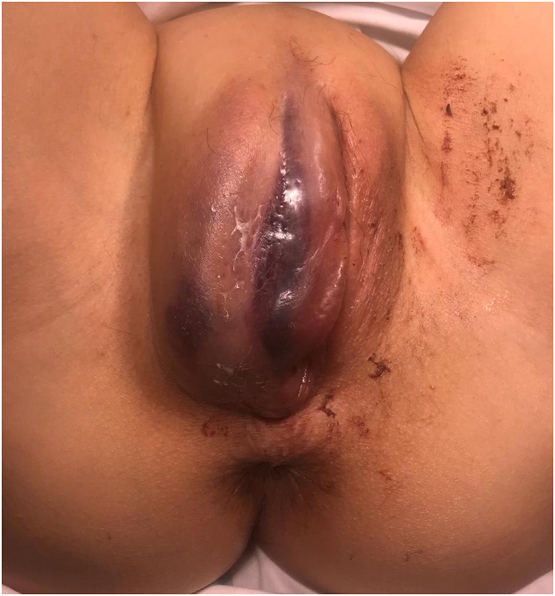
Preoperative image of the swollen vulva, with a clotted blood mass on the right labia minora and lateral deviation of the left labia majora.
A preoperative laboratory analysis indicated that hemoglobin, biochemistry and coagulations were all within normal levels, but that the patient had mild leukocytosis and neutrophilia. Opioid pain killers were administered intravenously. One hour later, given the persistence of pain, the difficulty in assessing the extent of the lesion and the patient's difficulty in urinating, it was decided to operate to drain the hematoma, which had progressively spread to other dependent areas. Antibiotic prophylaxis was administered. Under spinal and general anesthesia, after placing a urinary catheter and creating an aseptic surgical field, a 2 cm incision was applied to the area with the highest tension, on the base of the right labium majus. Multiple clots were extracted and mild debridement was performed so that they could be removed without causing further bleeding (Fig. 2). Then, the operative site was checked for active bleeding. A Penrose drain was placed and two simple stitches with polyglactin 2/0 were applied on both ends of the incision in order to reduce its size (Fig. 3).
Fig. 2.
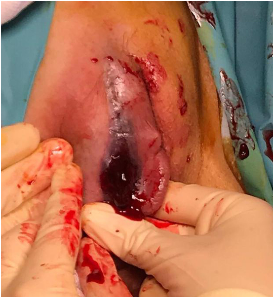
Intraoperative image of the extraction of multiple blood clots.
Fig. 3.
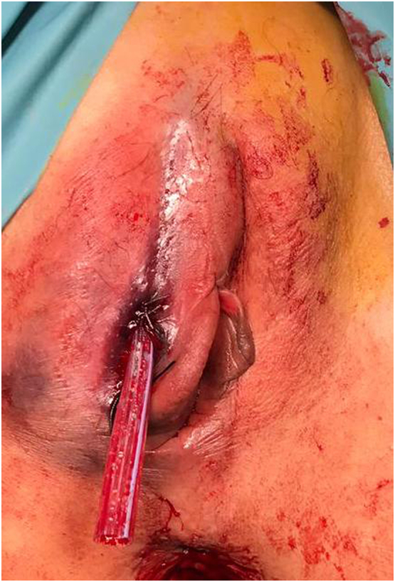
Postoperative image, with closure of dead space and a Penrose drain.
The postoperative period was uneventful. The patient had good oral tolerance and was able to urinate with much less difficulty. Eight hours after her admission, the patient was in a good general condition. She reported a very significant decrease of the pain, and ambulation with some discomfort, but possible. She also expressed her wish to be discharged from the hospital. The swelling had been greatly reduced; the wound had a good appearance and it drained a little hematic material. The patient was discharged and the drain was removed 24 h later. Amoxicillin/clavulanic acid was prescribed for 10 days together with an oral anti-inflammatory drug. She was given an appointment one week later but she failed to attend.
3. Discussion
Severe non-obstetric vulvar hematomas are rare and there are few reported cases in the literature. They have an incidence of 3.7% and represent 0.8% of all gynecological emergencies [3,4]. This partly explains the difficulty in deciding the most appropriate approach in each case, which depends on the size of the hematoma, the involvement of adjoining organs and hemodynamic stability. Traumatic vulvar hematomas are generally caused by falls on to or the straddling of objects, by vigorous coitus and by acts of physical aggression [5].
The vulva is made up of loose connective tissue and smooth muscle which is supplied by branches of the pudendal artery, which branches off the internal iliac artery. The injury to labial branches of the internal pudendal artery, which is located in the superficial fascia of the anterior and posterior pelvic triangle, may cause significant vulvar hematomas [1]. In our case, the patient fell on an object that compressed the soft tissue and the underlying pelvic fascia against the pelvic bones, which caused laceration and the formation of a hematoma. The swelling in such cases generally follows the cleavage planes and may become large size because the subcutaneous tissue offers low resistance [6]. If bleeding takes place, as in this case, beneath the pelvic fascia and the levator ani, the latter is separated from the perineum. On the other hand, if the hematoma is on the pelvic fascia, it can spread below Poupart's ligament and continue retroperitoneally to the renal fossae [5]. Although there is no anatomical explanation, 70% of all reported vulvar hematomas appear on the right labius (as in our case) [7].
There is no consensus on the management of non-obstetric vulvar hematomas. Propst et al. observed that in the absence of acute hematoma expansion, conservative management can yield good results [8]. However, Benrubi et al. found that conservative management of hematomas was associated with longer stays in hospital, an increased need for antibiotics and blood transfusion, and greater subsequent operative intervention [9].
Most cases can be treated conservatively, with rest, compression and local cold application, if lesions in adjacent organs (urethra, vagina and anus) and bone fractures have been ruled out [4]. If the hematoma interferes with urination because of its size, a vesical catheter must be placed and hemoglobin levels controlled. Bleeding lacerations must be sutured after thorough cleaning with liquids [10].
If the swelling keeps increasing, if the patient is unstable or if pain persists after intravenous treatment, surgical intervention will be necessary [3]. Because of the pressure they create, vulvar hematomas more than 4 cm in diameter may cause necrosis and require surgical assessment [10]. Currently, diagnostic techniques such as transperineal ultrasound may be used to assess the extension of the hematoma in greater detail [11].
With regard to the surgical approach, according to Hudock in 1955 [6] the incision is best applied over the point of maximum bulging, at the mucocutaneous junction or through the vaginal mucosa; these approaches produce similar results. All the blood clots must be removed to visualize the depth of the lesion, but without excessive manipulation of the traumatized area if there are bleeding points that need clotting. In most cases, the surface will show diffuse bleeding of venous origin. Although in our case it was not necessary to use any kind of hemostatic material or to ligate bleeding vessels, those are options. The removal of clots is important to reduce pressure necrosis and the risk of infection [12]. If drainage is applied, a Penrose drain must be placed in the most dependent area and then removed 24 h later.
Arterial embolization is emerging as an alternative to classic techniques [12,13], but its availability is still very limited.
Given that few cases of non-obstetric genital hematomas have been reported, there may be little need for guidelines on their management, but we have found it useful to review the literature.
4. Conclusions
Non-obstetric vulvar hematomas are a rare pathology in which damage of adjacent organs must always be ruled out. Most cases can be managed conservatively, at least initially, but if the pain does not disappear and if there is suspicion of an expanding hematoma, it will be necessary to examine the area with the patient under anesthesia and drainage in order to reduce the pain, accelerate the recovery and prevent secondary infection and necrosis.
Contributors
M. Victoria Lapresa Alcalde conceived the case report, was involved in patient care, analyzed the data, and drafted the paper.
Estrella Hernández Hernández conceived the case report, was involved in patient care, and drafted the paper.
Sandra Bustillo Alfonso conceived the case report, was involved in patient care, and drafted the paper.
María José Doyague Sánchez conceived the case report.
Conflict of Interest
The authors declare that they have no conflict of interest regarding the publication of this case report.
Funding
No funding was sought or secured in relation to this case report.
Patient Consent
Written informed consent was obtained from the patient for publication of this case report and the accompanying images.
Provenance and Peer Review
This case report was peer reviewed.
References
- 1.Schmidt A.B., Lykkebo A.W. Post-coital genital injury in healthy women: a review. Clin. Anat. 2015;28(3):331–338. doi: 10.1002/ca.22476. [DOI] [PubMed] [Google Scholar]
- 2.Ernest A., Knapp G. Severe traumatic vulva hematoma in teenage girl. Clin. Case Rep. 2015;3(12):975–978. doi: 10.1002/ccr3.395. [DOI] [PMC free article] [PubMed] [Google Scholar]
- 3.Papoutsis D., Haefner H.K. Large vulvar haematoma of traumatic origin. J. Clin. Diagn. Res. 2017;11(9):QJ01–QJ02. doi: 10.7860/JCDR/2017/30104.10542. [DOI] [PMC free article] [PubMed] [Google Scholar]
- 4.Jones I.S., O'Connor A. Non-obsetric vulval trauma. Emerg. Med. Australas. 2013;25(1):36–39. doi: 10.1111/1742-6723.12016. [DOI] [PubMed] [Google Scholar]
- 5.Vermesh M., Deppe G., Zbella E. Non-puerperal traumatic vulvar hematoma. Int. J. Gynaecol. Obstet. 1984;22(3):217–219. doi: 10.1016/0020-7292(84)90009-2. [DOI] [PubMed] [Google Scholar]
- 6.Hudock J.J., Dupayne N., McGeary J.A. Traumatic vulvar hematomas; report of six cases and review of the literature. Am. J. Obstet. Gynecol. Nov, 1955;70(5):1064–1073. [PubMed] [Google Scholar]
- 7.Shesser R., Schulman D., Smith J. A non puerperal traumatic vulvar hematoma. J. Emerg. Med. 1986;4(5):397–399. doi: 10.1016/0736-4679(86)90218-0. [DOI] [PubMed] [Google Scholar]
- 8.Propst A.M., Thorp J.M. Traumatic vulvar hematomas: conservative versus surgical management. South. Med. J. 1998;91(2):144–146. doi: 10.1097/00007611-199802000-00004. [DOI] [PubMed] [Google Scholar]
- 9.Benrubi G., Neuman C., Nuss R.C., Thompson R.J. Vulvar and vaginal hematomas: a retrospective study of conservative versus operative management. South. Med. J. 1987;80(8):991–994. doi: 10.1097/00007611-198708000-00014. [DOI] [PubMed] [Google Scholar]
- 10.Hernández-Tiria M., Navarro-Devia A., Osorio-Ruiz A. Lesión vulvar y perineal secundaria a trauma pelviperineal complejo: presentación de un caso y revisión de la literatura. Rev. Colomb. Obstet. Ginecol. 2015;66(4):297–305. [Google Scholar]
- 11.Sherer D.M., Stimphil R., Hellmann M., Abdelmalek E., Zinn H., Abulafia O. Transperineal sonography of a large vulvar hematoma following blunt perineal trauma. J. Clin. Ultrasound. 2006;34(6):309–312. doi: 10.1002/jcu.20188. [DOI] [PubMed] [Google Scholar]
- 12.Egan E., Dundee P., Lawrentschuk N. Vulvar hematoma secondary to spontaneous rupture of the internal iliac artery: clinical review. Am. J. Obstet. Gynecol. 2009;200(1):e17–e18. doi: 10.1016/j.ajog.2008.09.024. [DOI] [PubMed] [Google Scholar]
- 13.Machado-Linde F., Capel-Alemán A., Sánchez-Ferrer M.L., Cascales-Campos P., Pérez-Carrión A., Ortiz-Vera C. Major post-traumatic non-obstetric large haematoma: transarterial embolisation. Eur. J. Obstet. Gynecol. Reprod. Biol. 2011;154(1):118–119. doi: 10.1016/j.ejogrb.2010.08.012. [DOI] [PubMed] [Google Scholar]