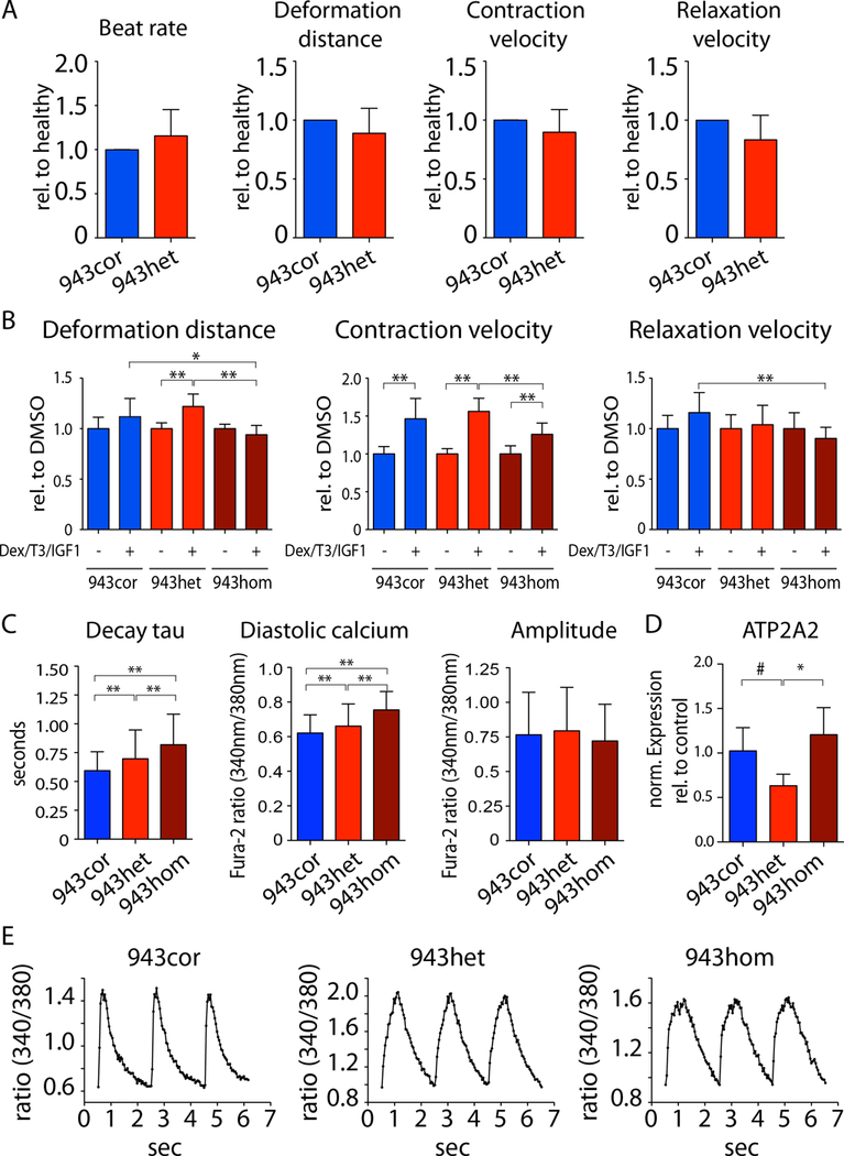
Figure 2: Functional evaluation of HCM iPSC-CMs.

(A) Contraction analysis of iPSC-CM monolayers using a vector-based imaging approach. The relative differences of disease (943het) vs. healthy (943cor) were calculated for each respective experiment (n=5). (B) iPSC-CMs from 943cor, 943het, and 943hom were treated with dexamethasone (Dex), triiodothyronine (T3), and insulin like growth factor 1 (IGF1) for 72 hours prior to contractility analysis (n=3 batches). Analysis was performed using vector-based imaging approach. (C) Analysis of calcium handling properties in isogenic iPSC-CMs (943cor, 943het, 943hom) using ratiometric measurements of fluorescence after Fura-2 staining under continuous field potential stimulation (n=4/4/6 differentiation batches each, 40 cells per batch). (D) Relative expression of ATP2A2 in iPSC-CMs (943cor, 943het, 943hom; n=3–6 differentiation batches each). (E) Representative raw traces of intracellular calcium signaling. # p<0.1, * p<0.05; ** p<0.01.
