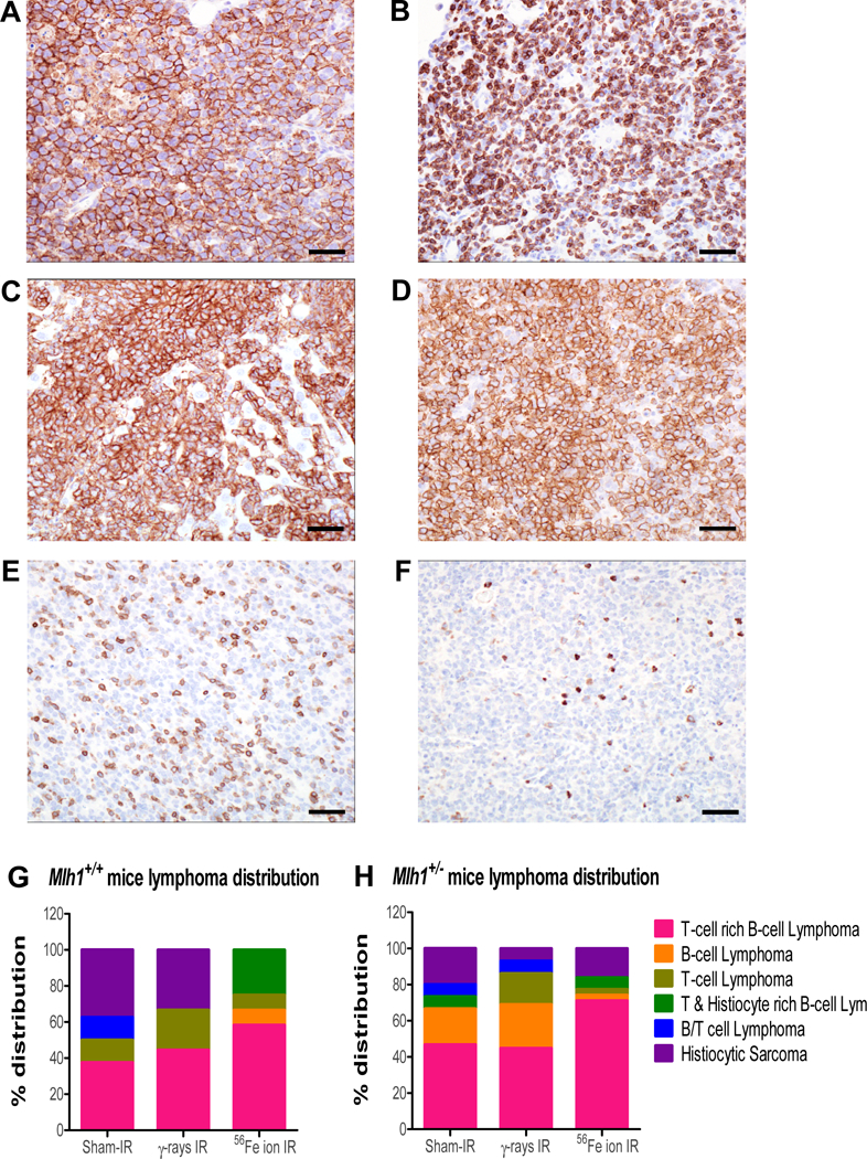
Figure 3: Immunohistochemistry of lymphomas from Mlh1+/+ and Mlh1+/− mice.

(A) B-cell lymphoma in a mesenteric lymph node shows diffuse and strong positive membrane immunoreactivity for B220 antibody. (B) T-cell lymphoma in mesenteric lymph node shows diffuse membrane and cytoplasmic immunoreactivity to CD3 antibody. (C) Histiocytic sarcoma in the liver shows strong and diffuse membrane immunoreactivity to F4/80 antibody. (D-F) The majority of neoplasms had an immunophenotype of T-cell rich, B-cell lymphomas, characterized by a dominant population of neoplastic B cells immunoreactive to B220 antibody (D), with a minority population of well-differentiated T-cells immunoreactive to CD3 antibody (E), and only a few resident macrophages illustrated by F4/80 immunoreactivity (F). (A-F) 40X, bar = 20um. (G) Distribution, based on immunohistochemistry, of lymphomas collected from Mlh1+/+ mice treated with sham-, γ-, or 56Fe ion irradiation. (H) Distribution, based on immunohistochemistry, of lymphomas collected from Mlh1+/− mice treated with sham-, γ-rays, or 56Fe ion irradiation. IHC was performed on 8–12 lymphomas of Mlh1+/+ origin and 15–31 lymphomas of Mlh1+/− origin.
