Abstract
Objective:
Triple A syndrome is a rare autosomal recessive disorder caused by mutations in the AAAS gene on chromosome 12q13. Its main clinical features are alacrima, achalasia and adrenal insufficiency, with most patients also having neurological symptoms and autonomic dysfunction. The neurologic manifestations are less well-understood, especially in children. Here we examine two siblings found to have a novel mutation in the AAAS gene, and who were found to have subtle, but important, neurologic findings.
Design:
This is a case report of two siblings.
Results:
We discuss two siblings exhibiting different signs and the disorder including neurologic dysfunction found at varying ages. Genetic analysis revealed that both patients have the same compound heterozygous mutations in the AAAS gene consisting of one novel mutation (c.500 C>A, A167E), and one previously described mutation (c.1331+1G> A/IVS14+1 G>A). A diagnosis of triple A syndrome was reached based on their clinical and genetic findings.
Conclusions:
The unique characteristic of these two cases is the novel mutation in the AAAS gene, which is likely pathogenic. In addition, they showcase the genotype-phenotype variability of the disease, as well as the importance of early identification of the neurologic abnormalities, which can result in early intervention and possibly improved outcomes.
Keywords: Triple A Syndrome, AAAS, Allgrove Syndrome, novel variant
Introduction:
Triple A syndrome (Allgrove syndrome) is an autosomal recessive disorder that was first described in 1978 by Allgrove and his colleagues.1 It is a syndrome with significant phenotypic and genotypic heterogeneity; its main clinical features are alacrima, achalasia and adrenal insufficiency.2–4 The denomination 4A syndrome has recently been proposed, as most of the patients present with additional neurological symptoms, including autonomic dysfunction.
Alacrima is the most common and consistent symptom of the syndrome, with up to a 99% prevalence.5 It has been suggested as an early diagnostic sign since it is present at birth in almost all cases.6,7 The pathophysiology of alacrima is thought to be due to autonomic denervation of the lacrimal gland and therefore, may be a result of the autonomic dysfunction in the disease and not a separate clinical feature.8
Achalasia usually develops during childhood, in the first decade of life, but may also occur later. It has an up to a 93% prevalence and presents with vomiting, dysphagia, failure to thrive and chronic cough.5
Adrenal insufficiency (AI) is the third clinical feature of triple A syndrome, that typically develops later in the course of the disease, either in the first decade of life, in adolescence or even adulthood.9 It is caused by resistance of the adrenals to ACTH secretion, which results in very high serum levels of ACTH and low serum cortisol levels. It presents with the classic symptoms of AI (fatigue, weakness, abdominal pain), progressive hyperpigmentation in many patients and significant hypoglycemic episodes.6 Cases of mineralocorticoid insufficiency have also been reported, although it is not commonly seen in triple A syndrome.2
The fourth and most phenotypically heterogenic clinical manifestation of Allgrove syndrome is the neurological dysfunction, which can be central, peripheral or autonomic. It usually occurs later in life and is progressive in nature, while it is the main clinical feature in adult triple A syndrome.10–12 Less is known about the neurologic manifestations presenting in childhood. Other features of the syndrome that have been described are microcephaly, nasal speech, plantar and palmar hyperkeratosis, low bone mineral density and osteoporosis, short stature and xerostomia.6,13,14
Though cases of triple A syndrome have been reported in pediatric patients, there continues to be significant phenotypic heterogeneity among affected individuals. In this article, we present two siblings with the same compound heterozygous mutations consisting of one novel mutation and one previously described mutation in the AAAS gene. Although they have the same genotype, they exhibit different signs and symptoms presenting at varying times. These cases also highlight the possible subtlety of the neurologic phenotype and the importance of a detailed neurologic exam to identify signs early.
Case Reports:
We describe two siblings diagnosed with Triple A syndrome. The first subject is a twelve-year-old boy who was born full term after an uncomplicated pregnancy and presented with alacrima at birth. He has a history of gastroesophageal reflux disease beginning in infancy and persisting through childhood despite medical treatment. He underwent esophageal manometry at age eleven and was diagnosed with achalasia immediately after his sister received the same diagnosis. He was then treated with a Heller esophageal myotomy without fundoplication which vastly improved his symptoms. Given the combination of alacrima and achalasia, and considering that his sister had similar symptoms, the possibility of Triple A syndrome was raised. Subsequently, he underwent genetic testing which identified two biallelic pathogenic variants in the AAAS gene and the diagnosis was confirmed. The parents were tested, and each carried one allele. The variants identified were:
AAAS: c.1331+1G> A/IVS14+1 G>A which affects a conserved splice site and is considered pathogenic and has been previously described.15,16
AAAS: c.500C>A, pAla167Glu. This specific mutation has not been previously reported but is considered likely pathogenic.
Two months later, he presented with fatigue, somnolence and abdominal pain. He underwent work up for adrenal insufficiency, including 8am serum cortisol and ACTH levels and an ACTH stimulation test. His 8am cortisol level was 6.8 mcg/dL (Reference range (RR): 4.8–19.5 mcg/dL) and his ACTH level was significantly elevated at 881.7 pg/ml (RR: 7–63 pg/ml). His cortisol levels after the ACTH stimulation were low, measuring 6 mcg/dL and 8 mcg/dL at 30 and 60 minutes respectively (normal > 18 mcg/dL). The diagnosis of adrenal insufficiency was made, and he was started on hydrocortisone replacement.
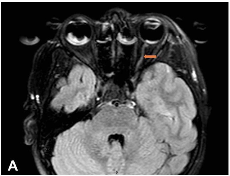
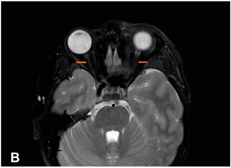
He presented to the NIH ten months later for further evaluation, including a focus on the neurologic components of his syndrome. In this context, he underwent an orbit MRI and an ophthalmology exam which showed thinning of his optic nerves bilaterally as well as anisocoria (Figure 1). A brain MRI showed no defects. He also had a neurology exam, which showed anisocoria, mild decreased distal bilateral lower extremity vibratory sensation, mild decreased bulk of the intrinsic muscles of the hand and 3+ deep tendon reflexes. On history it was found that he had a history of significant sweating. The family also stated that he may suffer from learning disabilities as he requires special help in social studies and math. He had a delayed bone age with estimated bone age of 10 years while his chronological age was 12 years and 2 months. His height is at 3rd percentile and weight at 36th percentile for age.
Figure 1:
MRI images of the orbits showing thinning of the optic nerves in sibling one. A. FLAIR image. B. T2-weighted image
His sibling is a developmentally appropriate five-year-old girl, born full term after an uncomplicated pregnancy, but also found to have alacrima at birth. She began to develop severe post-prandial vomiting around eighteen months of age that worsened over time leading to weight loss and failure to thrive despite multiple dietary manipulations. She was started on nasojejunal tube feeds around three years of age which she initially tolerated, but subsequently developed persistent post-prandial vomiting. An esophagogastroduodenoscopy was therefore performed, which revealed a severely dysfunctional esophagus and irregular contractions concerning for achalasia. Esophageal manometry was obtained which revealed aperistalsis and incomplete lower esophageal relaxation consistent with achalasia. She then underwent Heller esophagomyotomy and posterior Toupet fundoplication. Since the procedure, she has been able to tolerate food intake without emesis and is gaining weight well. She has not had any symptoms concerning for adrenal insufficiency at this point. She underwent genetic testing after her brother’s results were found, and she was identified to have the same two biallelic pathogenic variants (c. 1331+1G>A/IVS14+1 G>A and c.500C>A) in the AAAS gene.
She presented to the NIH with her brother for both neurologic evaluation and evaluation of adrenal insufficiency. Her 8am serum cortisol was 11.7 mcg/dL and within the normal range, but her ACTH level was elevated at 162pg/mL (RR: 5.0–46.0pg/mL). After the ACTH stimulation test, her cortisol levels were found to be suboptimal, measuring 16.8 and 16.8 mcg/dL at 30 and 60 minutes respectively. She was then diagnosed with early adrenal insufficiency and started on low dose hydrocortisone replacement therapy, given the assumption that it would continue to progress. Her neurological exam was grossly normal except that she was slightly hyperreflexic. Her ophthalmologic exam also showed mild thinning of the nerve fiber rim of the optic nerves bilaterally. She also had a delayed at bone age, with an estimated age of 4 years and 2 months at a chronological age of 5 years and 7 months. Her height was at 2nd percentile and her weight was at the 1st percentile for age.
The family history is significant for a paternal first cousin once removed who is wheelchair-bound and has a history of alacrima and GERD suspicious for Triple A syndrome but has never had genetic testing. No one else in the family has similar symptoms.
Discussion
The classic features of triple A syndrome (alacrima, achalasia, adrenal insufficiency and neurologic dysfunction) were found in varying degrees in these siblings who were found to have the same compound heterozygous mutations. One of the mutations in the AAAS gene (IVS14+I G>A), has been previously reported as a pathogenic mutation.15,16 However, these patients also carried a novel mutation in the second allele, not previously described in the literature (c.500 C>A, A167E). We believe this variant to also be pathogenic, as they both exhibit the constellation of clinical findings present in Triple A syndrome. Although both patients carry the same genotype, the severity of each of their presentations differ with the younger sister having much more severe achalasia and no evident neurologic symptoms at this time compared to the brother.
The AAAS mutation is inherited in an autosomal recessive pattern in triple A syndrome, thus the syndrome is rarely found in the family history. This disorder is caused by mutations in the AAAS gene on chromosome 12q13 in a maximum genetic interval of 6 cM between loci D12S1629 and D12S312.17 It encodes the 547 amino acid-containing protein called ALADIN, which stands for “alacrima - achalasia- adrenal insufficiency neurologic disorder.”16 ALADIN belongs to WD-repeat family of regulatory proteins and was identified as a component of nuclear pore complex (NPC), which mainly mediates nucleocytoplasmic transport in multiple cellular processes.16,18 In triple A syndrome, the altered ALADIN protein mislocalizes mainly in cytoplasm leading to defects in NPCs function,19 but the specific molecular basis of this protein still remains unknown. A variety of disease-associated mutations have been found scattered throughout the AAAS gene, including missense, nonsense, frameshift and splice-site mutations. The disease can manifest from homozygous or compound heterozygous mutations.16,20–22 The gene is ubiquitously expressed, but it is reported to have higher expression signals in endocrine and neuroendocrine derivatives, indicating its role in endocrine and neurological development.16,20
These siblings were both found to be compound heterozygotes and harbor the same mutations. One of the mutations they carry (c.1331+1G> A/IVS14+1 G>A) affects a conserved splice site and is considered pathogenic and has been previously described.15,16 A second mutation in the AAAS gene was found in both siblings (c.500C>A, pAla167Glu). This missense mutation leads to a substitution of alanine to glutamate at position 167 in the protein. This variant of A167E in the AAAS gene results in a non-conservative amino acid substitution, which likely impacts secondary protein structure. This position is conserved across species. A mutation affecting the same codon (A167V) has been reported in another23 patient with Triple A syndrome.
Another interesting aspect of their presentation is the mild neurologic abnormalities. The older sibling exhibited mild muscle wasting, decreased vibratory sensation, anisocoria, bilateral thinning bilateral of the optic nerves, hyperreflexic deep tendon reflexes and a history of significant sweating. The neurologic exam was reportedly normal for the second subject except for mild hyperreflexia and ophthalmologic finding of optic nerve thinning. A spectrum of neurologic dysfunction has been described in patients with Triple A syndrome including intellectual disability, global developmental delay, a myriad of motor abnormalities, peripheral sensorimotor polyneuropathies and autonomic dysfunction.6,10,24,25 Because neurologic dysfunction can present in numerous ways in Triple A syndrome, it is particularly important to pay close attention to any neurologic dysfunction when you have a patient with the other classical signs of Triple A syndrome. As we know well in pediatrics, early detection of neurodevelopmental disorders is imperative in order to obtain early intervention, which will provide the best chance of improved function. Although many of the neurologic symptoms associated with Triple A disease may be mild or subtle initially, they can be progressive. Therefore, it is extremely important to actively screen for neurologic abnormalities when the other classic findings of Triple A syndrome are present, in order to identify them and intervene early. Further investigation into whether or not intervention at the first signs of neurologic abnormalities can improve neurologic and developmental outcomes is warranted in this unique population.
In conclusion, we hope that this report can promote more awareness of this rare disorder and contribute to the growing body of knowledge regarding this syndrome, particularly in the field of pediatrics.
Acknowledgments
Research funding: This research was supported in part by the Intramural Research Program of the National Institutes of Health (NIH), Eunice Kennedy Shriver National Institute of Child Health and Human Development, protocol 1997-CH-0076 (ClinicalTrials.gov Identifier: NCT00001595).
Footnotes
Disclosures: The authors have nothing to disclose and there are no conflicts of interest.
References
- 1.Allgrove J, Clayden GS, Grant DB, Macaulay JC. Familial glucocorticoid deficiency with achalasia of the cardia and deficient tear production. Lancet. 1978;1(8077):1284–1286. [DOI] [PubMed] [Google Scholar]
- 2.Brooks BP, Kleta R, Stuart C, et al. Genotypic heterogeneity and clinical phenotype in triple A syndrome: a review of the NIH experience 2000–2005. Clin Genet. 2005;68(3):215–221. [DOI] [PubMed] [Google Scholar]
- 3.Lovrecic L, Pelet A, Peterlin B. Heterogeneity of the triple A syndrome and assessment of a case. Genet Couns. 2006;17(2):191–195. [PubMed] [Google Scholar]
- 4.Barat P, Goizet C, Tullio-Pelet A, Puel O, Labessan C, Barthelemy A. Phenotypic heterogeneity in AAAS gene mutation. Acta Paediatr. 2004;93(9):1257–1259. [DOI] [PubMed] [Google Scholar]
- 5.Patt H, Koehler K, Lodha S, et al. Phenotype-genotype spectrum of AAA syndrome from Western India and systematic review of literature. Endocr Connect. 2017;6(8):901–913. [DOI] [PMC free article] [PubMed] [Google Scholar]
- 6.Tibussek D, Ghosh S, Huebner A, Schaper J, Mayatepek E, Koehler K. “Crying without tears” as an early diagnostic sign-post of triple A (Allgrove) syndrome: two case reports. BMCPediatr. 2018;18(1):6. [DOI] [PMC free article] [PubMed] [Google Scholar]
- 7.Brown B, Agdere L, Muntean C, David K. Alacrima as a Harbinger of Adrenal Insufficiency in a Child with Allgrove (AAA) Syndrome. Am J Case Rep. 2016;17:703–706. [DOI] [PMC free article] [PubMed] [Google Scholar]
- 8.Aragona P, Rania L, Roszkowska AM, et al. 4A syndrome: ocular surface investigation in an Italian young patient. BMC Ophthalmol. 2014;14:155. [DOI] [PMC free article] [PubMed] [Google Scholar]
- 9.Thomas J, Subramanyam S, Vijayaraghavan S, Bhaskar E. Late onset adrenal insufficiency and achalasia in Allgrove syndrome. BMJ Case Rep. 2015;2015. [DOI] [PMC free article] [PubMed] [Google Scholar]
- 10.Sanyal D, Bhattacharjee S. A case of late-onset allgrove syndrome presenting with predominant autonomic dysfunction. Ann Indian Acad Neurol. 2013;16(2):266–268. [DOI] [PMC free article] [PubMed] [Google Scholar]
- 11.Houlden H, Smith S, De Carvalho M, et al. Clinical and genetic characterization of families with triple A (Allgrove) syndrome. Brain. 2002;125(Pt 12):2681–2690. [DOI] [PubMed] [Google Scholar]
- 12.Dumic M, Barisic N, Kusec V, et al. Long-term clinical follow-up and molecular genetic findings in eight patients with triple A syndrome. Eur J Pediatr. 2012;171(10):1453–1459. [DOI] [PubMed] [Google Scholar]
- 13.Dumic M, Putarek NR, Kusec V, Barisic N, Koehler K, Huebner A. Low bone mineral density for age/osteoporosis in triple A syndrome-an overlooked symptom of unexplained etiology. OsteoporosInt. 2016;27(2):521–526. [DOI] [PubMed] [Google Scholar]
- 14.Dumic M, Mravak-Stipetic M, Kaic Z, et al. Xerostomia in patients with triple A syndrome--a newly recognised finding. Eur J Pediatr. 2000;159(12):885–888. [DOI] [PubMed] [Google Scholar]
- 15.Kallabi F, Ben Rebeh I, Felhi R, et al. Molecular Analysis of Libyan Families with Allgrove Syndrome: Geographic Expansion of the Ancestral Mutation c.1331+1G>A in North Africa. Horm Res Paediatr. 2016;85(1):18–21. [DOI] [PubMed] [Google Scholar]
- 16.Tullio-Pelet A, Salomon R, Hadj-Rabia S, et al. Mutant WD-repeat protein in triple-A syndrome. Nat Genet. 2000;26(3):332–335. [DOI] [PubMed] [Google Scholar]
- 17.Weber A, Wienker TF, Jung M, et al. Linkage of the gene for the triple A syndrome to chromosome 12q13 near the type II keratin gene cluster. Hum Mol Genet. 1996;5(12):2061–2066. [DOI] [PubMed] [Google Scholar]
- 18.Cronshaw JM, Krutchinsky AN, Zhang W, Chait BT, Matunis MJ. Proteomic analysis of the mammalian nuclear pore complex. J Cell Biol. 2002;158(5):915–927. [DOI] [PMC free article] [PubMed] [Google Scholar]
- 19.Cronshaw JM, Matunis MJ. The nuclear pore complex protein ALADIN is mislocalized in triple A syndrome. Proc Natl Acad Sci U S A. 2003;100(10):5823–5827. [DOI] [PMC free article] [PubMed] [Google Scholar]
- 20.Handschug K, Sperling S, Yoon SJ, Hennig S, Clark AJ, Huebner A. Triple A syndrome is caused by mutations in AAAS, a new WD-repeat protein gene. Hum Mol Genet. 2001;10(3):283–290. [DOI] [PubMed] [Google Scholar]
- 21.Nakamura K, Yoshida K, Yoshinaga T, et al. Adult or late-onset triple A syndrome: case report and literature review. J Neurol Sci. 2010;297(1–2):85–88. [DOI] [PubMed] [Google Scholar]
- 22.Kurnaz E, Duminuco P, Aycan Z, et al. Clinical and genetic characterisation of a series of patients with triple A syndrome. Eur JPediatr. 2018;177(3):363–369. [DOI] [PubMed] [Google Scholar]
- 23.Stenson PD, Mort M, Ball EV, Shaw K, Phillips A, Cooper DN. The Human Gene Mutation Database: building a comprehensive mutation repository for clinical and molecular genetics, diagnostic testing and personalized genomic medicine. Hum Genet. 2014;133(1):1–9. [DOI] [PMC free article] [PubMed] [Google Scholar]
- 24.Grant DB, Barnes ND, Dumic M, et al. Neurological and adrenal dysfunction in the adrenal insufficiency/alacrima/achalasia (3A) syndrome. Arch Dis Child. 1993;68(6):779–782. [DOI] [PMC free article] [PubMed] [Google Scholar]
- 25.Koehler K, Brockmann K, Krumbholz M, et al. Axonal neuropathy with unusual pattern of amyotrophy and alacrima associated with a novel AAAS mutation p.Leu430Phe. Eur J Hum Genet. 2008;16(12):1499–1506. [DOI] [PubMed] [Google Scholar]


