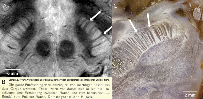
The “direct” and “indirect” pathways play crucial roles in movement disorder pathophysiology. Both traverse from the striatum to the internal pallidum and substantia nigra, the latter detouring to external pallidum and subthalamic nucleus. Anatomically, the pathways manifest within the striatofugal bundle that passes radially through the pallidum in the form of pencil-like tracts (first described by Wilson1; figure 1) before leaving the pallidum toward the substantia nigra in the form of a comb described by Edinger in 18962 (figure 2). A century later, these structures can be visualized in the living human brain (figures 1D and 2A).
Figure 1. Wilson pencils.
(A) Histologic depiction (image courtesy of Dr. Michael Bonert, McMaster University, CCBY-SA3.0). (B) Polarized light imaging in vervet monkey. (C) First description by Wilson (Brain), reproduced with permission from S.A. Kinnier Wilson. An experimental research into the anatomy and physiology of the corpus striatum. Brain 1914;36:427–492. By permission of Oxford University Press, available at: academic.oup.com/brain/article/36/3-4/427/309802?searchresult=1. For permissions, please email journals.permissions@oup.com. (D) Cardiac-gated T2*-weighted fast low angle shot sequence acquired using 7T MRI shows Wilson pencils.
Figure 2. Edinger comb.
(A) Cardiac-gated fast low angle shot sequence shows Edinger comb. (B) First description: “The pedunculus cerebri is traversed by striatal fibers that enter dorsally and connect peduncle and tegmentum—bundle between peduncle and tegmentum, comb system of the peduncle.” (C) Axial histologic section in dark-field microscopy demonstrates the human comb system.
Footnotes
Teaching slides links.lww.com/WNL/A850
Author contributions
A. Horn: drafting/revising the manuscript, data acquisition, study concept or design, analysis or interpretation of data, accepts responsibility for conduct of research and final approval, acquisition of data, study supervision. Siobhan G. Ewert: drafting/revising the manuscript, data acquisition, accepts responsibility for conduct of research and final approval, acquisition of data. Eduardo Joaquim Lopes Alho: drafting/revising the manuscript, accepts responsibility for conduct of research and final approval, acquisition and analysis of the dark field microscopy footage. M. Axer: data acquisition, accepts responsibility for conduct of research and final approval, acquisition of data, interpretation of measurements. H. Heinsen: drafting/revising the manuscript, data acquisition, analysis or interpretation of data, accepts responsibility for conduct of research and final approval, acquisition of data. Erich Talamoni Fonoff: drafting/revising the manuscript, data acquisition, accepts responsibility for conduct of research and final approval, acquisition of data. J.R. Polimeni: drafting/revising the manuscript, data acquisition, accepts responsibility for conduct of research and final approval, acquisition of data, study supervision, obtaining funding. T.M. Herrington: data acquisition, drafting/revising the manuscript, study concept or design, accepts responsibility for conduct of research and final approval, study supervision.
Study funding
No targeted funding reported.
Disclosure
A. Horn, S. Ewert, E. Alho, M. Axer, H. Heinsen, and E. Fonoff report no disclosures relevant to the manuscript. J. Polimeni reports funding by NIH NIMH R01-MH111438 and NIBIB P41-EB015896 and by the Athinoula A. Martinos Center for Biomedical Imaging. T. Herrington reports funding by NINDS grant K23NS099380 and an American Academy of Neurology/American Brain Foundation Clinical Research Training Fellowship. Go to Neurology.org/N for full disclosures.
References
- 1.Wilson SAK. An experimental research into the anatomy and physiology of the corpus striatum. Brain 1914;36:427–492. [Google Scholar]
- 2.Edinger L. Vorlesungen über den Bau der nervösen Centralorgane des Menschen und der Thiere. Für Ärzte und Studirende. Leipzig: F.C.W. Vogel; 1896. [Google Scholar]


