Abbreviations
- FDA
US Food and Drug Administration
- RBC
red blood cell
Indications
Therapeutic phlebotomy is indicated as an integral component of the treatment of several medical conditions. It is the cheapest and most effective method for removing excess iron in nonanemic patients.
Hereditary Hemochromatosis, Iron Overload
Hereditary hemochromatosis is one of the most common genetic disorders among Caucasians of northern European descent. The prevalence of the disorder in that region is approximately one in 250 people.1 Hereditary hemochromatosis and it's much rarer variant ‘the ferroportin disease’ are described in the recent issue of CLD devoted to Heavy Metals and the liver.1, 2
Acquired Iron Overload
Patients receiving chronic blood transfusions for disorders such as sickle cell anemia and Thalassemia may develop iron overload and subsequent iron deposition in various organs, which if untreated may lead to multiple organ failure.
Polycythemia Vera
This is a clonal progressive myeloproliferative disorder and is associated with significant erythrocytosis. Therapeutic phlebotomy is the best choice for the initial therapy.3
Other Medical Conditions
Many medical disorders may result in erythrocytosis and polycythemia. Such conditions include congenital erythrocytosis, which is associated with erythropoietic receptor gene mutation, as well as cyanotic heart disease and respiratory insufficiency. It is also associated with living at high altitudes.
Physiologic Mechanisms of Therapeutic Phlebotomy
In response to the phlebotomy, bone marrow stem cells are stimulated to make new red blood cells (RBC). To produce new RBC, iron is transported in the form of ferritin from body stores to make more hemoglobin. Consequently, patient's the overall iron level is reduced.
Treatment Regimen
In general, patient factors need to be considered when prescribing the phlebotomy regimen. These factors include age, sex, weight, comorbidities, overall health status, and the likelihood of compliance. The criteria for initiating therapeutic phlebotomy4 are outlined in Table 1. Half unit (250 ml) can be removed at a time in patients with small body mass, anemia, or cardiac or pulmonary disorder. In general, each unit of blood (500 ml) that is removed represents about 200 to 250 mg of iron, depending on the hemoglobin.
Hereditary hemochromatosis: weekly until mild hypoferritinemia (ferritin = 50‐100 ng/ml), hemoglobin < 11 mg/dl4, 5
Acquired iron overload: The regimen varies depending on the etiology.
Polycythemia vera: Weekly to monthly phlebotomy is recommended until the iron store is depleted. It is suggested that hematocrit levels should be maintained at < 50%.6, 7
Table 1.
Criteria for Initiating Therapeutic Phlebotomy
| Patient | Serum Ferritin (ng/ml) |
|---|---|
| < 18 years regardless of gender | >200 |
| Women | |
| Childbearing age, not pregnant | >500 |
| Childbearing age, pregnant | >200 |
| Men | |
| >18 years | >300 |
Patient Management and Monitoring
Serum ferritin is the most reliable method of monitoring patients who are receiving therapeutic phlebotomy. Patients with significantly high initial ferritin levels are greater than 1000 ng/ml and or more frequent phlebotomies should have their serum ferritin monitored every 2 to 3 months. Hemoglobin levels should be checked during each phlebotomy visit. Patients with a prephlebotomy hemoglobin concentration of less than 11 g/dl are more likely to have symptoms of hypovolemia and anemia. Therapeutic phlebotomy, when hemoglobin levels are less than 11 gm/dl, is less efficient at depleting iron stores in the body. In addition, patients with chronic hemolytic anemia and iron overload may not tolerate phlebotomy very well. In general, sound clinical judgment and careful monitoring are essential when managing patients with therapeutic phlebotomy.
It is important to encourage patients to drink fluids before and after each treatment. It is also helpful to inform patients to avoid strenuous physical activities for 24 hours after each treatment.
Phlebotomy Procedure
Therapeutic phlebotomy is performed in a medically supervised environment. It is commonly performed in a blood donor center, apheresis unit, physician office, or at home by a well‐trained phlebotomist, but home phlebotomy is rarely performed.8
The prescription for phlebotomy should contain the following elements:
Patient's name
Diagnosis
Date of birth or medical record number
Laboratory tests to be performed
Amount of blood to be drawn: The volume to be removed is established by the ordering physician. Any amount of blood to be removed over two units should be cleared by the requesting physician.
Frequency of phlebotomy: Usually, no more than one to two units of blood are removed in a 24‐hour period.
Hematocrit parameter
Postphlebotomy care instructions
Prior to initiating therapeutic phlebotomy, the following check‐list should be completed.
Blood pressure
Pulse
Respiration
Temperature
Hematocrit
Arm inspection
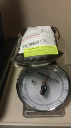
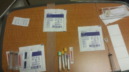
Obtain informed consent. Order appropriate laboratory tests. Affix labels to ethylene diamine tetraacetic acid or citrate‐containing specimen tubes. Transport specimens to the lab area after collection. Assemble all the necessary equipment, supplies, and reagents, as outlined in Table 2, Table 3, and Figure 1. Label transfer bag by affixing “Therapeutic Phlebotomy” label and include patient's disease and date. Place labeled transfer bag on a scale (Figure 2). Place scale lower than the patient's arm. To select and prepare venipuncture site, a vein in the antecubital is selected on the basis of it prominence, size and tonicity. The use of US Food and Drug Administration (FDA)‐approved disinfecting agents to provide surgical cleanliness is required to prepare the cubital fossa and the phlebotomy site. Blood should be collected into the approved transfer bag with a single venipuncture. The average time of collecting a unit (500 mL) of blood is less than 10 minutes. Finally, monitor the volume of blood drawn. Normal blood volume is between 477 to 530 g.
Table 2.
Equipment
| Clinical thermometer |
| Spacelabs blood pressure cuff |
| Dietary scale |
| Sebra heat sealer |
Table 3.
Reagents and Supplies
| Paperwork/Electronic documentation |
| Therapeutic phlebotomy order |
| Therapeutic phlebotomy consent |
| Procedure log |
| Product disposition log |
| Self‐sealing capillary tubes (2) |
| Chloraprep single swabstick applicator |
| 4 × 4 sterile gauze |
| Coflex |
| 600‐cc transfer packs with needle adapter |
| 17‐gauge hemodialysis Terumo needle |
| Tape |
| Personal protective devices (gloves, mask gown, eyewear) |
| Diatek thermometer probe cover |
| Blood bag label: therapeutic phlebotomy |
| Alcohol swabs 70% isopropyl |
Figure 1.

Blood transfer bag is placed on a scale to monitor blood draw volume.
Figure 2.

Reagents and supplies for performing therapeutic phlebotomy.
Clamp the C‐clamp on the tubing attached to the hemodialysis needle. Heat‐seal three times on the transfer pack tubing. Take appropriate samples. Release the blood pressure cuff. Take postvital signs. Remove the needle. Separate the blood bag and needle at weld. Place the needle and transfer pack in a properly labeled biohazard sharps container.
Observe the patient for reactions. If any reaction occurs, notify the transfusion services medical director and/or primary physician. Document the type of reaction and the resolution of symptoms. Offer refreshments and instruct patients to wait at least 15 minutes before returning to normal activity.
Allogenic Use of Collected Blood
There is a significant variation of opinions regarding the allogenic use of blood units collected from therapeutic phlebotomy patients. Many countries, including the United States, permit the allogenic use of blood units collected from hemochromatosis patients. However, the FDA requires that the units be visibly labeled to indicate that the donor had hemochromatosis.9 Because of the increased risk for developing leukemia, blood units from polycythemia vera are not used for transfusion, although the risk is very negligible.
Conclusion
Therapeutic phlebotomy is relatively safe and efficient at depleting iron stores in the body; thus, this procedure is effective for treating patients with hemochromatosis, polycythemia vera, and related conditions.
Potential conflict of interest: nothing to report.
References
- 1. DHG Crawford. Hereditary hemochromatosis types 1, 2 and 3. Clinical Liver Disease. 2014;5:96–97. doi: 10.1002/cld.339. [DOI] [PMC free article] [PubMed] [Google Scholar]
- 2. A Pietrangelo. The ferroportin disease. Clinical Liver Disease 2014;5:98–100. doi: 10.1002/cld.340. [DOI] [PMC free article] [PubMed] [Google Scholar]
- 3. Barton JC, McDonnell SM, Adams PC, et al. Management of hemochromatosis. Hemochromatosis Management Working Group . Ann Intern Med 1998;129:932–939. [DOI] [PubMed] [Google Scholar]
- 4. Niederau C, Fischer R, Sonnenberg A, Stremmel W, Trampisch HJ, Strohmeyer G. Survival and causes of death in cirrhotic and in noncirrhotic patients with primary hemochromatosis. N Engl J Med 1985;313:1256–1262. doi: 10.1056/nejm198511143 132004. [DOI] [PubMed] [Google Scholar]
- 5. Yang HS, Joe SG, Kim JG, Park SH, Ko HS. Delayed choroidal and retinal blood flow in polycythaemia vera patients with transient ocular blindness: a preliminary study with fluorescein angiography. Br J Haematol 2013;161:745–747. doi: 10.1111/ bjh.12290. [DOI] [PubMed] [Google Scholar]
- 6. Pearson TC, Wetherley‐Mein G. Vascular occlusive episodes and venous haematocrit in primary proliferative polycythaemia. Lancet 1978;2:1219–1222. doi: 10.1111/bjh.12290. [DOI] [PubMed] [Google Scholar]
- 7. Ram Kakaiya CAAaJJ. Whole blood collection and component processing at blood collection centers In: Roback J, ed. Technical Manual. Bethesda, MD: AABB; 2011:189. [Google Scholar]
- 8. Tan L, Khan MK, Hawk JC 3rd. Use of blood therapeutically drawn from hemochromatosis patients. Council on Scientific Affairs, American Medical Association . Transfusion 1999;39(9):1018–1026. [DOI] [PubMed] [Google Scholar]


