Figure 5.
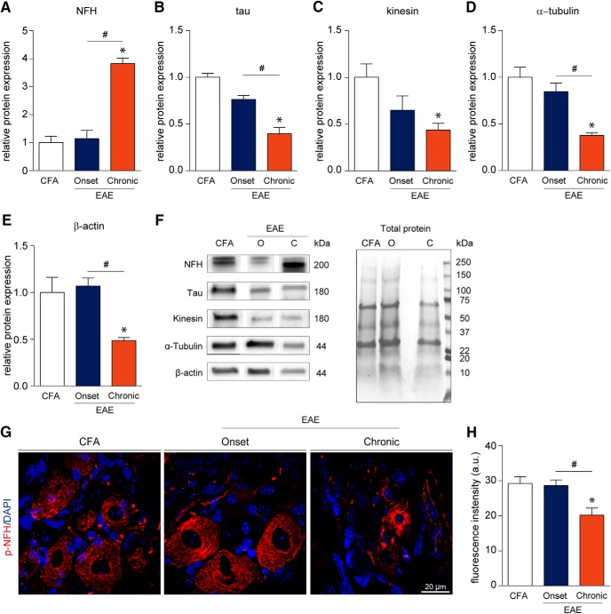
Cytoskeletal disruption of DRG neurons occurs late in the EAE disease course. A–F, Western blotting data suggest that cytoskeletal proteins remain intact at the onset of EAE symptoms and become impaired at the chronic time point. We observe a significant elevation in the level of the non-phosphorylated isoform of heavy-chain NFH at the chronic stage. On the contrary, a significant reduction in the levels of tau, kinesin, α-tubulin, and β-actin was detected chronically. G, H, Immunofluorescence staining of lumbar DRGs for the p-NFH revealed a significant reduction in fluorescence intensity in chronic samples (20.23 ± 1.976 a.u.) as compared to CFA control (29.26 ± 1.951 a.u.) and EAE onset samples (28.68 ± 1.581 a.u.). Furthermore, p-NFH staining in the soma of chronic DRG neurons displays aberrant morphology. a.u. = arbitrary units. Bars indicate mean ± SEM. O = onset; C = chronic; *,#p < 0.05, one-way ANOVAs with Tukey’s post hoc analysis. A–F, CFA, n = 5; onset, n = 4; chronic, n = 4. G, H, CFA, n = 4; onset, n = 6; chronic, n = 6.

