Abstract
Objective:
Root resorption due to orthodontic tooth movement may adversely affect the root-crown (R/C) ratios of permanent teeth, especially in patients with Short Root Anomaly (SRA), a poorly understood disorder affecting root development. Evaluation of SRA R/C ratios to normal dentition would facilitate diagnosis and orthodontic treatment planning. However, reference values are not available for all ethnicities. Our goal was to determine R/C ratios of permanent teeth and their relationship to gender and ethnicity.
Setting/Sample:
A retrospective study of 333 patients (109 Caucasians, 112 African Americans and 112 Hispanics) from the University of Alabama at Birmingham School of Dentistry.
Materials/Methods:
Root lengths and crown heights were measured from panoramic radiographs of 6,241 teeth using modified Lind’s method. A linear mixed model was used to compare the R/C ratios of teeth among subgroups (gender, ethnicity).
Results:
The mean R/C ratios varied from 1.80–2.21 for the maxillary teeth and 1.83–2.49 for the mandibular teeth. Gender differences in R/C ratios were found to be significant only for the lower central incisors (P<0.05). Hispanics showed significantly lower ratios for most teeth compared to the other two groups (P<0.05). There were significant differences in R/C ratios between African Americans and Caucasians in the upper lateral incisors, lower central incisors and lower first premolars (P<0.05).
Conclusion:
Our results suggest that ethnicity is an important factor in determining the R/C ratios of permanent teeth. Therefore, when diagnosing developmental conditions such as SRA, ethnic group-specific reference values should be considered.
Keywords: ethnic differences, permanent dentition, radiographic evaluation, root-crown ratios, short root anomaly
Introduction
A reduced root-crown (R/C) ratio may complicate dental treatment and adversely affect individual tooth prognosis. Patients with short roots often face an increased risk of root resorption during orthodontic treatment1, 2. Resorption of the natural root length in excess of 2mm may result in unfavorable R/C ratios3. Altered R/C ratios also affect the prosthodontic diagnosis and treatment planning of patients. Prosthodontic devices, such as removable or fixed partial dentures, can cause stresses on the already compromised teeth and worsen the prognosis of abutment teeth4. Therefore, the R/C ratios can dictate the methods and forms in which a patient is best treated orthodontically and prosthodontically.
Although root resorption from trauma and periapical inflammation contributes to short dental roots, genetic variations may also result in unfavorable R/C ratios3, 5. Short Root Anomaly (SRA) is a poorly understood genetic disease first described by Lind as a dental disorder affecting tooth root development, resulting in short roots with rounded apices and reduced crown to root ratios1, 6. SRA affects teeth bilaterally with a preference for maxillary incisors followed by maxillary and mandibular premolars2, 7. Orthodontic treatment of SRA patients with compromised R/C ratios may result in major adverse clinical outcomes such as severe root resorption and tooth loss1, 2, 8, 9. Thus, the ability to compare R/C ratios of new patients to normal dentition would facilitate SRA diagnosis.
Decreased R/C ratio due to root resorption is an adverse clinical outcome of orthodontic tooth movement10–12. Although root resorption may occur in individuals who have never undergone orthodontic treatments, the incidence among treated individuals is high, about one-third of patients showing signs of resorption4.
Subjective grading has often been used in the assessment of root shortening. This relies largely on clinician’s experience and makes it impossible to accurately compare treatment plans. Thus, the R/C ratios of normal dentition may provide reference values that can facilitate diagnosis and treatment planning for orthodontic, prosthodontic, and surgical procedures. Earlier studies have successfully used panoramic radiographs to assess the R/C ratios of permanent dentition in Finnish, Korean, and Iranian populations13–15. However, reference values are not available for all ethnic groups and no comparisons of the R/C ratios have been made among different ethnicities.
In this study, we examined the panoramic radiographs of individuals from three ethnic groups in the U.S.: Caucasian, African American, and Hispanic. The purpose of this study was to determine R/C ratios of fully developed permanent teeth measured from panoramic radiographs and evaluate their relationship to gender and ethnicity in healthy populations. These data may provide reference values for the R/C ratio, which can help guide the clinical diagnosis and treatment planning of patients, and enhance our understanding of the ethnic differences as they relate to dentition.
Materials and Methods
This retrospective study was approved by the University of Alabama at Birmingham Institutional Review Board (IRB Protocol Number X160428005).
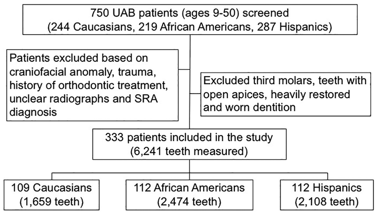
Panoramic radiographs were obtained from patients at the University X School of Dentistry. The patients ranged in age from 9 to 50 years. Only fully-developed permanent teeth were included in the study. Patients with craniofacial anomalies, evidence of prior orthodontic treatment, history of trauma or diagnosis of SRA were excluded (Figure 1). Third molars, heavily restored or worn teeth, or radiographs presenting unclear reference points were also excluded. In total, 333 patient radiographs were examined in this study and 6,241 teeth were analyzed.
Figure 1.
Flow diagram of the study
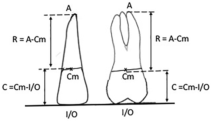
A method developed by Lind1 and modified by Holtta et al.14 was used to measure root lengths and crown heights with the MiPACS software tools (Medicor Imaging, Charlotte, NC). As described by Lind1, a midpoint was visually determined on panoramic radiographs along a line bisecting the mesial and distal cemento-enamel junctions (CEJ) (Figure 2). For single-rooted teeth, each root was measured from the apex to the corresponding midpoint. For multi-rooted teeth, the root was measured from an equilibrium point between the buccal roots to the corresponding midpoint. All crown heights were measured from the CEJ midpoints perpendicular to the incisal/occlusal reference line (formed tangent to incisal edge or buccal cusps). Data were compiled and each root length was divided by the respective crown height to calculate the root/crown ratio for each tooth.
Figure 2.
Method used to measure the root and crown lengths. Cm, midpoint along the bisecting line of the mesial and distal cemento-enamel junctions; A, root apex; I, incisal edge; O, occlusal surface; R, root length; C, crown height.
To assess the inter-examiner reproducibility, 35 panoramic radiographs were measured by two trained examiners so that two measurements were obtained for each tooth (measurement A and B). The intraclass correlation coefficient (ICC) between measurements A and B was calculated16.
The patients’ gender and ethnicity were summarized and the mean and median age calculated. The universal numbering system for adult teeth was adopted in this study and data were analyzed based on each tooth location in the arch. When examining the correlation between each tooth type from the left and right quadrants of the same arch in a patient, a linear mixed model was used to compare the crown/root ratios among subgroups (gender, ethnicity). Given a significant model (F test, p<0.05), the pairwise group comparison was tested using two-sample t tests. The significance level was defined as p < 0.05. The False Rate Correction was also calculated. All analyses were conducted using SAS 9.4 (Cary, NC).
Results
Reproducibility of the method
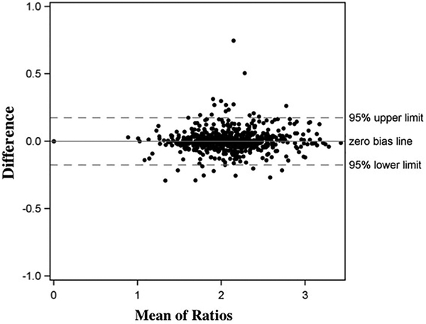
The ICC between measurements A and B was 0.996, which indicated high agreement between the two examiners and good reproducibility for the method (Figure 3). Furthermore, there were no systematic tendency, as shown by the Bland-Altman plot.
Figure 3.
Bland-Altman plot to assess the inter-examiner reproducibility.
Sample demographics
Radiographs from 333 patients (109 Caucasians, 112 African Americans and 112 Hispanics; 47.4% males and 52.6% females) were analyzed. The mean age of the patients examined was 28.0 years old for females and 28.6 years old for males (Table 1).
Table 1.
Sample Demographics
| Female | Male | Both genders | |
|---|---|---|---|
| Race/Ethnicity | |||
| Caucasian | 50 (44.6%) | 59 (55.4%) | 109 (32.73%) |
| African American | 68 (61%) | 44 (39%) | 112 (33.63%) |
| Hispanic | 57 (51%) | 55 (49%) | 112 (33.63%) |
| All ethnicities | 175 (52.6%) | 158 (47.4%) | 333 |
| Age (years) | |||
| Mean (SD) | 28.0 (10.4) | 28.6 (10.4) | 28.3 (10.4) |
| Median (range) | 27 (9 – 50) | 28 (11 – 50) | 28 (9 – 50) |
SD: standard deviation
Gender difference in the R/C ratios
The R/C ratios of fully-developed permanent teeth are presented in Table 2. The mean R/C ratios varied from 1.80–2.21 for the maxillary teeth and from 1.83–2.49 for the mandibular teeth. The mean R/C ratios measured from 1.84 to 2.49 in males and from 1.80 to 2.49 in females. In both genders, the lowest ratios were recorded for the upper central incisors (1.84 in males and 1.80 in females) and the highest ratios were in the lower second premolars (2.49 in both males and females). Gender differences in the R/C ratios were found to be significant only in the lower central incisors, with a lower value in females (1.83) compared to males (1.94).
Table 2.
Comparison of the mean root to crown ratios in males and females
| Teeth | Gender | N | Mean | SD | 95% CI | P value | FDR |
|---|---|---|---|---|---|---|---|
| 8,9 | F | 329 | 1.80 | 0.26 | 1.77–1.82 | 0.1098 | 0.3074 |
| M | 293 | 1.84 | 0.28 | 1.80–1.87 | |||
| 7,10 | F | 286 | 2.02 | 0.30 | 1.99–2.06 | 0.0749 | 0.2706 |
| M | 252 | 2.09 | 0.35 | 2.04–2.13 | |||
| 6,11 | F | 242 | 2.21 | 0.36 | 2.16–2.25 | 0.0557 | 0.2706 |
| M | 190 | 2.12 | 0.36 | 2.07–2.18 | |||
| 5,12 | F | 152 | 2.13 | 0.35 | 2.08–2.19 | 0.0773 | 0.2706 |
| M | 140 | 2.05 | 0.31 | 2.00–2.11 | |||
| 4,13 | F | 200 | 2.15 | 0.38 | 2.10–2.21 | 0.9929 | 0.9929 |
| M | 190 | 2.15 | 0.39 | 2.09–2.20 | |||
| 3,14 | F | 212 | 2.04 | 0.32 | 2.00–2.08 | 0.3006 | 0.5793 |
| M | 174 | 1.98 | 0.33 | 1.93–2.03 | |||
| 2,15 | F | 231 | 2.11 | 0.32 | 2.07–2.15 | 0.5673 | 0.722 |
| M | 192 | 2.09 | 0.33 | 2.04–2.14 | |||
| 24,25 | F | 290 | 1.83 | 0.30 | 1.80–1.87 | 0.0040* | 0.056 |
| M | 224 | 1.94 | 0.34 | 1.89–1.98 | |||
| 23,26 | F | 279 | 1.97 | 0.33 | 1.93–2.01 | 0.4381 | 0.6133 |
| M | 217 | 1.99 | 0.34 | 1.95–2.04 | |||
| 22,27 | F | 240 | 2.17 | 0.40 | 2.12–2.22 | 0.2641 | 0.5793 |
| M | 181 | 2.21 | 0.40 | 2.16–2.27 | |||
| 21,28 | F | 274 | 2.35 | 0.39 | 2.31–2.40 | 0.3724 | 0.5793 |
| M | 220 | 2.30 | 0.44 | 2.24–2.36 | |||
| 20,29 | F | 247 | 2.49 | 0.39 | 2.44–2.53 | 0.7127 | 0.8315 |
| M | 216 | 2.49 | 0.41 | 2.43–2.54 | |||
| 19,30 | F | 193 | 2.19 | 0.24 | 2.16–2.23 | 0.9684 | 0.9929 |
| M | 164 | 2.20 | 0.34 | 2.15–2.26 | |||
| 18,31 | F | 216 | 2.10 | 0.32 | 2.06–2.14 | 0.3659 | 0.5793 |
| M | 197 | 2.07 | 0.31 | 2.03–2.12 |
P<0.05. F: female; M: male; SD: standard deviation; CI: confidence interval; FDR: false discovery rate
Ethnic differences in the R/C ratios
We then analyzed and compared the R/C ratios among different ethnic groups (Table 3). Notably, Hispanic patients had lower mean R/C ratios in most teeth compared to both the Caucasians and African Americans. Significant differences in the R/C ratios were also observed between African Americans and Caucasians for the upper lateral incisors, lower central incisors and lower first premolars.
Table 3.
The effects of ethnicity on the root to crown ratios
| Teeth | African American | Hispanic | Caucasian | F test | P Values | ||||||
|---|---|---|---|---|---|---|---|---|---|---|---|
| Mean | SD | Mean | SD | Mean | SD | FDR | AA vs H | AA vs C | H vs C | ||
| 8,9 | 1.83 | 0.25 | 1.76 | 0.26 | 1.86 | 0.29 | 0.0051* | 0.0143 | 0.0215* | 0.3901 | 0.0017* |
| 7,10 | 2.01 | 0.28 | 2.00 | 0.30 | 2.17 | 0.39 | <.0001* | 0.0005 | 0.7423 | <.0001* | <.0001* |
| 6,11 | 2.22 | 0.35 | 2.09 | 0.38 | 2.18 | 0.35 | 0.0321* | 0.056 | 0.0093* | 0.4459 | 0.1014 |
| 5,12 | 2.17 | 0.35 | 2.03 | 0.30 | 2.07 | 0.33 | 0.0175* | 0.0368 | 0.0053* | 0.0774 | 0.3212 |
| 4,13 | 2.17 | 0.38 | 2.14 | 0.39 | 2.13 | 0.39 | 0.7203 | 0.7757 | |||
| 3,14 | 2.00 | 0.35 | 2.08 | 0.33 | 1.95 | 0.26 | 0.0360* | 0.056 | 0.0586 | 0.4057 | 0.0149* |
| 2,15 | 2.11 | 0.31 | 2.13 | 0.37 | 2.06 | 0.28 | 0.3947 | 0.4605 | |||
| 24,25 | 1.88 | 0.29 | 1.79 | 0.33 | 1.98 | 0.34 | 0.0003* | 0.0011 | 0.0250* | 0.0303* | <.0001* |
| 23,26 | 1.98 | 0.31 | 1.93 | 0.34 | 2.04 | 0.36 | 0.0442* | 0.0619 | 0.1887 | 0.1629 | 0.0127* |
| 22,27 | 2.17 | 0.38 | 2.15 | 0.38 | 2.26 | 0.45 | 0.0857 | 0.1091 | |||
| 21,28 | 2.37 | 0.38 | 2.20 | 0.40 | 2.48 | 0.43 | <.0001* | 0.0005 | 0.0013* | 0.0226* | <.0001* |
| 20,29 | 2.52 | 0.35 | 2.37 | 0.43 | 2.61 | 0.38 | <.0001* | 0.0005 | 0.0058* | 0.0807 | <.0001* |
| 19,30 | 2.20 | 0.30 | 2.18 | 0.24 | 2.21 | 0.32 | 0.8777 | 0.8777 | |||
| 18,31 | 2.09 | 0.29 | 2.02 | 0.33 | 2.17 | 0.31 | 0.0184 | 0.0368 | |||
P<0.05. SD: standard deviation; AA: African American; H: Hispanic; C: Caucasian; FDR: false discovery rate.
Interaction of gender and ethnicity in the R/C ratios
Finally, the analysis of the R/C ratios was stratified by both ethnicity and gender (Supplemental Table 1 and 2). Ethnicity had a significant effect on the differences in the ratios in males and females. Interestingly, the differences in the ratios between African Americans and Hispanics were mainly due to the differences in females, as no significant variations were found in males.
Discussion
Radiographic measurements of dental hard tissues are typically evaluated from panoramic, periapical, or Cone Beam Computed Tomography (CBCT) images13, 17. Most studies of the R/C ratios use panoramic radiographs, which are relatively inexpensive, accessible, and a part of most new patient exams13–15, 18. Although panoramic radiographs may have some limitations in terms of the accuracy of measurements compared with CBCT, they provide adequate information and are more practical for the assessment of changes in large number of teeth without additional exposure to radiation18. While the tooth angulation may affect the absolute linear length of the tooth, the change in R/C ratio is negligible19. In addition, in this study, the palatal roots of the maxillary molars were not measured due to their diverging inclination compared to the crown, which may result in proportionately greater enlargement than that seen in the buccal roots20.
To address possible inter-observer variability, we used the ICC method to test the reliability of the quantitative measurements performed by the two examiners16. The ICC value of 0.996 indicates excellent agreement between these examiners. In addition, a Bland-Altman plot showed that the mean differences between examiners was distributed uniformly across the zero bias line and fell within the 95% limit, demonstrating that there were no systematic differences between the measurements of the two examiners.
It is also important to distinguish between the two different methods that can be used to measure the R/C values. These include the measurement of clinical R/C ratios15, where the alveolar bone level is used as a reference point, and the measurement of anatomical R/C ratios14, where the crown is measured from the CEJ. A limitation of using the clinical R/C ratios is that they change over time. Patients with altered passive eruption will have higher clinical R/C ratios, while those with periodontal disease will exhibit decreased ratios. The anatomical R/C ratios, on the other hand, are based on intrinsic tooth measurements and do not rely on alveolar bone levels. Our study used a modified Lind’s method, where the teeth were measured from the CEJ, and is therefore determining the anatomical R/C ratios1, 14.
In his paper, Lind only measured the maxillary central incisors, which were reported to have an average R/C ratio of 1.6 regardless of gender. However, our overall mean ratios for these teeth were higher (1.80 and 1.84 in females and males, respectively) and similar to those reported by Holtta et al.14. By using the modifications of Lind’s method described by Holtta et al.14, we were able to record the R/C ratios of all permanent teeth (excluding third molars). Our data are comparable to studies evaluating the R/C ratios of permanent dentition in Finnish and Iranian populations13, 14. Similar to these studies, our results showed that the mandibular premolars had the highest R/C ratios, while the maxillary central incisors had the lowest ratios of all permanent teeth examined. However, while gender-related differences were significant in the Finnish study, our present study and that of the Iranian patients suggested that gender had only a limited effect on the R/C ratios of fully-developed permanent teeth.
Moreover, these prior studies13, 14, similar to ours, reported much higher R/C values than the ones recorded in the Korean population15. However, that study used the clinical method to measure the ratios. Since these lower ratios in the Korean population may at least in part be due to the measurement method used, a true comparison cannot be made with our values.
While the previous studies have reported the R/C ratios of permanent dentition in Finnish, Iranian, and Korean populations13–15, ours is the first to establish and compare the R/C reference values for three major ethnic groups in the United States: Caucasians, African Americans, and Hispanics. Our Caucasian R/C ratios were similar to those described in the Finnish patients (another Caucasian population)14. To the best of our knowledge, there have been no previous studies of the values in African American and Hispanic patients. Of note, we found that African Americans and Caucasians differ significantly in the R/C ratios in three tooth types (upper laterals, lower centrals and lower first premolars). Hispanics, on the other hand, exhibited lower ratios for most teeth compared to the other two ethnic groups. These significant differences suggest that ethnicity has a stronger influence on the tooth morphology in Hispanic patients. This may be due in part to ethnic differences in the single nucleotide polymorphisms (SNPs) of genes important for normal root development21.
As mentioned earlier, low R/C ratios may result from either the resorption of fully-developed roots, or from extrinsic or genetic causes that interfere with normal root formation13, 22. Orthodontic treatment can also contribute to root shortening14. SRA, described by Lind as a genetic disorder of root development, further increases the risk of root resorption with orthodontic treatment1, 2, 7. Thus, patients with SRA who require orthodontic treatment must be closely monitored in order to avoid unfavorable outcomes, such as tooth loss or restoration failure.
Since the incidence of SRA varies with ethnicity1, 2, 6, its effects on the overall R/C ratios in the permanent dentition will also differ. To avoid this limitation in our study, we examined all of the fully-developed teeth and excluded patients with SRA. More research is needed to determine the prevalence of this dental anomaly in the different ethnic groups and to define its influence on the R/C ratios.
Conclusions
In this study, we established specific R/C reference values for African Americans, Hispanics, and Caucasians in the U.S. Our findings suggest that ethnicity may play a more important role in the R/C ratios than gender. Our data demonstrate, for the first time, that ethnicity is an important factor in tooth development, as indicated by the R/C ratios of permanent teeth. Therefore, when diagnosing developmental conditions such as SRA, ethnic group-specific reference values should be used.
Supplementary Material
Acknowledgements
This work was supported by AAOF, NIDCR and NCATS.
Footnotes
Conflict of Interest
The authors have no conflicts of interest to declare.
References
- 1.Lind V Short Root anomaly. Scand J Dent Res. 1972;80:85–93. [DOI] [PubMed] [Google Scholar]
- 2.Puranik C, Hill A, Henderson Jeffries K, et al. Characterization of short root anomaly in a Mexican cohort–hereditary idiopathic root malformation. Orthod Craniofac Res. 2015;18:62–70. [DOI] [PubMed] [Google Scholar]
- 3.Brezniak N, Wasserstein A. Orthodontically induced inflammatory root resorption. Part II: the clinical aspects. Angle Orthod. 2002;72:180–184. [DOI] [PubMed] [Google Scholar]
- 4.Taithongchai R, Sookkorn K, Killiany DM. Facial and dentoalveolar structure and the predictionof apical root shortening. Am J Orthod Dentofacial Orthop. 1996;110:296–302. [DOI] [PubMed] [Google Scholar]
- 5.Hartsfield J Jr, Everett ET, Al-Qawasmi R. Genetic factors in external apical root resorption and orthodontic treatment. Crit Rev Oral Biol Med. 2004;15:115–122. [DOI] [PubMed] [Google Scholar]
- 6.Apajalahti S, Arte S, Pirinen S. Short root anomaly in families and its association with other dental anomalies. Eur J Oral Sci. 1999;107:97–101. [DOI] [PubMed] [Google Scholar]
- 7.Lamani E, Feinberg KB, Kau CH. Short root anomaly-A potential “Landmine” for orthodontic and orthognathic surgery treatment of patients. Ann Maxillofac Surg. 2017;7:296. [DOI] [PMC free article] [PubMed] [Google Scholar]
- 8.Jakobsson R, Lind V. Variation in root length of the permanent maxillary central incisor. Eur J Oral Sci. 1973;81:335–338. [DOI] [PubMed] [Google Scholar]
- 9.Marques LS, Generoso R, Armond MC, Pazzini CA. Short-root anomaly in an orthodontic patient. Am J Orthod Dentofacial Orthop. 2010;138:346–348. [DOI] [PubMed] [Google Scholar]
- 10.Blake M, Woodside D, Pharoah M. A radiographic comparison of apical root resorption after orthodontic treatment with the edgewise and Speed appliances. Am J Orthod Dentofacial Orthop. 1995;108:76–84. [DOI] [PubMed] [Google Scholar]
- 11.Janson GR, de Luca Canto G, Martins DR, Henriques JFC, de Freitas MR. A radiographic comparison of apical root resorption after orthodontic treatment with 3 different fixed appliance techniques. Am J Orthod Dentofacial Orthop. 2000;118:262–273. [DOI] [PubMed] [Google Scholar]
- 12.Mavragani M, Vergari A, Selliseth NJ, Bøe OE, Wisth PJ. A radiographic comparison of apical root resorption after orthodontic treatment with a standard edgewise and a straight-wire edgewise technique. Eur J Orthod. 2000;22:665–674. [DOI] [PubMed] [Google Scholar]
- 13.Haghanifar S, Moudi E, Abbasi S, et al. Root-crown ratio in permanent dentition using panoramic radiography in a selected Iranian population. J Dent (Shiraz). 2014;15:173–179. [PMC free article] [PubMed] [Google Scholar]
- 14.Holtta P, Nystrom M, Evalahti M, Alaluusua S. Root-crown ratios of permanent teeth in a healthy Finnish population assessed from panoramic radiographs. Eur J Orthod. 2004;26:491–497. [DOI] [PubMed] [Google Scholar]
- 15.Yun HJ, Jeong JS, Pang NS, Kwon IK, Jung BY. Radiographic assessment of clinical root-crown ratios of permanent teeth in a healthy Korean population. J Adv Prosthodont. 2014;6:171–176. [DOI] [PMC free article] [PubMed] [Google Scholar]
- 16.Lachin JM. The role of measurement reliability in clinical trials. Clin Trials. 2004;1:553–566. [DOI] [PubMed] [Google Scholar]
- 17.Choi SH, Kim JS, Kim CS, Yu HS, Hwang CJ. Cone-beam computed tomography for the assessment of root-crown ratios of the maxillary and mandibular incisors in a Korean population. Korean J Orthod. 2017;47:39–49. [DOI] [PMC free article] [PubMed] [Google Scholar]
- 18.Dudic A, Giannopoulou C, Leuzinger M, Kiliaridis S. Detection of apical root resorption after orthodontic treatment by using panoramic radiography and cone-beam computed tomography of super-high resolution. Am J Orthod Dentofacial Orthop. 2009;135:434–437. [DOI] [PubMed] [Google Scholar]
- 19.Brook A, Holt R. The relationship of crown length to root length in permanent maxillary central incisors. Paper presented at: Proceedings of the British Paedodontic Society, 1978. [PubMed] [Google Scholar]
- 20.Thanyakarn C, Hansen K, Rohlin M, Akesson L. Measurements of tooth length in panoramic radiographs. 1. The use of indicators. Dentomaxillofac Radiol. 1992;21:26–30. [DOI] [PubMed] [Google Scholar]
- 21.Huang T, Shu Y, Cai Y-D. Genetic differences among ethnic groups. BMC genomics. 2015;16:1093. [DOI] [PMC free article] [PubMed] [Google Scholar]
- 22.Al-Jamal GA, Hazza’a AM, Rawashdeh MA. Crown-root ratio of permanent teeth in cleft lip and palate patients. Angle Orthod. 2010;80:1122–1128. [DOI] [PMC free article] [PubMed] [Google Scholar]
Associated Data
This section collects any data citations, data availability statements, or supplementary materials included in this article.


