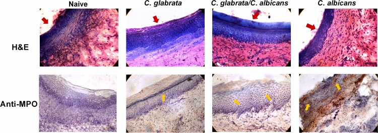
FIG 4.
Histological analysis of palatal inflammation in rats inoculated with C. albicans and/or C. glabrata. Rats fitted with dentures were inoculated 3 times at 3-day intervals with 1 × 10⁹ CFU C. albicans, C. glabrata, or both species together (5 × 10⁸ CFU each). Palate tissue was harvested at 4 weeks postinoculation. Frozen tissue sections were stained with hematoxylin and eosin (H&E) for histopathological analysis or with anti-myeloperoxidase (MPO, brown-red) or isotype control (mouse IgG1) antibodies. Red arrows indicate the apical surface of the palate epithelium. Yellow arrows represent cells positively stained for MPO. Figure shows a representative result of 2 independent experiments. Magnification, ×400.

