Abstract
Pharmaceutical effluents contain toxic xenobiotics capable of contaminating aquatic environments. Untreated effluents are illegally discharged into aquatic environment in most developing countries. Pharmaceutical effluent induced alterations in biomarkers of genetic and systemic damage on rodents. However, information is relatively scarce on the possible cytogenotoxicity and systemic toxicity of this effluent on aquatic vertebrates. The study herein assessed the cytogenotoxic, hematological and histopathological alterations induced by pharmaceutical effluent in Clarias gariepinus. 96 h acute toxicity of the effluent was determined after C. gariepinus was exposed to six different concentrations (10 - 60 %) of the effluent. Subsequently, fish was exposed to sub-lethal concentrations (2.18 - 17.41 %) obtained from the 96 h LC50 for 7 and 14 days after which micronucleus (MN) and nuclear abnormalities (NAs) in peripheral erythrocytes were assessed as cytogenotoxic biomarkers, alterations in hematological indices and histopathological lesions were also examined. Fish, concurrently exposed to dechlorinated tap water and benzene (0.01 mL/L), served as negative and positive controls respectively. The derived 96 h LC50 of 17.41 % which was 1.89 times more toxic than the 24 h LC50 (32.95 %) showed that the effluent induced concentration-dependent mortality according to exposure duration. The effluent caused significant (p<0.05) time-dependent increase in the frequency of MN and abnormal nuclear erythrocytes compared to the negative control. Also, there was decrease in total erythrocyte counts, hemoglobin and hematocrit concentrations and increase in leucocyte and lymphocyte counts. The effluent induced pathological lesions on gills, liver and kidneys of treated fish. Higher physicochemical parameters than standard permissible limits in the effluent are capable of inducing genomic instability and systemic damage in fish. Pharmaceutical effluent can increase micropollutants in aquatic environmental and health risks to aquatic biota. There is need to promulgate stringent laws against illegal discharge of effluents into aquatic environment.
Keywords: acute toxicity, African catfish, hematology, histopathology, micronucleus assay, untreated pharmaceutical effluent
Introduction
Increasing contamination of the aquatic environment worldwide has been associated with improper discharge of solid wastes, industrial, medical and agricultural effluents (Ibekwe et al., 2012[32]; Larsson, 2014[38]; Alimba et al., 2017[5]). This is raising concern in most countries worldwide, due to the deteriorating effects on surface and underground water and sediment qualities of most aquatic environment, and hence the deleterious health effects on biotic communities (Chigor et al., 2012[14]; Othman et al., 2012[49]). Effluents from pharmaceuticals and personal care products (PPCPs), along with metals and metalloids are increasingly being released directly into aquatic environment via untreated and or poorly treated wastewater and sewage treatment facilities (Nikolaou et al., 2007[45]; Blair et al., 2013[13]; Larsson, 2014[38]). This increase is linked with the quest for quality life expectancy of the rising human and animal populations that depend on pharmaceuticals for their sustenance.
Occurrence of pharmaceuticals in the aquatic environment has been the research focus for over three decades now (Ternes, 1998[56]; Daughton and Ternes, 1999[19]). There are several reports on the presence of pharmaceuticals and their metabolites in most aquatic environments worldwide (Zuccato et al., 2000[64]; Nikolaou et al., 2007[45]; Kookana et al., 2014[36]). Many of these reports showed that these pharmaceuticals were commonly observed in aquatic environments of most high income nations, with very scanty information from low- and middle-income countries (Kookana et al., 2014[36]). This may be attributed to low environmental awareness or ignorance among developing countries on the occurrence of drugs in aquatic environment. Also inability to afford high standard equipment for analysis of drugs in the aquatic matrix, a common phenomenon in most low- and middle-income nations, may be a contributing factor. However, in recent times, there is an increase in awareness among researchers from Africa and Asia on the need to investigate pharmaceuticals and their metabolites in most surface and underground water bodies. For instance, Agunbiade and Moodley (2016[2]) reported eight acidic pharmaceuticals; four antipyretics (Ibuprofen, Ketoprofen, Diclofenac and Aspirin), three antibiotics (Ampicillin, Ciprofloxacin and Nalidixic acid), and one lipid regulator (Bezafibrate) in wastewater, surface water, and sediments from Msunduzi River in the province of KwaZulu-Natal, South Africa. Also, Olarinmoye et al. (2016[47]) observed thirty seven pharmaceuticals classified as antibiotics, estrogens and lipid-lowering drugs in surface water and industrial, domestic and hospital sewage sludge from Lagos State, Southwest, Nigeria. The presence of these drugs in waterways from low- and middle-income nations was attributed to poor enforcement of regulation and laws restricting indiscriminate discharge of untreated pharmaceutical, hospital and sewage wastewaters directly into the aquatic environment. Also some pharmaceutical industries have shifted from high income countries to low- or middle-income countries mainly due to reduced production costs (Kookana et al., 2014[36]). This is expected to accelerate industrialization and population increase, which will invariably increase wastewater generation and discharge into the environment (Agunbiade and Moodley, 2016[2]).
Pharmaceuticals in aquatic environment occur in concentrations ranging from Limits of Detection (LOD = 8.84 µg/L) to levels exceeding the ecotoxicological predicted no-effect concentrations (PNEC) (Kookana et al., 2014[36]). Most of these pharmaceuticals are active chemicals synthesized to exert specific physiological change(s) on targeted organ(s) of species (mostly mammals). However, they can also affect non-targeted aquatic biota and adversely cause disturbance on the integrity of body systems even at low concentrations (Hernando et al., 2006[28]; Maselli et al., 2015[41]). Pharmaceutical effluents contain mixture of various classes of organic and inorganic micropollutants that are capable of inducing toxicological effects on aquatic organisms. In Nigeria, due to high cost of effluent treatment, most industries illegally discharge untreated effluent directly into aquatic environment. This act is worrisome and has elicited public concern due to increasing occurrence of drugs and metals in coastal waters via Nigerian waterways (Olarinmoye et al., 2016[47]). The individuals and interactions of mixture constituents of the effluents are eliciting adverse impacts on aquatic life (Alimba et al., 2015[7]). Micropollutants in pharmaceutical effluents can cause somatic mutations and systemic toxicity in fish which may cause tumor formation, biodiversity loss and mortality (Viarengo et al., 2007[60]).
Laboratory designed experiment to simulate the genotoxicity and systemic toxicity of pharmaceutical effluents in aquatic environment using fish is scarce. Available reports have shown that pharmaceuticals elicited acute toxicity on different aquatic invertebrates (Zounková et al., 2007[63]). Pharmaceutical effluent is capable of causing alterations in gene expressions and enzyme activities of plasma vitellogenin which may lead to intersex in fish species (Larsson, 2014[38]). Also, there is evidence that pharmaceutical effluents significantly induced somatic and germ line genotoxicity in rodents (Zhao et al., 2007[62]; Bakare et al., 2009[11]; Adeoye et al., 2015[1]). The use of cytogenetic markers in the routine monitoring of industrial effluents for the presence of xenobiotics is important since DNA damage in aquatic biota may reduce survival by affecting prompt reproduction and increase pollutant-induced stress syndromes (Malins et al., 1988[40]).
The study herein aims at increasing knowledge on the genotoxicity and systemic toxicity induced by untreated pharmaceutical effluent in Clarias gariepinus. To achieve this aim, acute toxicity (96 h LC50), one of the preliminary toxicological screening bioassay for xenobiotics (OECD, 1992[46]; Akhila et al., 2007[3]), was used to determine the concentration of the pharmaceutical effluent that caused 50 % mortality to the animal model, juvenile Clarias gariepinus. Subsequently, the formation of micronucleated (MN) and nuclear abnormalities (NAs) in erythrocytes, alterations in the histological architecture of the liver, gill and kidney, as well as changes in the hematological indices were assessed following sub-chronic exposure to sub-lethal concentrations (96 h LC50) of the effluent.
Materials and Methods
Effluent collection, physico-chemical and heavy metals analysis
Pharmaceutical effluent used in the study herein was obtained from a pharmaceutical industry located at Satellite Town, Lagos State, Nigeria. Composite mixture of the untreated effluent was collected into a 25 L transparent high density polyethylene plastic container. The pH was measured and transported in a dark condition using poly-fiber container with cold ice to the animal house unit of the Department of Cell Biology and Genetics, University of Lagos for further processing. The physical and chemical parameters; biochemical oxygen demand (BOD), chemical oxygen demand (COD), dissolved oxygen (DO), turbidity, alkalinity, chlorides, sulphates, nitrates, ammonia and phosphates were analyzed using standard methods (APHA, 2005[9]). Metals and metalloids; arsenic (As), cadmium (Cd), chromium (Cr), lead (Pb), copper (Cu), iron (Fe) and manganese (Mn), were also analyzed in compliance with standard method (APHA, 2005[9]; US EPA, 2014[58]) using Perkin-Elmer A3100 atomic absorption spectrophotometer for the metals.
Clarias gariepinus collection and acclimatization for the experimental study
Juvenile C. gariepinus with mean ± SD body weight, 12.05 ± 2.30 g and length 9.40 ± 1.50 cm, obtained from Fish farm along Badagry, Lagos State, were used for the study. They were acclimated to laboratory conditions of 26.1 ± 3.0 oC and 12/12 hour dark/light modes for 14 days prior to the experimental set-up. They were stocked at a population density of 10 fish per 25 L aquarium and fed 10 % of their body weight with basal diet containing 35 % crude proteins, rice bran, fish meal and mustard oil-cake (Coppens 2 mm floating feed®), twice daily. Guide for care and use of laboratory animals published by the US National Institutes of Health (NIH Publication No. 85-23, revised in 1996) (Gad, 2007[26]; CIOMS, 2012[15]), and approved by the ethical committee, University of Lagos for the use of animals in experimental studies was carefully adhered to during the study.
Acute toxicity testing and assessment of quantal response (mortality)
After series of range finding tests, 10 juvenile C. gariepinus per group, were randomly distributed into seven experimental groups containing 0.0, 10.0, 20.0, 30.0, 40.0, 50.0 and 60.0 % (v/v; effluent / dechlorinated tap water) for a period of 4 days to determine the 96 h acute toxicity (LC50) of the effluent. Safe concentration of the wastewater at 96 h was obtained by multiplying the 96 h LC50 by a factor of 0.1 in accordance with EIFAC (1998[21]). Toxicity factor (TF) for 24 hourly relative potency measurements of the effluent were also determined. During the acute toxicity testing, the fish were not fed and the test effluent not renewed (non-renewable static bioassay). Also mortality and behavioral patterns of exposed fish in each experimental group were recorded every 24 h in accordance with the guidelines of the Organization for Economic Cooperation and Development (OECD, 1992[46]). Fish was assumed dead when there was no body or operculum movement, even when prodded with a glass rod.
Sub-chronic exposure to sub-lethal concentrations of the 96 h LC50 effluent
Ten juvenile C. gariepinus were randomly distributed into 25 L tanks containing sub-lethal concentrations: 2.18, 4.35, 8.71 and 17.41 % (v/v, effluent / dechlorinated tap water) (corresponding to 1/8, 1/4, 1/2 and x1 of the 96 h LC50 respectively) of the effluent for 14 days. Similar treatment was given to fish in 0.01 mL/L (v/v, Benzene / dechlorinated tap water) and dechlorinated tap water as positive and negative controls respectively. The fish was fed three times daily with 10 % feed per body weight and the water for the treatment and control groups replaced every 48 h to minimize volatilization of less stable components of the effluent so as to maintain concentration of the effluent, and reduce accumulation of metabolic wastes and remains of food particles. On days 7 and 14 of the exposure period, peripheral blood was collected from 5 fish, per sampling time, from each of the treatment and control groups via caudal vein into EDTA bottles for micronucleus assay and hematological analysis. The fish was then sacrificed and dissected, and the gills, liver and kidney collected and fixed in Bouin fluid fixative for 48 h prior to histopathological analysis.
Micronucleus, hematological and histological analysis
Thin smear from an aliquot of the peripheral blood was made on three pre-cleaned slides per fish. They were air dried, fixed in absolute methanol for 30 min, and counterstained with 10 % May-Grunwald and 5 % Giemsa stains in accordance with standard procedure (Alimba et al., 2015[4]; Alimba and Bakare, 2016[6]). Three thousand erythrocytes per fish were scored for MN induction at 1000x magnification. Cells scored for nuclear abnormalities (NAs) include those with small MN that is still connected to the nucleus (nuclear bud, Nbud), cells with two nuclei (binucleated, BN) and cells with vacuoles in their nuclei (vacuolated nuclei, VN). Only cells with intact plasma and nuclear membranes were scored (Alimba et al., 2015[4]; Alimba and Bakare, 2016[6]).
Aliquot of the blood in EDTA bottle was analyzed to determine the concentration of the fish hemogram including Red blood cell (RBC) count, hemoglobin (Hb) content, percentage hematocrit (Ht), mean corpuscle hemoglobin concentration (MCHC), mean corpuscle volume (MCV), mean corpuscle hemoglobin (MCH), platelets, total white blood cell (WBC) count and lymphocytes in the treatment and control groups using automated analyzer (Abbott Hematology Analyzer Cell-Dyn 1700, Abbott Laboratories, Abbott Park, Illinois, USA).
The fixed sections of the gills, liver and kidney from the treated and control fish were dehydrated by passing through ascending order of ethyl alcohol-water concentrations, cleared in xylene and embedded in paraffin wax using rotary microtome. Four μm thick sections of the tissues were prepared on slides, stained with Hematoxylin-Eosin (H&E) and mounted in neutral DPX medium for microscopic examination at x400 magnification by trained pathologist.
Statistical analysis
Data from the acute toxicity (mortality) were analyzed using probit analysis with SPSSTM version 17.0, and presented as LC5 (lethal concentration that caused 5 % mortality), LC50 (lethal concentrations that caused 50 % mortality) and LC95 (lethal concentration that caused 95 % mortality) at their corresponding 95 % confidence interval. Frequencies of micronucleated and abnormal nuclear (NA) erythrocytes were presented as mean ± SE (standard error). Significant difference among the various treatment and control groups was determined using One-way ANOVA. Dunnett multiple post hoc test was used to compare the degree of significance (p<0.05) of each treatment group with the negative control.
Results
Physicochemical and heavy metal analysis of the effluent
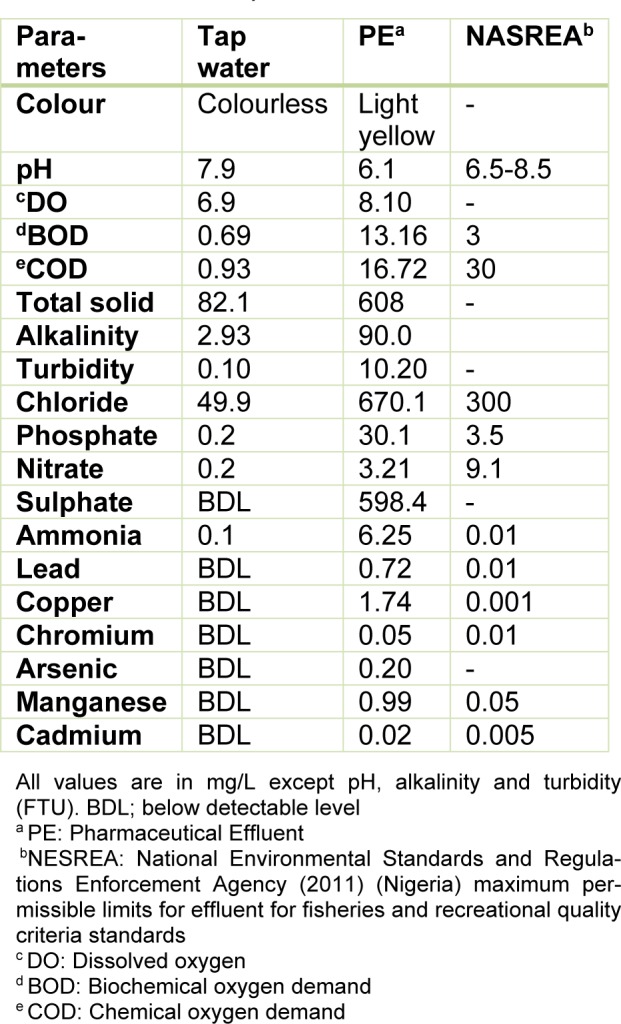
Table 1(Tab. 1) presents the results of the physico-chemical parameters, metals and metalloids analyzed in the effluent. The light yellow coloured effluent which was slightly acidic (pH=6.1) contained high concentrations of the analyzed physicochemical parameters and metals than respective values in the negative control (dechlorinated tap water) and National Environmental Standards and Regulations Enforcement Agency (NESREA, Nigeria) allowable limits for effluent quality criteria standards. However, nitrate and COD were lower than permissible limits but higher than the negative (dechlorinated tap water) control.
Table 1. Physicochemical parameter and metal analyses of the pharmaceutical effluent (PE), tap water and national permissible standards.
Acute toxicity (mortality) induced by the effluent to juvenile C. gariepinus
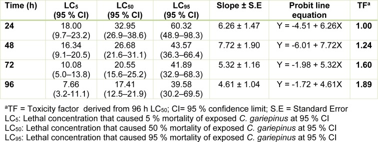
The toxicity indices (daily LC50) obtained from the concentration-mortality data decreased according to increase in exposure duration: 32.95 % (24 h LC50) < 26.68 % (48 h LC50) < 20.55 % (72 h LC50) < 17.41 % (96 h LC50). The daily LC50 along with the LC5 and LC95 values for the 24 - 96 h acute toxicity (Table 2(Tab. 2)), showed that the pharmaceutical effluent induced concentration-dependent and exposure related mortality of the juvenile C. gariepinus. The toxicity factor computed from the 96 h LC50 (TF = 1.89) showed that the effluent was highly toxic to C. gariepinus (Table 2(Tab. 2)). The safe concentration of the waste-water to the juvenile C. gariepinus at 96 h exposure period is 1.74 %.
Table 2. 96 h acute toxicity determination of pharmaceutical effluent (PE) using Clarias gariepinus.
Micronucleated and abnormal nuclear erythrocyte formation in C. gariepinus
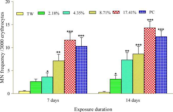
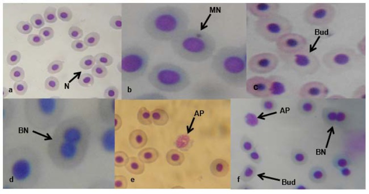
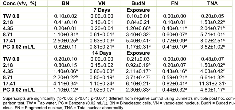
Figure 1(Fig. 1) presents the results of the cytogenetic analysis in C. gariepinus exposed to sub-lethal concentrations of the tested effluent. The effluent elicited concentration-dependent significant (p<0.05) increase in MN (Figure 2b(Fig. 2)). The induced MN which was also exposure related, showed 4.07, 7.10, 13.96 and 22.82 (corresponding to 2.18, 4.35, 8.71 and 17.41 % concentrations of the effluent respectively) fold increase compared to the negative control during day 7 of exposure, and 10.83, 25.24, 29.72 and 49.28 fold increase than the corresponding negative control during day 14 of exposure. There was significant (p < 0.001) increase in the frequencies of nuclear bud (Nbud) (Figure 2c(Fig. 2)), binucleated (BN) erythrocytes (Figure 2d(Fig. 2)), vacuolated (VN) and fragmented nucleus (AP) (Figure 2e(Fig. 2)) formed in the peripheral erythrocytes of effluent exposed fish compared to the negative control. The induced NAs which was concentration-dependent and exposure duration related was in the order; Nbud > BN > VN > AP (Table 3(Tab. 3)).
Figure 1. Frequency of micronucleated erythrocytes in C. gariepinus exposed to pharmaceutical effluent. ∗p<0.05; ∗∗p< 0.01; ∗∗∗p< 0.001 are significantly different from the negative control (tap water) using Dunnett's multiple post hoc comparison test. PC= Benzene (0.01 mL/L) positive control, TW= tap water (negative control).
Figure 2. Micronucleated and abnormal nuclear erythrocytes in the effluent treated C. gariepinus: (a) normal peripheral erythrocyte (N). (b) Micronucleated erythrocyte (MN). (c) erythrocyte with budded nucleus (Nbud). (d) binucleated erythrocyte (BN). (e) fragmented erythrocyte (AP). (f) AP, BN and Nbud erythrocytes (x1000).
Table 3. Mean (± SE) of nuclear abnormalities (NAs)/3000 peripheral erythrocytes of C. gariepinus exposed to pharmaceutical effluent for 7 and 14 days.
Hematological profile of Clarias gariepinus exposed to pharmaceutical effluent
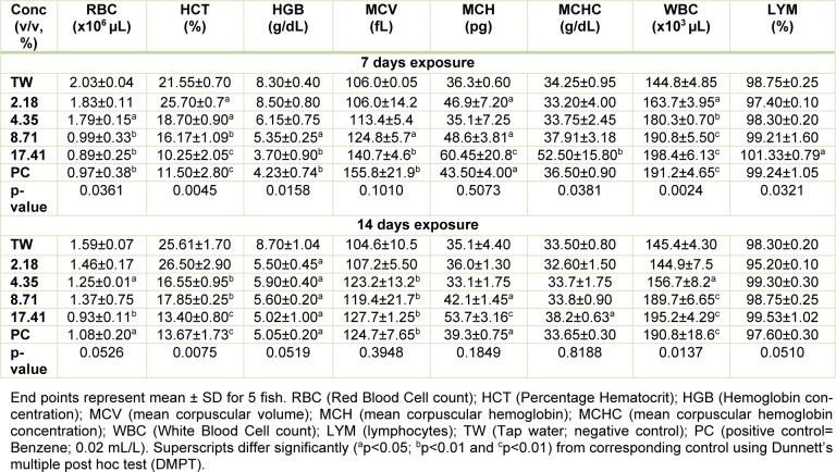
The sub-lethal concentrations of the pharmaceutical effluent significantly reduced red blood cells (RBC), percentage hematocrit (HCT), hemoglobin (HGB) concentration, but increased white blood cells (WBC) and lymphocytes (except at 2.18 % concentration of the effluent where there was insignificant decrease in lymphocytes compared to the negative control) in a concentration-dependent pattern during 7 day exposure. However, the mean corpuscular volume (MCV), mean corpuscular hemoglobin (MCH) and mean corpuscular hemoglobin concentration (MCHC) were insignificantly (p > 0.05) increased in the treated fish compared to the negative control during 7 day exposure. For 14 day exposure period, the tested effluent significantly (p < 0.05) reduced red blood cells (RBC), percentage hematocrit (HCT), hemoglobin (HGB) concentration, but increased white blood cells (WBC) (except at the 2.18 % concentration of the effluent where there was insignificant decrease in the leucocyte counts compared to the negative control) and lymphocytes. The altered hematological indices in the exposed fish were not concentration-dependent. The mean corpuscular volume (MCV), mean corpuscular hemoglobin (MCH) and mean corpuscular hemoglobin concentration (MCHC) data from fish exposed for 14 days were insignificantly (p > 0.05) increased compared to the negative control and in a concentration-independent pattern (Table 4(Tab. 4)).
Table 4. Hematological profile of C. gariepinus exposed to the pharmaceutical effluent for 7 and 14 days.
Histological alterations in tissues of C. gariepinus exposed to the effluent
Gills collected from fish exposed to dechlorinated tap water (negative control) presented apparently normal filaments and lamellae (Figure 3a(Fig. 3)). However, gills from fish exposed to sub-lethal concentrations of the effluents for 7 and 14 days revealed some histopathological lesions which include severe congestion of the blood capillaries and thickening of the filaments (Figure 3b(Fig. 3)). Also gill lamellae were absent from the sections of some fish from the treatment groups, and the covering epithelium of the operculum markedly separated from the central cartilaginous core by sparse amounts of loose connective tissues (Figure 3c(Fig. 3)).
Figure 3. a. Gill from a control group showing apparently normal gill filament and gill lamellae.
b. There is severe congestion (C) of the blood capillaries; necrosis (N) thickening (T) of the gill filament, disorganization of the gill lamella.
c. Loss of gill lamellae (arrow); the covering epithelium of the operculum is markedly separated from the central cartilaginous core by sparse amounts of loose connective tissues. Mag. x400
Kidney sections from fish exposed to tap water (negative control) showed apparently normal tubular (TC) and hematopoietic compartment (HC), with the TC consisting of closely packed blood capillaries and glomeruli (Figure 4a(Fig. 4)). There were multiple foci of tubular degeneration and severe depletion of the TC and HC in the effluent treated fish (Figure 4b-c(Fig. 4)).
Figure 4. a. Sections of the kidney from the control fish showing apparently normal tubular and hematopoietic compartment. The tubular is a closely packed tubule with glomeruli.
b. Section of kidney from effluent treated fish showing severe depletion of the tubular (T) and hematopoietic (H) compartments thus appearing more prominent. Mag. x400
c. Section from the effluent treated fish showing tubules that are widely separated from each other with decrease in the tubular (T) compartment and accompanying increase in the hematopoietic (H) compartment; there are multiple foci of degenerated tubules (D). Mag. x400
The histological presentation of the hepatic sections of the tap water treated fish showed the normal architecture of fish hepatocytes with closely packed hepatic plates without cytoplasmic vacuoles (Figure 5a(Fig. 5)). However, some histological lesions including congestions of central veins, large prominent bile ducts and multiple moderate-sized vacuoles were observed in the liver tissue of the effluent treated fish (Figure 5b and 5c(Fig. 5)).
Figure 5. a. Section of the liver of a negative control fish showing closely packed hepatic plates with the hepatocytes not showing visible cytoplasmic vacuoles and with intact bile ducts.
b. The hepatocytes of effluent treated fish contain multiple moderate-sized vacuoles (V), large prominent bile duct with a moderately congested (C) of the central veins and disarrayed hepatocytes.
c. In the effluent treated fish, there are widespread multiple foci of hepatocytes with clear cytoplasmic vacuoles. Also there are widespread large multiple cytoplasmic vacuoles within the hepatocytes, and the central veins moderately congested with disarrayed hepatocytes. Mag. x400.
Discussion
Increasing utilization of pharmaceuticals in order to improve quality of health and extend life expectancy in humans and domesticated animals has an inevitable consequence of increasing surface and ground water contamination with biologically active chemicals including toxic metals (Corcoran et al., 2010[16]; Olarinmoye et al., 2016[47]). Despite the possible adverse effects this may pose on aquatic wildlife, studies are scanty that have characterized the possible health effects of pharmaceutical effluents on aquatic vertebrates (including fish).
The 96 h LC50 (17.41 %) which was 1.89 (TF=1.89) times more toxic than the 24 h LC50 (32.95 %) showed that the effluent was highly toxic to the juvenile stage of C. gariepinus. The induced mortality was concentration-dependent and exposure duration related. In comparison with previous studies from our laboratory where similar sized juvenile C. gariepinus served as bio-indicator, the 96 h LC50 obtained for Aba Eku (43.29 %) and Olusosun (34.52 %) landfill leachates (Alimba and Bakare, 2016[6]) showed that the tested effluent herein was more toxic to the fish than solid waste landfill leachates, but was less toxic than abattoir effluent (96 h LC50 = 6.28 %) (Alimba et al., 2015[7]), textile effluent (96 h LC50 = 8.02 %) (Ayoola et al., 2012[10]) and hospital effluent (96 h LC50 = 1.30 %) (Alimba et al., 2017[5]). This observation showed that juvenile C. gariepinus responded differently to mortality and deleterious effects elicited by different industrial effluents. Hence, it can be concluded that C. gariepinus is a suitable bio-indicator for monitoring the toxicity of effluents. The derived safe concentration of 1.74 % of the effluent for the juvenile C. gariepinus used herein is significantly low compared to the volume of untreated wastewater that is directly discharged into aquatic environment in most low- and middle-income nations (Sogbanmu et al., 2016[55]). This may suggest a higher level of morbidity and eventual death of fish and many other lower vertebrates that inhabit such wastewater contaminated aquatic environment. The analyzed physicochemical parameters, toxic metals and metalloids (Table 1(Tab. 1)), in the effluent possibly interacted synergistically, antagonistically or additively to induce mortality in the fish (Magdaleno et al., 2014[39]). High total dissolved solids analyzed in the effluent may be due to inert solids and particulate matters. These solids are capable of clogging the gill system and may suffocate the fish to death (FAO, 1991[23]).
The selected sub-lethal concentrations of the tested pharmaceutical effluent did not lead to immediate mortality of the fish, however it elicited alterations in the biomarkers of somatic mutations and systemic damage examined. MN assay, which has been used for over 30 years as biomarker of cytogenetic damage in fish (Al-Sabti, 1986[8]), due to its reliability, sensitivity and cost effectiveness, showed that the tested effluent is cytogenotoxic. Significant increase in micronucleated erythrocytes in the effluent treated C. gariepinus indicated that the constituents of the effluent are clastogenic and/or aneugenic to fish genetic materials. The observed genome instability elicited by the tested effluents in C. gariepinus herein, had been previously reported in mice and rat wherein pharmaceutical effluents increased frequency of chromosome aberrations and micronucleated polychromatic erythrocytes in bone marrow erythrocytes of mice and rats (Bakare et al., 2009[11]; Adeoye et al., 2015[1]), and increased germ line mutation in mice (Zhao et al., 2007[62]; Bakare et al., 2009[11]). Scoring NAs along with MN in C. gariepinus exposed to mixture of xenobiotics in leachates and effluents is an efficient and reliable biomarker of cytogenetic damage (Ayoola et al., 2012[10]; Bakare et al., 2013[12]; Alimba et al., 2015[7][4], 2017[5]; Alimba and Bakare, 2016[6]). Significant increase in total nuclear abnormalities in the effluent exposed C. gariepinus compared with the negative control suggests that the constituents of the effluent induced perturbation in cell cycle and DNA synthesis during hematopoiesis in the fish (Udroiu, 2006[57]). Nuclear bud formation is associated with the entrapment of extra-chromosomally amplified DNA during S-phase and is related to genotoxicity events (Fenech et al., 2011[25]). Binucleated cells (biomarker of cytotoxicity) are formed possibly due to the presence of cytotoxins in the effluents which altered cytokinesis during M phase of the cell cycle. Increased fragmented (apoptotic) and vacuolated nuclei observed in the erythrocytes of effluent exposed C. gariepinus may be linked to alterations in p53 protein expression which led to the activation of antioxidant genes associated with apoptotic cell formation (Verlhac and Gabaudan, 1994[59]), and or damaged erythrocytes during hematopoitic process, which were eliminated by programmed cell death (apoptosis) (Pulido and Parrish, 2003[50]). There is paucity of information on the possible cytogenotoxic effects of pharmaceutical effluents on aquatic vertebrate including fish. However, it is important to note that significant increase in frequencies of MN and NAs are associated with genome instability which has been strongly correlated with genetic related syndromes; different pathogenesis, reproductive dysfunctions and cancer formation in fish (Malins et al., 1988[40]; Kurelec, 1993[37]; Alimba et al., 2015[7]; Daiwile et al., 2015[18]). The analyzed metals (Table 1(Tab. 1)) in the effluent are potent genotoxins in both in vivo and in vitro test systems and may account for the observed MN and NAs induction in the effluent exposed fish (Jiraungkoorskul et al., 2008[34]; Guedenon et al., 2015[27]).
Considering the importance of blood during body and systemic circulation in fish, alteration in hematological indices in response to water contamination is considered a sensitive biomarker of fish health (Seriani et al., 2015[51]). Usually changes in hematological indices appear first before the onset of any morphological and degenerative damage in fish (Mazon et al., 2002[42]). Decrease in RBC, hemoglobin concentrations and percentage hematocrit in C. gariepinus during both 7 and 14 day exposures to the effluent suggests anemic condition in the exposed fish. This may be due to the deleterious effects of the effluent constituents on the hemotopoietic system (mainly the kidney system) by inhibiting erythropoiesis via transferrin dysfunction (Javed et al., 2016[33]). Increase in MCV, MCH and MCHC in the effluent exposed fish, although not statistically significant, may suggest that the anemic condition induced by the effluent is macrocytic hyperchromic anemia. It is possible that the critical hematotoxic conditions on the fish are in tandem with the high mortality observed in the acute toxicity (Table 2(Tab. 2)). Macrocytic hyperchromic anemia has been associated with defects in DNA synthesis during erythropoiesis (Hoffman et al., 2009[30]). Total white blood cell (WBC) and differentials in fish form components of the non-specific immune cells (da Silva Correa et al., 2016[17]). Leukocytosis and lymphocytosis observed herein may indicate physiological and immunological (inflammatory) challenges in response to the toxic effects of the effluent (Duthie and Tort, 1985[20]). Leukocytosis is directly related to severity of damage and stress induced by metals with a consequential result of immunological defense stimulation (Javed et al., 2016[33]). Similar trends of alteration in hematological indices were reported in Cyprinus carpio exposed to Cr(VI) (Shaheen and Akhtar, 2012[52]), in Labeo rohita exposed to effluents from paint, dye and petroleum industries (Zutshi et al., 2010[65]), in Oreochromis mossambicus exposed to Cd (Shalaby, 2001[53]) and in rats exposed to pharmaceutical effluent for 28 days (Adeoye et al., 2015[1]). These may be linked to individual and/or interactive effects of the effluent constituents on the metabolic and hemotopoietic activities in the fish.
Changes in the histology of tissues in fish have been widely used, both in laboratory regulated experiments (Mela et al., 2007[43]) and field studies (Alimba et al., 2015[7]) to assess fish health. Histopathology, as a biomarker of toxicity, is relevant in monitoring organ specific damage induced by environmental pollutants and relates the damage organs to specific physiological function in the fish body (Evans, 1987[22]). Furthermore, histopathological findings corroborate functional biomarkers to provide specific information on the acute and chronic effects of toxicants on targeted organs (Evans, 1987[22]; Mela et al., 2007[43]).
Gill epithelium of teleost, apart from being the gaseous exchange site, is also responsible for ionic regulation, acid-base balance, and nitrogenous waste excretion in fishes (Hoar and Randall, 1984[29]). The observed severe congestion of the blood capillaries, necrosis and thickening of the gill filament and disorganization of the lamellae in effluent exposed C. gariepinus (Figure 3b and 3c(Fig. 3)) showed that constituents of the effluent caused structural alterations on the gills. The effluent constituents come in direct contact with the gills and considering that the gill filaments and lamellae have increase surface area of exposure to contaminants, makes gills the most critical site of toxicity (Evans, 1987[22]; Wood et al., 2002[61]). Also considering that the gills are highly metabolically active, they are readily prone to damage from environmental micropollutants, mostly the toxic metals via oxidative stress induction (Farombi et al., 2007[24]). The observed histopathological lesions in the effluent exposed C. gariepinus had been similarly reported in Oreochromis niloticus which were exposed to petroleum refinery effluent (Onwumere and Oladimeji, 1990[48]) and in free ranging Synodontis clarias caught from toxic metal and organic compounds polluted Lekki Lagoon and Ogun River in Nigeria (Alimba et al., 2015[7]).
It is well known that the liver and kidneys are among the major organs that readily respond to toxic effects of a wide variety of environmental micropollutants owning to their involvement in absorption and bio-concentration of pollutants (Hook, 1980[31]; Koca et al., 2008[35]). The liver is mainly involved in metabolism, detoxification, storage, and excretion of xenobiotics and their metabolites, while the kidneys serve as the main hematopoietic organ of most teleost (Mela et al., 2007[43]). These functions make them targets for xenobiotics induced pathophysiological injuries (Mela et al., 2007[43]; Koca et al., 2008[35]; Alimba et al., 2015[7]). That these organs readily bio-accumulate toxic metals and metabolize organic toxicants (Silva and Martinez, 2007[54]), may account for the pathological lesions observed in the treated fish compared to the negative control. The effluent induced hepatic and renal damage to the exposed fish. The observed lesions may be attributed to the direct or indirect deleterious actions of the effluent constituents on the tissues following the attempt by the fish to detoxify and eliminate the toxic metals and other xenobiotics from the body. The observed pathological lesions in the exposed fish herein have been similarly reported in rats orally exposed to pharmaceutical effluent for 28 days (Adeoye et al., 2015[1]). This suggests that the constituents of untreated pharmaceutical effluent pose great health challenge to both terrestrial and aquatic biota. Multiple foci of degenerated tubules and severe depletion of the hematopoietic compartment of the kidney, the organ of blood production in teleost, corroborate the observed alterations in the fish hemogram and micronucleus and abnormal nuclear formation in the erythrocytes.
The findings herein showed that pharmaceutical effluent contains varying concentrations of toxic metals, metalloids and physicochemical parameters higher than standard permissible limits. The effluent increased frequency of MN and NAs in erythrocytes, altered hematological indices and induced histopathological lesions in gills, liver and kidney in C. gariepinus. This suggests the constituents of pharmaceutical effluents as emerging carcinogens and mutagens that are capable of increasing genome instability, altering blood cell indices and causing pathological lesions in fish tissues. This is a threat to the functioning aquatic ecosystems and the survival of aquatic biota.
Acknowledgement
The authors appreciate Angela I. Chikeluba for her technical assistance during the study.
Funding
The authors declare that the study herein was not funded by any funding body.
Conflict of interest
The authors declare that no form of conflict of interest exists.
References
- 1.Adeoye GO, Alimba CG, Oyeleke OB. The genotoxicity and systemic toxicity of a pharmaceutical effluent in Wistar rats may involve oxidative stress induction. Toxicol Rep. 2015;2:1265–72. doi: 10.1016/j.toxrep.2015.09.004. [DOI] [PMC free article] [PubMed] [Google Scholar]
- 2.Agunbiade F, Moodley B. Occurrence and distribution pattern of acidic pharmaceuticals in surface water, wastewater, and sediment of the Msunduzi River, KwaZulu-Natal, South Africa. Environ Toxicol Chem. 2016;35:36–46. doi: 10.1002/etc.3144. [DOI] [PubMed] [Google Scholar]
- 3.Akhila JS, Deepa S, Alwar MC. Acute toxicity studies and determination of median lethal dose. Curr Sci. 2007;93:917–920. [Google Scholar]
- 4.Alimba CG, Ajayi EO, Hassan T, Sowunmi AA, Bakare AA. Cytogenotoxicity of abattoir effluent in Clarias gariepinus (Burchell, 1822) using micronucleus test. Chinese J Biol. 2015;2015 [Google Scholar]
- 5.Alimba CG, Ajiboye RD, Fagbenro OS. Dietary ascorbic acid reduced micronucleus and nuclear abnormalities in Clarias gariepinus (Burchell 1822) exposed to hospital effluent. Fish Physiol Biochem. 2017;43:1325–1335. doi: 10.1007/s10695-017-0375-y. [DOI] [PubMed] [Google Scholar]
- 6.Alimba CG, Bakare AA. In vivo micronucleus test in the assessment of cytogenotoxicity of landfill leachates in three animal models from various ecological habitats. Ecotoxicology. 2016;25:310–9. doi: 10.1007/s10646-015-1589-3. [DOI] [PubMed] [Google Scholar]
- 7.Alimba CG, Saliu JK, Ubani-Rex OA. Cytogenotoxicity and histopathological assessment of Lekki lagoon and Ogun River in Synodontis clarias (Linnaeus, 1758) Toxicol Environ Chem. 2015;97:221–34. [Google Scholar]
- 8.Al-Sabti K. Comparative micronucleated erythrocyte cell induction in three cyprinids by five carcinogenic-mutagenic chemicals. Cytobios. 1986;47(190-191):147–54. [PubMed] [Google Scholar]
- 9.APHA, American Public Health Association. Standard methods for the examination of water and wastewater. 21st ed. Washington, DC: American Public Health Association; 2005. [Google Scholar]
- 10.Ayoola SO, Bassey BO, Alimba CG, Ajani EK. Textile effluent induced genotoxic effects and oxidative stress in Clarias gariepinus. Pakistan J Biol Sci. 2012;15:804–2. doi: 10.3923/pjbs.2012.804.812. [DOI] [PubMed] [Google Scholar]
- 11.Bakare AA, Alabi O, Adetunji OA, Jenmi HB. Genotoxicity assessment of a pharmaceutical effluent using four bioassays. Genet Mol Biol. 2009;32:373–81. doi: 10.1590/S1415-47572009000200026. [DOI] [PMC free article] [PubMed] [Google Scholar]
- 12.Bakare AA, Alabi OA, Gbadebo AM, Ogunsuyi OI, Alimba GC. In vivo cytogenotoxicity and oxidative stress induced by electronic waste leachate and contaminated well water. Challenges. 2013;4:169–87. [Google Scholar]
- 13.Blair BD, Crago JP, Hedman CJ, Klape RD. Pharmaceuticals and personal care products found in the Great Lakes above concentrations of environmental concern. Chemosphere. 2013;93:2116–23. doi: 10.1016/j.chemosphere.2013.07.057. [DOI] [PubMed] [Google Scholar]
- 14.Chigor VN, Umoh VJ, Okuofu CA, Ameh JB, Igbinosa EO, et al. Water quality assessment: surface water sources used for drinking and irrigation in Zaria, Nigeria are a public health hazard. Environ Monit Assess. 2012;184:3389–400. doi: 10.1007/s10661-011-2396-9. [DOI] [PubMed] [Google Scholar]
- 15.CIOMS International guiding principles for biomedical research involving animals. 2012. [02 February 2019]. Available from: https://olaw.nih.gov/sites/default/files/Guiding_Principles_2012.pdf. [PubMed]
- 16.Corcoran J, Winter MJ, Tyler CR. Pharmaceuticals in the aquatic environment: A critical review of the evidence for health effects in fish. Crit Rev Toxicol. 2010;40:287–304. doi: 10.3109/10408440903373590. [DOI] [PubMed] [Google Scholar]
- 17.da Silva Correa SA, de Souza Abessa DM, dos Santos LG, da Silva EB, Seriani R. Differential blood counting in fish as a non-destructive biomarker of water contamination exposure. Toxicol Environ Chem. 2016;99:482–491. [Google Scholar]
- 18.Daiwile AP, Naoghare PK, Giripunje MD, Rao PDP, Ghosh TK, Krishnamurthi K, et al. Correlation of melanophore index with a battery of functional genomic stress indicators for measurement of environmental stress in aquatic ecosystem. Environ Toxicol Pharmacol. 2015;39:489–95. doi: 10.1016/j.etap.2014.12.006. [DOI] [PubMed] [Google Scholar]
- 19.Daughton C, Ternes T. Pharmaceuticals and personal care products in the environment: agents of subtle change? Environ Health Perspect. 1999;107:907–9. doi: 10.1289/ehp.99107s6907. [DOI] [PMC free article] [PubMed] [Google Scholar]
- 20.Duthie GG, Tort L. Effect of dorsal aortic cannulation on the respiration and haematology of the Mediterranean dog-fish Scyliorhinus canicula. Comp Biochem Physiol. 1985;81A:879–83. [Google Scholar]
- 21.EIFAC, European Inland Fisheries Advisory Commission. Revised report on fish toxicology testing procedures. Rome: FAO; 1998. (EIFAC technical paper, No. 24). [Google Scholar]
- 22.Evans DH. The fish gill: site of action and model for toxic effects of environmental pollutants. Environ Health Perspect. 1987;71:47–58. doi: 10.1289/ehp.877147. [DOI] [PMC free article] [PubMed] [Google Scholar]
- 23.FAO, Food and Agricultural Organization. African fisheries and the environment. Accra, Ghana: FAO Regional Office; 1991. [Google Scholar]
- 24.Farombi EO, Adelowo OA, Ajimoko YR. Biomarkers of oxidative stress and heavy metal levels as indicators of environmental pollution in African cat fish (Clarias gariepinus) from Nigeria Ogun River. Int J Environ Res Public Health. 2007;4:158–65. doi: 10.3390/ijerph2007040011. [DOI] [PMC free article] [PubMed] [Google Scholar]
- 25.Fenech M, Kirsch-Volders M, Natarajan AT, Surralles J, Crott JW, Parry J, et al. Molecular mechanisms of micronucleus, nucleoplasmic bridge and nuclear bud formation in mammalian and human cells. Mutagenesis. 2011;26:125–32. doi: 10.1093/mutage/geq052. [DOI] [PubMed] [Google Scholar]
- 26.Gad SC. Animal models in toxicology. 2nd ed. Boca Raton, FL: CRC Press; 2007. [Google Scholar]
- 27.Guedenon P, Alimba CG, Segbo JG, Edorh AP. Experimental approaches to cytotoxicity and genotoxicity assessment of cadmium and mercury and their mixtures on Clarias gariepinus. Proceedings of 7th International Toxicology Symposium in Africa, South Africa; 2015. pp. 59–60. [Google Scholar]
- 28.Hernando MD, Mezcua M, Fernandez-Alba AR, Barcelo D. Environmental risk assessment of pharmaceutical residues in wastewater effluents, surface waters and sediments. Talanta. 2006;69:334–42. doi: 10.1016/j.talanta.2005.09.037. [DOI] [PubMed] [Google Scholar]
- 29.Hoar WS, Randall DJ. Fish physiology. Vol. XI: The physiology of developing fish, Part B: Viviparity and posthatching juveniles. Orlando, FL: Academic Press; 1984. [Google Scholar]
- 30.Hoffman R, Benz EJ, Furie B, Shattil SJ. Hematology: basic principles and practice. Philadelphia, PA: Churchill Livingstone; 2009. [Google Scholar]
- 31.Hook JB. Toxic responses of the kidney. In: Doull J, Klaassen CD, Amdur MO, editors. Casarett and Doull's toxicology. The basic science of poisons. New York: MacMillan; 1980. pp. 232–245. [Google Scholar]
- 32.Ibekwe AM, Leddy MB, Bold RM, Graves AK. Bacterial community composition in low-flowing River water with different sources of pollutants. FEMS Microbiol Ecol. 2012;79:155–66. doi: 10.1111/j.1574-6941.2011.01205.x. [DOI] [PubMed] [Google Scholar]
- 33.Javed M, Ahmad I, Ahmad A, Usmani N, Ahmad M. Studies on the alterations in haematological indices, micronuclei induction and pathological marker enzyme activities in Channa punctatus (spotted snakehead) Perciformes, Channidae exposed to thermal power plant effluent. SpringerPlus. 2016;5(1):761. doi: 10.1186/s40064-016-2478-9. [DOI] [PMC free article] [PubMed] [Google Scholar]
- 34.Jiraungkoorskul W, Sahaphong S, Kangwanrangsan N, Zakaria S. The protective influence of ascorbic acid against the genotoxicity of waterborne lead exposure in Nile tilapia, Oreochromis niloticus (L.) J Fish Biol. 2008;73:355–66. [Google Scholar]
- 35.Koca S, Koca YB, Yildiz S, Gurcu B. Genotoxic and histopathological effects of water pollution on two fish species, Barbus capito pectoralis and Chondrostoma nasus in the Buyuk Menderes River, Turkey. Biol Trace Element Res. 2008;122:276–91. doi: 10.1007/s12011-007-8078-3. [DOI] [PubMed] [Google Scholar]
- 36.Kookana RS, Williams M, Boxall ABA, Larsson DGJ, Gaw S, Choi K, et al. Potential ecological footprints of active pharmaceutical ingredients: an examination of risk factors in low-, middle- and high-income countries. Philos Transact Royal Soc B. 2014;369:1–16. doi: 10.1098/rstb.2013.0586. [DOI] [PMC free article] [PubMed] [Google Scholar]
- 37.Kurelec B. The genotoxic disease syndrome. Marine Environ Res. 1993;35:341–8. [Google Scholar]
- 38.Larsson DGJ. Pollution from drug manufacturing: review and perspectives. Philos Transact Royal Soc B. 2014;369:1–7. doi: 10.1098/rstb.2013.0571. [DOI] [PMC free article] [PubMed] [Google Scholar]
- 39.Magdaleno A, Juárez AB, Dragani V, Saenz ME, Paz M, Moretton J. Ecotoxicological and genotoxic evaluation of Buenos Aires City (Argentina) hospital wastewater. J Toxicol. 2014;2014:248461. doi: 10.1155/2014/248461. [DOI] [PMC free article] [PubMed] [Google Scholar]
- 40.Malins DC, McCain BB, Landahl JT, Myers MS, Krahn MM, Brown DW, et al. Neoplastic and others diseases in fish in relation to toxic chemicals: an overview. Aquat Toxicol. 1988;11:43–67. [Google Scholar]
- 41.Maselli BS, Luna LAV, Palmeira JO, Tavares KP, Barbosa S, Beijo LA, et al. Ecotoxicity of raw and treated effluents generated by a veterinary pharmaceutical company: a comparison of the sensitivities of different standardized tests. Ecotoxicology. 2015;24:795–804. doi: 10.1007/s10646-015-1425-9. [DOI] [PubMed] [Google Scholar]
- 42.Mazon AF, Monteiro EAS, Pinheiro GHD, Fernandes MN. Hematological and physiological changes induced by short-term exposure to copper in the freshwater fish Prochilodus scrofa. Brazilian J Biol. 2002;62:621–31. doi: 10.1590/s1519-69842002000400010. [DOI] [PubMed] [Google Scholar]
- 43.Mela M, Randi MAF, Ventura DF, Carvalho CEV, Pelletier E, Oliveira Ribeiro CA. Effects of dietary methylmercury on liver and kidney histology in the neotropical fish, Hoplias malabaricus. Ecotoxicol Environ Safety. 2007;68:426–35. doi: 10.1016/j.ecoenv.2006.11.013. [DOI] [PubMed] [Google Scholar]
- 44.NESREA, National Environmental Standards and Regulations Enforcement Agency. National environmental (surface and groundwater quality) regulations. Abuja, Nigeria: NESREA; 2011. [Google Scholar]
- 45.Nikolaou A, Meric S, Fatta D. Occurrence patterns of pharmaceuticals in water and wastewater environments. Anal Bioanal Chem. 2007;387:1225–34. doi: 10.1007/s00216-006-1035-8. [DOI] [PubMed] [Google Scholar]
- 46.OECD, Organization for Economic Cooperation and Development. Guidelines for testing of chemicals. Guideline no. 203: Fish, acute toxicity test. Paris: OECD; 1992. [Google Scholar]
- 47.Olarinmoye O, Bakare A, Ugwumba O, Hein A. Quantification of pharmaceutical residues in wastewater impacted surface waters and sewage sludge from Lagos, Nigeria. J Environ Chem Ecotoxicol. 2016;8(3):14–24. [Google Scholar]
- 48.Onwumere BG, Oladimeji AA. Accumulation of metals and histopathology in Oreochromis niloticus exposed to treated NNPC Kaduna (Nigeria) petroleum refinery effluent. Ecotoxicol Environ Safety. 1990;19:123–34. doi: 10.1016/0147-6513(90)90061-9. [DOI] [PubMed] [Google Scholar]
- 49.Othman FMEA, Mohamed I. Trend analysis of a tropical urban River water quality in Malaysia. J Environ Monit. 2012;14:3164–3173. doi: 10.1039/c2em30676j. [DOI] [PubMed] [Google Scholar]
- 50.Pulido MD, Parrish AR. Metal-induced apoptosis: mechanisms. Mutat Res. 2003;533:227–41. doi: 10.1016/j.mrfmmm.2003.07.015. [DOI] [PubMed] [Google Scholar]
- 51.Seriani R, Franca JG, Lombardi JV, Brito JM, Ranzani-Paiva MJT. Hematological changes and cytogenotoxicity in the Tilapia, Oreochromis niloticus caused by Sub-chronic exposures to mercury and selenium. Fish Physiol Biochem. 2015;41:311–22. doi: 10.1007/s10695-014-9984-x. [DOI] [PubMed] [Google Scholar]
- 52.Shaheen T, Akhtar T. Assessment of chromium toxicity in Cyprinus carpio through hematological and biochemical blood markers. Turk J Zool. 2012;36:682–90. [Google Scholar]
- 53.Shalaby AM. Protective effect of ascorbic acid against mercury intoxication in Nile tilapia (Oreochromis niloticus) J Egypt Acad Soc Environ Dev Stud. 2001;2:79–97. [Google Scholar]
- 54.Silva CS, Martinez M. Histopathology of gills, kidney and liver of a Neotropical fish caged in an Urban stream. Environ Toxicol Pharmacol. 2007;23:185–92. doi: 10.1016/j.etap.2006.08.009. [DOI] [PubMed] [Google Scholar]
- 55.Sogbanmu TO, Nagy E, Phillips DH, Arlt V, Otitoloju AA, Bury NR. Lagos lagoon sediment organic extracts and polycyclic aromatic hydrocarbons induce embryotoxic, teratogenic and genotoxic effects in Danio rerio (zebrafish) embryos. Environ Sci Pollut Res. 2016;23:14489–501. doi: 10.1007/s11356-016-6490-y. [DOI] [PMC free article] [PubMed] [Google Scholar]
- 56.Ternes T. Occurrence of drugs in German sewage treatment plants and rivers. Water Res. 1998;32:3245–60. [Google Scholar]
- 57.Udroiu I. The micronucleus test in piscine erythrocytes. Aquat Toxicol. 2006;79:201–4. doi: 10.1016/j.aquatox.2006.06.013. [DOI] [PubMed] [Google Scholar]
- 58.US EPA, United States Environmental Protection Agency. The 2013 annual effluent guidelines review report. 2014. Available from: http://www.epa.gove/sites/production/files/2015-06/documents/2013-annual-effluent-guideline-review-report.pdf.
- 59.Verlhac V, Gabaudan J. Influence of vitamin C on the immune system of salmonids. Aqua Fish Manage. 1994;25:21–36. [Google Scholar]
- 60.Viarengo A, Lowe D, Bolognesi C, Fabbri E, Koehler A. The use of biomarkers in biomonitoring: a 2-tier approach assessing the level of pollutant-induced stress syndrome in sentinel organisms. Comp Biochem Physiol C. 2007;146:281–300. doi: 10.1016/j.cbpc.2007.04.011. [DOI] [PubMed] [Google Scholar]
- 61.Wood CM, Kelly SP, Zhou B, Fletcher M, O’Donnell M, Eletti B, et al. Cultured gill epithelia as models for the freshwater fish gill. Biochem Biophy Acta. 2002;1566:72–83. doi: 10.1016/s0005-2736(02)00595-3. [DOI] [PubMed] [Google Scholar]
- 62.Zhao D, Zhu C, Sun S, Yu H, Zhang L, Pan W, et al. Toxicity of pharmaceutical wastewater on male reproductive system of Mus musculus. Toxicol Ind Health. 2007;23:47–54. doi: 10.1177/0748233707077446. [DOI] [PubMed] [Google Scholar]
- 63.Zounková R, Odráška P, Doležalová L, Hilscherová K, Maršálek B, Bláha L. Ecotoxicity and genotoxicity assessment of cytostatic pharmaceuticals. Environ Toxicol Chem. 2007;26:2208–14. doi: 10.1897/07-137R.1. [DOI] [PubMed] [Google Scholar]
- 64.Zuccato E, Calamari D, Natangelo M, Fanelli R. Presence of therapeutic drugs in the environment. Lancet. 2000;355:1789–90. doi: 10.1016/S0140-6736(00)02270-4. [DOI] [PubMed] [Google Scholar]
- 65.Zutshi B, Prasad SGR, Nagaraja R. Alteration in hematology of Labeo rohita under stress of pollution from Lakes of Bangalore, Karnataka, India. Environ Monit Assess. 2010;168:11–9. doi: 10.1007/s10661-009-1087-2. [DOI] [PubMed] [Google Scholar]


