Figure 3.
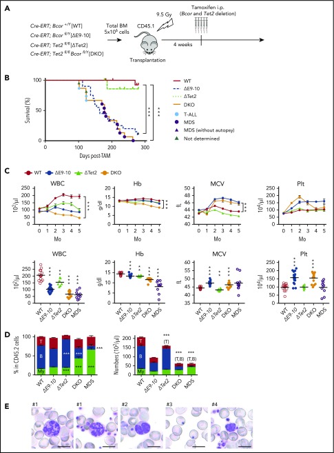
Concurrent deletion of Bcor and Tet2 causes MDS-like BM failure. (A) Strategy for analyzing hematopoietic cells with conditional knockout alleles for Tet2 and Bcor exons 9 and 10. Total BM cells from Cre-ERT control (WT), Cre-ERT;BcorflE9-10/y, Cre-ERT;Tet2fl/fl, and Cre-ERT;Tet2fl/flBcorflE9-10/y CD45.2 male mice were transplanted into lethally irradiated CD45.1 recipient mice. After engraftment, Tet2 and Bcor exons 9 and 10 were deleted by intraperitoneal injections of tamoxifen at 4 weeks posttransplantation. (B) Kaplan-Meier survival curves of WT (n = 15), ΔE9-10 (n = 20), ΔTet2 (n = 10), and DKO (n = 15) mice after the injection of tamoxifen; data from 2 independent experiments were combined. ***P < .001 by the log-rank test. The causes of death in DKO mice are indicated by circles or triangles with different colors. (C) PB cell counts in WT, ΔE9-10, ΔTet2, and DKO mice. WBC, hemoglobin (Hb), mean corpuscular volume (MCV), and platelet (Plt) counts in PB from WT (n = 15), ΔE9-10 (n = 19), ΔTet2 (n = 8), and DKO (n = 14) mice up to 5 months after the injection of tamoxifen are shown (upper panels). Data of moribund DKO MDS mice (n = 10) are shown with those of WT (n = 15), ΔE9-10 (n = 19), ΔTet2 (n = 8), and DKO (n = 14) mice 3 months after the injection of tamoxifen in the lower panels. (D) The proportions of myeloid (My) (Mac-1+ and/or Gr-1+), B220+ B cells, and CD4+ or CD8+ T cells among CD45.2+ donor-derived hematopoietic cells and their absolute numbers in PB from WT (n = 15), ΔE9-10 (n = 19), ΔTet2 (n = 8), and DKO (n = 14) mice 3 months after the injection of tamoxifen and moribund DKO MDS mice (n = 10). (E) Smear preparation of PB from moribund DKO MDS mice 6 months after the deletion of Bcor after May-Giemsa staining. Hypersegmented neutrophils (#1), hyposegmented neutrophils consistent with a pseudo Pelger-Huët anomaly (#2), Howell-Jolly bodies (#3), and giant platelets (#4) are shown. Bars represent 10 μm. Data are shown as the mean ± SEM in panels C and D. Statistical significance is shown relative to WT. *P < .05; **P < .01; ***P < .001 by the Student t test.

