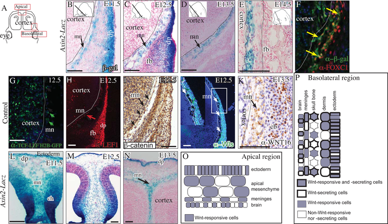
Fig. 1. Meningeal mesenchyme transduces Wnt signaling preceding differentiation of meningeal layers.
Schematic illustration of coronal embryonic mouse head sections at the eye level along the dorsoventral axis in the forebrain (A). β-galactosidase staining and counterstain with eosin Y showing canonical Wnt signaling in meningeal mesenchyme progenitors (mn) between E11.5–14.5 (basolateral, B-E; apical L-N). Indirect immunofluorescence with DAPI counterstain (F, G, H, J) and immunohistochemistry with hematoxylin counterstain (I, K) showing canonical Wnt signaling (G, H, I) and Wnt ligand secretion capacity (J, K) in the meningeal mesenchyme (F) and neighboring cranial lineages. Summary schematic illustration of the cranial mesenchyme lineages and meninges in the apical region (O) and basolateral (P) regions at E12.5. Other abbreviations: dermal progenitors (dp) mesenchyme, and frontal bone (fb) primordia, cartilage base (cb), cortical hem (ch). All arrows point to the meningeal mesenchyme. Dotted line demarcates the frontal cortex of the brain from the meningeal mesenchyme. Scale bars represent 100μm.

