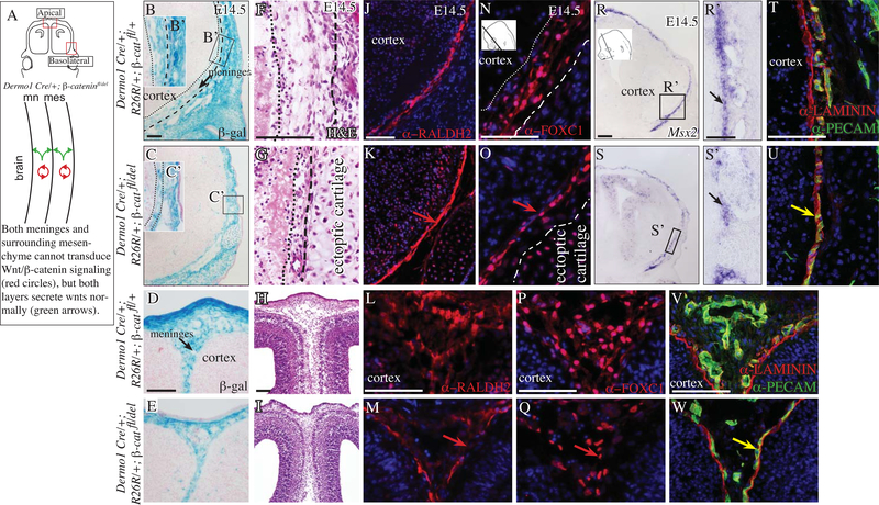
Fig 4. Conditional deletion of cranial and meningeal mesenchyme β-catenin results in thinner meninges.
Diagram of coronal embryonic mouse head sections at the eye level in the forebrain showing the regions of interest and simplified schematic of genetic model (A) Coronal mouse embryonic sections at eye level (B-W) in the basolateral region (top two rows) and apical region (bottom two rows). β-galactosidase expression counterstained with eosin Y (B-E), hematoxylin and eosin stain (F-I). Indirect immunofluorescence for meningeal markers (J-Q), basement membrane (BM) LAMININ (red, T-W), CD31/PECAM+ endothelial cells (green, T-W). Red arrow points to compaction of meninges marker expressing cells (K, O) and diminished domain of expression in the apical site in the mutant (M, Q). Yellow arrow points to overlap of BM LAMININ and PECAM layers in the mutant (red and green, U, W). Scale bars represent 100μm.

