



Abstract
Background:
It is now widely recognized that endogenous, picomolar concentrations of the 42 amino acid long peptide, amyloid-β (Aβ42) is secreted under normal physiological conditions and exerts important functional activity throughout neuronal intracellular compartments. Transgenic animal models that overexpress Aβ42 and its precursor, amyloid precursor protein (APP), have not provided predictive value in testing new treatments for Alzheimer’s disease (AD), resulting in failed clinical trials. While these results are discouraging, they underscore the need to understand the physiological roles of Aβ42 and APP under normal conditions as well as at early pre- symptomatic stages of AD.
New Method:
We describe the use of acrolein-perfusion in immunoelectron microscopy in combination with novel antibodies directed against endogenous murine Aβ42 and APP fragments to study abnormalities in the endolysosomal system at early stages of disease. The specific requirements, limitations and advantages of novel antibodies directed against human and murine Aβ42, APP and APP fragments are discussed as well as parameters for ultrastructural analysis of endolysosomal compartments.
Results:
Novel antibodies and a detailed protocol for immunoelectron microscopy using acrolein as a fixative are described. Acrolein is shown to preserve intraneuronal Aβ42 species, as opposed to paraformaldehyde fixed tissue, which primarily preserves membrane bound species.
Comparison with Existing Method(s):
Technology sensitive enough to detect endogenous Aβ42 under physiological conditions has not been widely available. We describe a number of novel and highly sensitive antibodies have recently been developed that may facilitate the analysis of endogenous Aβ42.
Conclusions:
Using novel and highly specific antibodies in combination with electron microscopy may reveal important information about the timing of aberrant protein accumulation, as well as the progression of abnormalities in the endolysosomal systems that sort and clear these peptides.
Introduction
In recent years, the amyloid hypothesis of Alzheimer’s disease (AD) has been heavily scrutinized as clinical trials of drugs successful in ameliorating amyloid load in mouse models of AD have failed to show efficacy in humans, and in some cases have had serious adverse side effects (Hardy 2009, Coric, Salloway et al. 2015). The negative outcomes of clinical trials has lead some leaders in the field to re-examine the Amyloid Cascade Hypothesis (Hardy 2009, Selkoe and Hardy 2016), while others believe the problem is a fundamental misunderstanding of the Aβ peptide and the importance of its physiological function (Puzzo, Gulisano et al. 2015). The latter is rooted in the idea that the complexity of AD, combined with preconceptions of amyloid-β (Aβ42) as an exclusively harmful protein have prevented investigators in the field from focusing on important facets of the disease state (Puzzo, Gulisano et al. 2015).
The ability to detect endogenous Aβ42 peptides in naïve rats and wildtype mice is a recent advancement enabled by the development of highly specific antibodies that detect the peptide at endogenous picomolar concentrations. Some conventional methods to study AD pathology include genetic models that overexpress human APP and/or presenilin genes, ultimately resulting in abundant production of the Aβ42 peptide that leads to rapid accumulation of the protein in the rodent brain. Other investigations have utilized the administration of exogenous Aβ42 peptides in high micromolar concentrations and in various conformational states, to recapitulate certain aspects of AD pathology. These methods have been critical in elucidating mechanisms of late disease stages, however, do not yield information regarding early, critical intervention stages of AD. Here, we advocate for the study of endogenous Aβ42 peptides, a novel route of investigation that may advance our knowledge regarding the transition from physiological to pathological states. This transition is thought to occur over a prolonged incubation period, termed the prodromal period, during which patients exhibit numerous co-morbid conditions including metabolic disorders such as diabetes, cardiovascular and psychiatric diseases. These disparate co-morbid diseases have an onset of symptoms before the onset of cognitive symptoms of dementia, and during the critical prodromal stage at which neuronal damage leading to AD is thought to occur.
The study of endogenous Aβ42 peptides in models of disease states that are co-morbid with Alzheimer’s disease may advance our understanding of pathological states that render individuals more susceptible to dementia with age. For example, our laboratory focuses on the locus coeruleus (LC) stress integrative system and has recently localized endogenous Aβ42 peptides in the noradrenergic (NE) cell bodies of the LC and axon terminals of the medial prefrontal cortex (mPFC) (see figures 2,3 and 5). The presence of endogenous Aβ42 within the LC-NE circuit may therefore have serious implications for conditions of chronic stress in which we hypothesize that aberrant neuronal activity of the LC may result in increased accumulation of the Aβ42 peptide.
Figure 2. Distinguished labeling patterns of 22C11, APP-β, and MOAB-2 antibodies in the naïve rat.
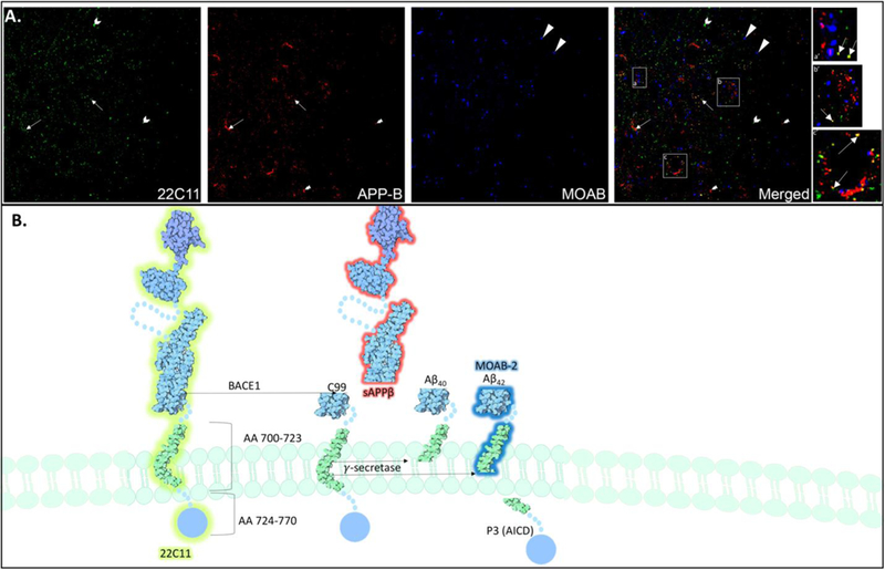
The epitopes labeled by three different antibodies within the APP protein readily distinguish three distinct fragments with unique distributions. The 22C11 (green) antibody labels an epitope that reveals full length APP, while the APP-β (red) antibody labels sAPP-β, the fragment resulting from BACE-1 cleavage of full-length APP, and MOAB-2 (blue) an antibody that labels the intracellular fragment that results from γ-secretase cleavage of the sAPP-β fragment. can be readily distinguished using low-resolution immunofluorescence techniques. Individually labeled puncta in close proximity (panel a,a’) may represent full length, membrane-bound APP and highlights the distinct region of the APP peptide in which the antibody-specific epitope is present; for example, 22C11 labeling (green) is the N-terminal extracellular region of APP, while MOAB-2 (blue) embedded within the membrane, and APP-β (red) that labels the C-terminus is facing the cytoplasmic side of the plasma membrane. There are several occurrences of co-localization, which primarily occur between the 22C11 and APP-β antibodies (yellow puncta), that indicate β-CTF labeling. Importantly, there are few occasions in which MOAB-2 labeling is co-localized with APP-β (magenta), which may be readily distinguished from γ-secretase cleavage products identified by MOAB-2 labeling alone. There were no occurrences of MOAB-2 immunoreactivity with 22C11 labeling. MOAB-2 is also more frequently visualized as individual puncta, and further away from 22C11 and APP-β immunoreactivity, alluding to the putative intracellular localization of Aβ42 peptides. However, it should be noted that electron microscopy is necessary to define the subcellular localization of these fragments with any certainty.
Figure 3: APP Processing and the Endolysosomal System.

The fate of APP is dictated largely by its subcellular localization, thus highlighting the importance of trafficking in parallel with proteolytic cleavage. Transmembrane proteins such as APP that are targeted for degradation enter the endosomal-lysosomal pathway by undergoing endocytosis, autophagy or phagocytosis. APP is internalized from the plasma membrane via endocytosis and further processed in endocytic, recycling and lysosomal compartments. In addition, once in the endosome, APP may be transported back to the TGN (G, blue) via retromer proteins (Vieira, Rebelo et al. 2010), following recognition by the sortilin related receptor (SORLA). Thus, transference of various forms of APP and its fragments occurs via the highly dynamic membrane enclosed vesicular structures that are compositionally and functionally distinct. These structures have been well characterized and include the early endosome, recycling endosome, late endosome (End, green) and lysosome (Lys, red) (Huotari and Helenius 2011). Arrow heads point to immunogold labeled Aβ42.
Figure 5. Aβ42 subcellular localization.

A. Immunoelectron micrographs of TH-immunoreactive dendrites (TH-d), one of which is dually labeled with immunogold Aβ42 (arrow heads). More specifically, Aβ42 is localized to a lysosomal (Lys) compartment within the dendrite, identified at the ultrastructural level. B. Example of an autolysosome that contains heterogeneous mixture of electron dense materials, including immunogold labeled Aβ42. C. Immunogold labeled Aβ42 is localized to axon terminals (at) presynaptic to TH immunolabeled dendrite. Here, immunogold labeled Aβ42 is associated with mitochondrial membranes (m). D. Immunoelectron micrograph of immunogold labeled Aβ42 localized to an axon terminal filled with dense core vesicles (dcv), a subcellular compartment derived from multivesicular bodies that frequently contain neuropeptides co-packaged with fast acting neurotransmitters that may be released from asynaptic sites. E. TH-immunolabeled cell body that contains several lysosomes with immunogold labeled Aβ42; immunogold labeled Aβ42 is also present on the cell surface, potentially indicating secretion into the extracellular space.
The physiological and pathological roles of Amyloid Precursor Protein (APP) and the products of its proteolytic cleavage remain poorly understood, thus, the emphasis of this review will be on studying the basic cellular biology of APP and its fragments using novel antibodies directed against APP and the products of its proteolytic cleavage. The use of these antibodies in combination with electron microscopy may facilitate tracking the location and accumulation of APP fragments in endolysosomal compartments of the cell. Such trafficking studies are emerging as an important area of investigation, as early abnormalities in the endolysosomal system have been identified as a cellular vulnerability in AD and a precipitating factor in the accumulation of Aβ42 peptides in models of the disease (Nixon, Wegiel et al. 2005, Yu, Cuervo et al. 2005, Jiang, Mullaney et al. 2010).
We start by describing the structure and processing of APP and then move into describing specific antibodies that may be used to detect fragments along the two diverging processing routes. We then provide an immunoelectron microscopy protocol and details regarding the ultrastructural identification of endolysosomal components under the transmission electron microscope. Future studies in this area may reveal a wealth of information about the timing of aberrant accumulation of these APP-derived fragments, as well as the progression of abnormalities in the endosomal and lysosomal systems that sort and clear these peptides.
APP Structure and Processing
APP is a type I transmembrane protein synthesized in the rough endoplasmic reticulum (ER) and exists in 3 major isoforms ranging from 695 to 770 amino acids in length. While APP770 has been chosen as the canonical sequence, the APP695 isoform is the predominant form in neuronal tissue. The APP695 isoform does not contain amino acids 290–364, also known as the Kunitz protease inhibitor (KPI) domain, distinguishing it from the APP770 isoform. Isoforms APP751 and APP770 are widely expressed in non-neuronal tissues (D. Goodsell 2015). APP is composed of several domains that have been characterized using X-ray diffraction and nuclear magnetic resonance (NMR) spectroscopy (Rossjohn, Cappai et al. 1999, Wang and Ha 2004). Full length APP includes three domains that extend outside the cell, one transmembrane domain and one cytoplasmic intracellular domain. The N-terminal domain, copper-binding domain (Barnham, McKinstry et al. 2003), and cell adhesion domains comprise the extracellular portions of the protein and include amino acids 18–699 counting residues by APP770. The transmembrane domain is a helical structure that is composed of amino acids 700–723, and the cytoplasmic domain is composed of amino acids 724–770 counting residues by APP770. The various domains of APP and its complex structure enable a wide variety of functions including that of a cell surface receptor, processes involved in neurite growth, adhesion and axonogenesis, as well as cell mobility (Rossjohn, Cappai et al. 1999, Hoffmann, Twiesselmann et al. 2000, Barnham, McKinstry et al. 2003, Muresan and Ladescu Muresan 2015). APP has also been implicated in the regulation of mitochondrial function, transcription, copper homeostasis and oxidative stress by modulating various protein-protein interactions (Barnham, McKinstry et al. 2003, Turner, O’Connor et al. 2003, Muresan and Ladescu Muresan 2015).
APP undergoes dynamic trafficking and processing resulting in the production of distinct fragments that are localized to different intracellular compartments and are thought to serve diverse functions (Figures 1 and 2). APP undergoes a number of post translational modifications that direct its trafficking throughout the cell. For example, phosphorylated APP species preferentially accumulate in filopodia, while modification by the peptidyl-prolyl cis-isomerase, Pin 1, or cytoplasmic glycosylation by O-glycNAcase control the trafficking and targeting/anchoring to specific destinations of APP (Muresan and Ladescu Muresan 2015). The proteolytic processing of APP generally occurs via two divergent pathways that have discrete outcomes in terms of the fragments produced, and the functional impact on neurons. The non-amyloidogenic pathway (Figure 1) is the most common route of processing under normal physiological conditions. Cleavage of APP by α-secretases embedded in the plasma membrane results in the formation of sAPPα composed of amino acids 18–687 and a C83 carboxyl terminal fragment (CTF)composed of amino acids 688–770. APP cleavage via α-secretases precludes the formation of Aβ40 and Aβ42 peptides, the final products of the amyloidogenic pathway (LaFerla, Green et al. 2007). The amyloidogenic pathway (Figure 1) represents a smaller portion of APP processing, although in the brain this is a significant processing pathway (Choi, Berger et al. 2009). However, the products of amyloidogenic processing may have important physiological roles that are an important area of ongoing investigation. In this pathway, APP undergoes proteolytic cleavage by the aspartic protease β-secretase (BACE-1), which cleaves APP on the luminal side of the membrane, releasing a soluble APPβ fragment (sAPPβ) (Figures 1 and 2) composed of amino acids 18–671, and C99 CTF composed of amino acids 672–770 (Vassar, Bennett et al. 1999). BACE-1 cleavage results in the formation of a new N-terminus with the first aspartic amino acid 672 of Aβ (LaFerla, Green et al. 2007). Subsequent cleavage of this fragment, typically between 38–43 amino acids downstream from the BACE-1 cleavage site by the γ-secretase results in the release of the Aβ peptides (Figures 1 and 2).
Figure 1: APP Fragments and Specific Antibody Recognition Sites.

The proteolytic processing of APP generally occurs via two divergent pathways. The non-amyloidogenic pathway is the most common route of processing in most cells, and results from cleavage of APP by α-secretases embedded in the plasma membrane. This cleavage results in the formation of sAPPα composed of amino acids 18–687 and a carboxyl terminal C83 fragment (α-CTF) composed of amino acids 688–770. The amyloidogenic pathway accounts for a smaller portion of APP processing. In this pathway, APP undergoes proteolytic cleavage by the aspartic protease β-secretase (BACE-1), which cuts APP on the luminal side of the membrane, releasing a soluble APPβ fragment (sAPPβ) composed of amino acids 18–671, and carboxyl terminal C99 fragment (β-CTF) composed of amino acids 672–770 (Vassar, Bennett et al. 1999). BACE-1 cleavage results in the formation of a new N-terminus with the first aspartic amino acid 672 of Aβ (LaFerla, Green et al. 2007), which is a neo-epitope detected by some antibodies. The C1/61 antibody binds the C-terminus of APP, thus detecting all full-length APP and related CTFs. Following BACE-1 cleavage, antibody JRF/N25 detects the C99 β-CTF. Subsequent cleavage of this β-CTF, at 38–43 amino acids downstream of this β-cleavage site by the γ-secretase, results in the release of the Aβ40, Aβ42, and to a lesser extent other Aβ peptides. To readily distinguish fragments Aβ40 and Aβ42, the N-terminal specific antibodies JRF/cAβ40/10 and JRF/cAβ 42/26 may be used, respectively. The structure of the APP molecule is attributed to David S. Goodsell and the RCSB Protein Data Bank (PDB), and was modified to depict fragments following proteolytic cleavage (D. Goodsell 2015).
Importantly, the fate of APP is dictated largely by its subcellular localization, thus highlighting the importance of trafficking in parallel with proteolytic cleavage (Figure 3). APP traffics through the plasma membrane alongside α-secretases, facilitating non-amyloidogenic processing. However, APP may also be internalized via endocytosis where it encounters the β- and γ-secretases that reside in acidic intracellular compartments particularly within the endolysosomal system, but may also include the trans Golgi network (TGN), the ER, and mitochondrial membranes (Mizuguchi, Ikeda et al. 1992, Xu, Greengard et al. 1995, Kinoshita, Shah et al. 2003), all of which may be sites for intracellular Aβ production (LaFerla, Green et al. 2007, Haass, Kaether et al. 2012). Typically, the internalization of APP favors amyloidogenic processing and the formation of Aβ, whereas its presence on the cell surface confers increased likelihood that it will encounter the α-secretase known to produce soluble APPα, and non-amyloidogenic C83 fragments (Hong, Huang et al. 2014). Once internalized, APP undergoes amyloidogenic processing in the early endosome followed by recycling in which Aβ peptides are released into the extracellular space, or alternatively, through subsequent endosomal-lysosomal compartments in which it may ultimately be degraded in the lysosome.
It has been observed that synaptic Aβ production may be significantly decreased by inhibiting clathrin-mediated endocytosis (Cirrito, Kang et al. 2008). Following neuronal depolarization and neurotransmitter release, synaptic vesicles must be formed and recycled in order to maintain an adequate number of vesicles for transmitter release during neuronal activation. Thus it is hypothesized that synaptic vesicle recycling during neuronal activation concurrently results in increased internalization of APP embedded in the plasma membrane, and therefore increases amyloidogenic processing by increasing the frequency of encountering β- and γ-secretases that reside in intracellular compartments.
The Endosomal-Lysosomal Pathway (Figure 3)
The endosomal-lysosomal pathway has become an active area of investigation in the context of neurodegenerative disorders characterized by an aberrant accumulation of proteins. Macromolecules and transmembrane proteins such as APP that are targeted for degradation may enter the endosomal-lysosomal pathway by undergoing endocytosis, autophagy or phagocytosis. As previously discussed, APP is commonly internalized from the plasma membrane via endocytosis and processed in the secretory and lysosomal pathways. APP synthesized in the ER and transported to the TGN can also traffic directly to the endosome by binding the adapter AP-4 (Burgos, Mardones et al. 2010). In addition, once in the endosome, APP may be transported back to the TGN via retromer proteins (Vieira, Rebelo et al. 2010), notably for APP by interacting with the sortilin related receptor (SORLA). Thus, transference of various forms of APP and its fragments occurs via the highly dynamic membrane enclosed vesicular structures that are compositionally and functionally distinct. These structures have been well characterized and include the early endosome, recycling endosome, late endosome and lysosome (Huotari and Helenius 2011).
The maturation model is based on the concept that endosomes go through defined stages as they mature, and that the compositional changes that occur during this process result in functionally distinct roles for endosomes in each stage of maturation. The early endosome is associated with the small GTPase rab5 and has a slightly acidic intraluminal pH, allowing for receptor cargo to readily dissociate and be sorted. This allows receptors to recycle back to the cell surface while ligands proceed to the late endosome and lysosome for degradation. As the early endosome accumulates an increasing number of intraluminal vesicles (ILVs) and increases intraluminal acidity, it also moves toward the microtubule organizing center and associates with different rab proteins. These alterations indicate a transition from the early endosome stage to the late endosome stage. The function of the late endosome is to generate ILVs and to serve as a sorting compartment for macromolecules destined for lysosomal degradation. Critical to this process, are the endosomal sorting complexes required for transport (ESCRT) machinery. ESCRT is comprised of complexes 0-III that bind ubiquinated cargo and generate intraluminal vesicles that are delivered to lysosomes and degraded (Edgar, Willen et al. 2015). Late endosomes are associated with the small GTPase rab 7, and are the stage in which multivesicular bodies (MVB) may form (Hu, Dammer et al. 2015). MVBs may fuse with the cell surface for the release of ILVs as exosomes, fuse with autophagosomes (Figure 5B) to generate amphisomes, or continue down the endosomal-lysosomal pathway. Facilitated by ESCRT machinery, MVBs or late endosomes may fuse with lysosomes, resulting in a highly acidic intraluminal environment in which hydrolases degrade cargo contents. Thus, the endolysosomal pathway is a complex and branching pathway with multiple points of entry and exit. Understanding the transport of APP through this complex system under normal physiological conditions and conditions that drive amyloidogenic processing in the rodent and the human is a crucial knowledge gap that must be investigated to leverage the intrinsic ability of neurons to remove and degrade Aβ42 and β-CTFs, and perhaps develop disease modifying therapeutics at increasing the efficacy of these systems in doing so.
Investigation of ESCRT machinery in the context of APP processing, and Aβ42 production have suggested that APP is trafficked in a subpopulation of MVBs that are destined to the lysosome for degradation, and further, suggests a role for early ESCRT components in limiting Aβ42 accumulation by promoting lysosomal targeting (Edgar, Willen et al. 2015). Closely paralleled with these observations, other investigators have found that autophagosomes are key organelles that connect with the MVB/lysosomal pathway for efficient turnover of CTFs. These investigators demonstrated that the fusion of autophagosomes with endolysosomal compartments cause an increase in C99 levels that accumulated primarily in structures resembling ILVs of MVBs, while activation of autophagosome formation enhanced lysosomal clearance of C99 CTFs. Thus supporting a model in which autophagosomes cooperate in the turnover of C99 and APP, forming an amphisome-like compartment by fusing with MVBs before fusion with lysosomes. Another pathway for removal of CTFs from the MVB is within exosomes secreted from the cell, which are enriched in β-CTFs in particular (Levy 2017). Further, conditions that compromise the fusion of autophagosomes to endosomal compartments accumulate C99 CTFs(Gonzalez, Munoz et al. 2017). Interestingly, another recent study identified Tetraspannin 6 (TSPAN6), a protein enriched in MVBs and ILVs, as a pivotal point in the pathway of ILV towards exosomes or lysosomal degradation. Heightened expression of TSPAN6 resulted in increased endosomal size, increased number of ILVs and increased secretion of exosomes containing APP CTFs (Guix, Sannerud et al. 2017). Electron microscopy and ultrastructural analysis indicated a higher number of endosomal compartments with non-degraded material in the lumen as well as a larger number of ILVs (Guix, Sannerud et al. 2017).
Protocol
Electron Microscopy (Figure 4)
Figure 4: Electron microscopy.

Following immunohistochemical procedures, tissues are prepared for visualization under the electron microscope with osmification, serial dehydration, flat-embedding, and tissue sectioning at 74 nm on an ultramicrotome (Commons, Beck et al. 2001). Sections are collected on copper mesh grids and examined using an electron microscope (Morgani, Fei Company, Hillsboro, OR). Digital images are viewed and captured using the AMT advantage HR HR-B CCD camera system (Advance Microscopy Techniques, Danvers, MA). Electron micrograph images are then prepared using Adobe Photoshop to adjust the brightness and contrast.
Selection of Antibodies and Experimental Design
Schmidt et al. (Schmidt, Jiang et al. 2005, Schmidt, Mazzella et al. 2012) have previously described tissue homogenate treatments to extract membrane-bound and soluble fragments of APP prior to Western Blot (WB), immunoprecipitation (IP) and enzyme-linked immunosorbent assay (ELISA), techniques that have been applied to mouse brain to characterized each APP fragment derived from the endogenously expressed APP (Morales-Corraliza, Mazzella et al. 2009). Here, we describe the antibodies routinely used in our laboratories that may be used on total homogenate or separate soluble and membrane fractions to reveal various fragments of APP. We also describe antibodies that may be used on tissue sections for immunohistochemistry. The C1/6.1 antibody binds the C-terminus of APP, thus detects all full-length APP and related CTFs. Following BACE-1 cleavage, antibody JRF/N25 can detect the C99 CTF in models expressing human APP, as well as subsequent fragments Aβ40 and Aβ42. Antibody m3.2 has similar reactivity against murine APP derived fragments. Subsequent cleavage of the C99 CTF, between 38–41 amino acids by the γ-secretase results in the release of the Aβ40 and Aβ42 peptides, respectively. To readily distinguish fragments Aβ40 and Aβ42, the N-terminal specific antibodies JRF/cAβ40/10 and JRF/cAβ 42/26 may be used, respectively [Figure 1, (Morales-Corraliza, Mazzella et al. 2009, Schmidt, Mazzella et al. 2012, D. Goodsell 2015)].
Tissue Preparation
Male and female rodents used for immunohistochemistry (IHC) were deeply anesthetized using isoflurane (Vedco, St. Joseph, MO) and subsequently transcardially perfused through the ascending aorta with the following fixatives: 10 ml of 1000 units/ml heparinized saline and 50 ml of 3.75% acrolein in 2% formaldehyde pH 7.4. Acrolein fixation yields optimal tissue preservation for ultrastructural analysis, and has been shown to preserve Aβ42 peptides within the intracellular compartments of the cell, in contrast to paraformaldehyde-only perfused tissues (Takahashi, Milner et al. 2002). The brains were removed, cut into 4–5 mm coronal blocks, stored in 2% paraformaldehyde fixative for 30 minutes and then sectioned (30–40 um) on a vibrating microtome (Vibratome; Pelco EasiSlicer, Ted Pella, Redding, CA). Brains were cut in 40 μm coronal sections, and sections from regions of interest (LC) were selected for IHC processing using the rat brain atlas of Paxinos and Watson (Paxinos, 1986).
Immunohistochemistry
Acrolein-perfused tissues were incubated in 1% sodium borohydride in 0.1 M PB to remove reactive aldehydes, and then blocked in 0.5% BSA in 0.1 M Tris-buffered saline (TBS). Tissue sections were incubated for 48 hours at 4°C in primary antibody in 0.1% BSA in 0.1 M TBS.
Secondary antibodies used for electron microscopy include biotinylated (1:400) and 1 nm gold particle conjugated IgG (1:50) (Electron Microscopy Science, Hatfield, PA). Immunoperoxidase labeling may be performed in addition to immunogold labeling for dual electron microscopy. The examples provided in figures 2 and 4 show immunoperoxidase labeling of tyrosine hydroxylase (TH) to identify cell bodies of the locus coeruleus, though investigators could use a variety of other antigens to identify other cell populations, or intracellular markers of interest. Electron-dense labeling is detected via silver intensification of immunogold particles using a silver enhancement kit (Aurion R-GENT SE-EM kit, Electron Microscopy Science). The examples provided in figures 2 and 4 show immunogold conjugated secondary antibodies that are bound to MOAB-2 primary antibody. Tissues were prepared for visualization under the electron microscope with osmification, serial dehydration, flat-embedding, and tissue sectioning at 74 nm on an ultramicrotome (Commons, Beck et al. 2001). Sections were collected on copper mesh grids and examined using an electron microscope (Morgani, Fei Company, Hillsboro, OR). Digital images were viewed and captured using the AMT advantage HR HR-B CCD camera system (Advance Microscopy Techniques, Danvers, MA). Electron micrograph images were then prepared using Adobe Photoshop to adjust the brightness and contrast.
Ultrastructural Analysis
Controls and Criteria
Adequate preservation of ultrastructural morphology is one of the criteria imposed when selecting tissue sections to be used for ultrastructural analysis. A minimum of 3 sections per region of each animal were used for analysis. At least 10 grids containing 4–7 thin sections each were collected from plastic-embedded sections of the regions of interest from each animal. Quantitative evaluation of immunoreactive elements was applied only to the outer 1–3 μm of the epon–tissue interface where penetration of antibodies is optimal. To prevent the inclusion of spurious labeling in quantification, only profiles with a minimum of 2 gold particles were considered immunoreactive and used for quantification. For dual labeling, only micrographs containing both peroxidase and gold–silver markers were used for the tissue analysis to ensure that the absence of one marker did not result from uneven penetration of markers (Leranth and Pickel, 1989).
For each animal, comparable levels of the region of interest were selected for ultra-thin sectioning. Dendritic and axon-terminal profiles were sampled from at least 5 copper grids of ultrathin tissue sections near the tissue-plastic interface. All profiles were scanned and selected for analysis based on the following criteria. Dendrites with a maximal cross-sectional diameter between 0.7 μm and 5 μm, and a mix of dendrites of sizes from across this range were included in the analysis. Large profiles were excluded to avoid the bias towards positive labeling of larger structures. Extremely small, large, longitudinal and irregularly shaped profiles were excluded from the analysis due to possibly higher perimeter/surface ratios and risk of biasing the silver grain counts towards the membrane. Any profiles containing large, irregularly shaped silver grains of more than 0.25 μm were excluded from the analysis. Cellular profiles that fail to meet any of the described criteria were excluded from analysis.
Trafficking and Identification of Organelles
Cellular elements were isolated and classified based on Fine Structure of the Nervous System (Peters 1991). Somata were identified by the presence of a nucleus, Golgi apparatus, and smooth endoplasmic reticulum. Proximal dendrites contain endoplasmic reticulum, were typically apposed to axon terminals, and were larger than 0.7 μm in diameter. Synapses were verified by the presence of a junctional complex, a restricted zone of parallel membranes with slight enlargement of the intercellular space, and/or associated postsynaptic thickening. A synaptic specialization was only designated to the profiles that form clear morphological characteristics of either Type I or Type II (Gray 1959). Asymmetric synapses were identified by thick postsynaptic densities (Gray’s Type I; Gray 1959), while symmetric synapses had thin densities both pre- and post-synaptically (Gray’s Type II; Gray 1959). An undefined synapse was defined as an axon terminal plasma membrane juxtaposed to a dendrite or soma devoid of recognizable membrane specializations and no intervening glial processes. Axon terminals were distinguished from unmyelinated axons based on synaptic vesicle presence and a diameter of greater than 0.1 μm.
The recognition of endolysosomal compartments at various stages of processing were recognized morphologically using electron microscopy (Figures 2, 5). Early endosomal compartments may be identified by the presence of a central vacuole of ∼100–500 nm diameter, that is often referred to as the sorting endosome. This structure is primarily electron lucent, or clear, and is frequently visualized with a cytoplasmic clathrin coat. Extending from the main sorting endosome, are smaller tubules structures that are part of the recycling endosomal system (Klumperman and Raposo 2014) (Figure 3, green). Occasionally, endosomes in this state contain a few ILVs, which range in size from 40 to 100 nm (Murk, Humbel et al. 2003, Klumperman and Raposo 2014). Late endosomal compartments are comprised of a vacuole 250–1000 nm in diameter, and budding compartments that connect to the TGN (Figure 3, blue). Various EM studies have used the increasing number of ILVs within these compartments to pinpoint the switch from the early endosome to the late endosome that is thought to occur in the range of five to eight ILVs (Mobius, van Donselaar et al. 2003, Murk, Humbel et al. 2003, Mari, Bujny et al. 2008). Thus, late endosomes are more readily distinguishable when there are greater numbers of ILVs, at which point they are frequently referred to as MVBs (Klumperman and Raposo 2014) or dense core vesicles (dcv) (see Figure 5D).
Lysosomes (Figure 5 A, E) are the terminal degradative compartment receiving cargo from the endosomal and autophagy pathways. They are spherical organelles with diameters between 200 nm and >1 μm, and typically have an electron dense lumen, reflecting high protein concentrations (Bainton 1981, Klumperman and Raposo 2014). When lysosomes are supplied with cargo from the autophagy pathway, autolysosomes are formed (Figure 5B). Autolysosomes are generally known to be larger and more irregularly shaped than lysosomes, with a highly variable content. Autophagosomes are known to have a double limiting membrane and the cytoplasm inside has the same appearance as the cytoplasm outside the autophagosome. As noted in a cautionary piece of literature on the morphological analysis of autophagy-related structures, this is frequently a source of inaccuracy, as empty vacuoles and sometimes enlarged mitochondria may be identified as endolysosomal and autophagy compartments based on the presence of their limiting membrane alone (Eskelinen 2008). For an extensive ultrastructural analysis of how these components are altered under conditions of degeneration, and how disruption of the endolysosomal system can influence the efficiency of autophagy we refer the reader to (Nixon, Wegiel et al. 2005).
Quantification
Quantification and analysis may be conducted as previously reported (Commons, Beck et al. 2001, Oropeza, Mackie et al. 2007). To further define the subcellular distribution of the APP fragments of interest and their trafficking under various treatment conditions, distribution ratios may be calculated for each subcellular compartment of interest for each animal in naïve and experimental treatment conditions, as we have previously reported (Oropeza, Mackie et al. 2007). To calculate the distribution ratio, investigators counted the number of immunogold particles within each subcellular compartment of interest. A ratio of immunogold particles identified within the compartment of interest to total immunogold particles per profile was computed. An average of ratios for each animal was taken. A one-way analysis of variance (ANOVA) was used to determine if there were within-group differences in the distribution ratio among animals receiving identical experimental conditions. If no differences were detected between animals within the same experimental group, data from these animals were pooled and an average distribution ratio from the animals under each experimental condition was calculated. ANOVA was used for within group comparisons to determine the presence of statistically significant shifts in the distribution of immunoreactivity. Tukey’s post-hoc tests were used for between group comparisons.
Discussion and Recommendations
Previous studies have utilized electron microscopy to identify the subcellular location of Aβ42 peptides in AD transgenic mice as well as in human brains, and have demonstrated that accumulation, which increases with age, occurs in MVB prior to plaque formation. Additionally, the cell bodies in which these MVB were formed had abnormal morphology and dystrophic neurites (Takahashi, Milner et al. 2002). In another electron microscopy study, this group went on to describe a subtle change in the distribution of oligomerized Aβ in vitro and in vivo. Monomeric Aβ42 aggregated on the outer membranes of endosomal vesicles and MVBs, while Aβ42 oligomers were distributed to the inner membranes of morphologically abnormal endosomal organelles and to microtubules (Takahashi, Almeida et al. 2004).
Combining the use of antibodies specific to various fragments of APP and electron microscopy may yield important information about subtle changes in the distribution of various fragments throughout the cell, as well as indicate signs of neuronal injury or altered morphology of the organelles to and from which the fragments are being trafficked. This may be particularly important for studies aiming to assess early markers of disease such as early-endosome abnormalities, altered cholesterol metabolism or other signs of neuronal injury and dysregulation of lysosomes such as the presence of lipofuscin, which is identified morphologically at the ultrastructural level using electron microscopy. Of note, we describe here a protocol that utilizes a combination of paraformaldehyde and acrolein as tissue fixatives, which has been shown to preserve intraneuronal Aβ species, in contrast to tissues perfused with paraformaldehyde alone which can show APP and its cleavage products primarily on the plasma membrane.
We have previously used various antibodies to successfully localize endogenous murine Aβ by EM: MOAB2 specifically recognizes intraneuronal 42 amino acid long Aβ, while D54D2 recognizes endogenous Aβ42, Aβ40, Aβ39, Aβ38, and Aβ37 that may result from variable γ-secretase cleavage. Each antibody was determined to be specific using APP KO mice and rat liver slices, where APP expression and Aβ are low (Ross, Reyes et al. 2017). Inclusion of negative controls either lacking APP expression or, for instance β-cleaved APP products such as β-CTFs and Aβ in a BACE-1 KO model, are valuable tools when assessing the specificity of an antibody immuno-EM signal. Additionally, antibodies such as those described in Table 1, can be used to detect APP itself as well as other APP fragments. For example, C1/6.1 binding will illustrate the distribution of APP and the CTFs within a cell. The distribution of additional APP fragments can be inferred by the localization of two antibody-binding patterns overlaid in a single section. For example, luminal JRF/N25 labeling in an endosome may be either β-CTFs or Aβ based upon the fragment-specificity of this antibody. Close juxtaposition of this signal with cytoplasmic C1/6.1 is consistent with β-CTF, which contains the C-terminal APP domain recognized by C1/6.1 but missing in the Aβ peptides. While these types of analyses are demanding, and require differentially directly-conjugated primary antibodies when both antibodies are mouse monoclonal antibodies, these are the approaches that are necessary to precisely define the subcellular localization of APP fragments. As noted above for Aβ identification, the addition of KO tissue where a specific APP cleavage event does not occur can be valuable. In the above scenario using JRF/N25 and C1/6.1 to identify β-CTFs, concurrent immunolabeling of BACE-1 KO would show the distribution of APP and α-CTFs detected by C1/6.1 in cells lacking any specific JRF/N25 binding and lacking β-CTFs.
Table 1:
Antibodies for IHC, IP, WB
| Antibody | Source | Epitope | Fragments Detected | Applications | References |
|---|---|---|---|---|---|
| 22C11 | Millipore (MAB348) | Amino acids 66–81 of N-terminal APP | All three isoforms of APP: immature ~110kDa, sAPP ~120kDa, and mature ~130kDa | ELISA, IHC, IP and WB | (Hoffmann, Twiesselmann et al. 2000) |
| C1/6.1 | Laboratory of Dr. Paul Mathews New York University & Nathan Klein Institute | C-Terminus residues 676 – 695 of APP695 | APP holoprotein but not sAPP | IP, ICC, IHC, WB | (Mathews, Jiang et al. 2002) (Jiang, Mullaney et al. 2010) |
| Alternative Commercially available: APP-β | Thermo Fisher (51–2700) | 22 amino acid residues of C-terminus | sAPP-β | WB, IF,IHC,ELISA | |
| M3.2 | Laboratory of Dr. Paul Mathews New York University & Nathan Klein Institute | residues 1–15 of the Aβ peptide | APP holoprotein, sAPPα, β-CTF and Aβ | ELISA, IHC, IP, WB | (Choi, Berger et al. 2009) (Morales-Corraliza, Mazzella et al. 2009) |
| Alternative Commercially available: D54D2 | Cell Signaling | N-terminus of Aβ | Aβ42, Aβ40, Aβ39, Aβ38, and Aβ37. | IHC 1:100 | (Ross, Reyes et al. 2017) |
| JRF/cAβ 42/26 | Laboratory of Dr. Paul Mathews New York University & Nathan Klein Institute | recognizes the C-terminus of Aβ42; does not detect Aβ40 or full-length APP | residues 33–42 of the Aβ 1–42 peptide | Aβ 1– 42 | (Vandermeeren, Geraerts et al. 2001, Mathews, Jiang et al. 2002, Schmidt, Nixon et al. 2005) (Janus, Pearson et al. 2000) |
| Alternative Commercially available: MOAB2 | Kerafast | Amino acids 1–4 of Aβ42 | Aβ42 | IHC; 1:1000 | (Youmans, Tai et al. 2012, Ross, Reyes et al. 2017) |
ACKNOWLEDGMENTS:
R01 DA202129 and R01 DA009082 to EJV., R21 AG058263 to EJV., P01 AG017617 and RF1 AG057517 to PM.
References
- Bainton DF (1981). “The discovery of lysosomes.” J Cell Biol 91(3 Pt 2): 66s–76s. [DOI] [PMC free article] [PubMed] [Google Scholar]
- Barnham KJ, McKinstry WJ, Multhaup G, Galatis D, Morton CJ, Curtain CC, Williamson NA, White AR, Hinds MG, Norton RS, Beyreuther K, Masters CL, Parker MW and Cappai R (2003). “Structure of the Alzheimer’s disease amyloid precursor protein copper binding domain. A regulator of neuronal copper homeostasis.” J Biol Chem 278(19): 17401–17407. [DOI] [PubMed] [Google Scholar]
- Burgos PV, Mardones GA, Rojas AL, daSilva LL, Prabhu Y, Hurley JH and Bonifacino JS (2010). “Sorting of the Alzheimer’s disease amyloid precursor protein mediated by the AP-4 complex.” Dev Cell 18(3): 425–436. [DOI] [PMC free article] [PubMed] [Google Scholar]
- Choi JH, Berger JD, Mazzella MJ, Morales-Corraliza J, Cataldo AM, Nixon RA, Ginsberg SD, Levy E and Mathews PM (2009). “Age-dependent dysregulation of brain amyloid precursor protein in the Ts65Dn Down syndrome mouse model.” J Neurochem 110(6): 1818–1827. [DOI] [PMC free article] [PubMed] [Google Scholar]
- Cirrito JR, Kang JE, Lee J, Stewart FR, Verges DK, Silverio LM, Bu G, Mennerick S and Holtzman DM (2008). “Endocytosis is required for synaptic activity-dependent release of amyloid-beta in vivo.” Neuron 58(1): 42–51. [DOI] [PMC free article] [PubMed] [Google Scholar]
- Commons KG, Beck SG, Rudoy C and Van Bockstaele EJ (2001). “Anatomical evidence for presynaptic modulation by the delta opioid receptor in the ventrolateral periaqueductal gray of the rat.” J Comp Neurol 430(2): 200–208. [PubMed] [Google Scholar]
- Coric V, Salloway S, van Dyck CH, Dubois B, Andreasen N, Brody M, Curtis C, Soininen H, Thein S, Shiovitz T, Pilcher G, Ferris S, Colby S, Kerselaers W, Dockens R, Soares H, Kaplita S, Luo F, Pachai C, Bracoud L, Mintun M, Grill JD, Marek K, Seibyl J, Cedarbaum JM, Albright C, Feldman HH and Berman RM (2015). “Targeting Prodromal Alzheimer Disease With Avagacestat: A Randomized Clinical Trial.” JAMA Neurol 72(11): 1324–1333. [DOI] [PubMed] [Google Scholar]
- Goodsell D, S. D., Zardecki C, Voigt M, Berman H, Burley S (2015). The RCSB PDB “Molecule of the Month”: Inspiring a Molecular View of Biology. . PLoS Biol. [DOI] [PMC free article] [PubMed] [Google Scholar]
- Edgar JR, Willen K, Gouras GK and Futter CE (2015). “ESCRTs regulate amyloid precursor protein sorting in multivesicular bodies and intracellular amyloid-beta accumulation.” J Cell Sci 128(14): 2520–2528. [DOI] [PMC free article] [PubMed] [Google Scholar]
- Eskelinen EL (2008). “To be or not to be? Examples of incorrect identification of autophagic compartments in conventional transmission electron microscopy of mammalian cells.” Autophagy 4(2): 257–260. [DOI] [PubMed] [Google Scholar]
- Gonzalez AE, Munoz VC, Cavieres VA, Bustamante HA, Cornejo VH, Januario YC, Gonzalez I, Hetz C, daSilva LL, Rojas-Fernandez A, Hay RT, Mardones GA and Burgos PV (2017). “Autophagosomes cooperate in the degradation of intracellular C-terminal fragments of the amyloid precursor protein via the MVB/lysosomal pathway.” FASEB J 31(6): 2446–2459. [DOI] [PubMed] [Google Scholar]
- Gray EG (1959). “Electron microscopy of synaptic contacts on dendrite spines of the cerebral cortex.” Nature 183(4675): 1592–1593. [DOI] [PubMed] [Google Scholar]
- Guix FX, Sannerud R, Berditchevski F, Arranz AM, Horre K, Snellinx A, Thathiah A, Saido T, Saito T, Rajesh S, Overduin M, Kumar-Singh S, Radaelli E, Corthout N, Colombelli J, Tosi S, Munck S, Salas IH, Annaert W and De Strooper B (2017). “Tetraspanin 6: a pivotal protein of the multiple vesicular body determining exosome release and lysosomal degradation of amyloid precursor protein fragments.” Mol Neurodegener 12(1): 25. [DOI] [PMC free article] [PubMed] [Google Scholar]
- Haass C, Kaether C, Thinakaran G and Sisodia S (2012). “Trafficking and proteolytic processing of APP.” Cold Spring Harb Perspect Med 2(5): a006270. [DOI] [PMC free article] [PubMed] [Google Scholar]
- Hardy J (2009). “The amyloid hypothesis for Alzheimer’s disease: a critical reappraisal.” J Neurochem 110(4): 1129–1134. [DOI] [PubMed] [Google Scholar]
- Hoffmann J, Twiesselmann C, Kummer MP, Romagnoli P and Herzog V (2000). “A possible role for the Alzheimer amyloid precursor protein in the regulation of epidermal basal cell proliferation.” Eur J Cell Biol 79(12): 905–914. [DOI] [PubMed] [Google Scholar]
- Hong L, Huang HC and Jiang ZF (2014). “Relationship between amyloid-beta and the ubiquitin-proteasome system in Alzheimer’s disease.” Neurol Res 36(3): 276–282. [DOI] [PubMed] [Google Scholar]
- Hu YB, Dammer EB, Ren RJ and Wang G (2015). “The endosomal-lysosomal system: from acidification and cargo sorting to neurodegeneration.” Transl Neurodegener 4: 18. [DOI] [PMC free article] [PubMed] [Google Scholar]
- Huotari J and Helenius A (2011). “Endosome maturation.” EMBO J 30(17): 3481–3500. [DOI] [PMC free article] [PubMed] [Google Scholar]
- Janus C, Pearson J, McLaurin J, Mathews PM, Jiang Y, Schmidt SD, Chishti MA, Horne P, Heslin D, French J, Mount HT, Nixon RA, Mercken M, Bergeron C, Fraser PE, St George-Hyslop P and Westaway D (2000). “A beta peptide immunization reduces behavioural impairment and plaques in a model of Alzheimer’s disease.” Nature 408(6815): 979–982. [DOI] [PubMed] [Google Scholar]
- Jiang Y, Mullaney KA, Peterhoff CM, Che S, Schmidt SD, Boyer-Boiteau A, Ginsberg SD, Cataldo AM, Mathews PM and Nixon RA (2010). “Alzheimer’s-related endosome dysfunction in Down syndrome is Abeta-independent but requires APP and is reversed by BACE-1 inhibition.” Proc Natl Acad Sci U S A 107(4): 1630–1635. [DOI] [PMC free article] [PubMed] [Google Scholar]
- Kinoshita A, Shah T, Tangredi MM, Strickland DK and Hyman BT (2003). “The intracellular domain of the low density lipoprotein receptor-related protein modulates transactivation mediated by amyloid precursor protein and Fe65.” J Biol Chem 278(42): 41182–41188. [DOI] [PubMed] [Google Scholar]
- Klumperman J and Raposo G (2014). “The complex ultrastructure of the endolysosomal system.” Cold Spring Harb Perspect Biol 6(10): a016857. [DOI] [PMC free article] [PubMed] [Google Scholar]
- LaFerla FM, Green KN and Oddo S (2007). “Intracellular amyloid-beta in Alzheimer’s disease.” Nat Rev Neurosci 8(7): 499–509. [DOI] [PubMed] [Google Scholar]
- Levy E (2017). “Exosomes in the Diseased Brain: First Insights from In vivo Studies.” Front Neurosci 11: 142. [DOI] [PMC free article] [PubMed] [Google Scholar]
- Mari M, Bujny MV, Zeuschner D, Geerts WJ, Griffith J, Petersen CM, Cullen PJ, Klumperman J and Geuze HJ (2008). “SNX1 defines an early endosomal recycling exit for sortilin and mannose 6-phosphate receptors.” Traffic 9(3): 380–393. [DOI] [PubMed] [Google Scholar]
- Mathews PM, Jiang Y, Schmidt SD, Grbovic OM, Mercken M and Nixon RA (2002). “Calpain activity regulates the cell surface distribution of amyloid precursor protein. Inhibition of calpains enhances endosomal generation of beta-cleaved C-terminal APP fragments.” J Biol Chem 277(39): 36415–36424. [DOI] [PubMed] [Google Scholar]
- Mizuguchi M, Ikeda K and Kim SU (1992). “beta-Amyloid precursor protein of Alzheimer’s disease in cultured bovine oligodendrocytes.” J Neurosci Res 32(1): 34–42. [DOI] [PubMed] [Google Scholar]
- Mobius W, van Donselaar E, Ohno-Iwashita Y, Shimada Y, Heijnen HF, Slot JW and Geuze HJ (2003). “Recycling compartments and the internal vesicles of multivesicular bodies harbor most of the cholesterol found in the endocytic pathway.” Traffic 4(4): 222–231. [DOI] [PubMed] [Google Scholar]
- Morales-Corraliza J, Mazzella MJ, Berger JD, Diaz NS, Choi JH, Levy E, Matsuoka Y, Planel E and Mathews PM (2009). “In vivo turnover of tau and APP metabolites in the brains of wild-type and Tg2576 mice: greater stability of sAPP in the beta-amyloid depositing mice.” PLoS One 4(9): e7134. [DOI] [PMC free article] [PubMed] [Google Scholar]
- Muresan V and Ladescu Muresan Z (2015). “Amyloid-beta precursor protein: Multiple fragments, numerous transport routes and mechanisms.” Exp Cell Res 334(1): 45–53. [DOI] [PMC free article] [PubMed] [Google Scholar]
- Murk JL, Humbel BM, Ziese U, Griffith JM, Posthuma G, Slot JW, Koster AJ, Verkleij AJ, Geuze HJ and Kleijmeer MJ (2003). “Endosomal compartmentalization in three dimensions: implications for membrane fusion.” Proc Natl Acad Sci U S A 100(23): 13332–13337. [DOI] [PMC free article] [PubMed] [Google Scholar]
- Nixon RA, Wegiel J, Kumar A, Yu WH, Peterhoff C, Cataldo A and Cuervo AM (2005). “Extensive involvement of autophagy in Alzheimer disease: an immuno-electron microscopy study.” J Neuropathol Exp Neurol 64(2): 113–122. [DOI] [PubMed] [Google Scholar]
- Oropeza VC, Mackie K and Van Bockstaele EJ (2007). “Cannabinoid receptors are localized to noradrenergic axon terminals in the rat frontal cortex.” Brain Res 1127(1): 36–44. [DOI] [PMC free article] [PubMed] [Google Scholar]
- Peters A, Palay SL (1991). The fine structure of the nervous system: neurons and their supporting cells, Oxford University Press. [Google Scholar]
- Puzzo D, Gulisano W, Arancio O and Palmeri A (2015). “The keystone of Alzheimer pathogenesis might be sought in Abeta physiology.” Neuroscience 307: 26–36. [DOI] [PMC free article] [PubMed] [Google Scholar]
- Ross JA, Reyes BAS, Thomas SA and Van Bockstaele EJ (2017). “Localization of endogenous amyloid-beta to the coeruleo-cortical pathway: consequences of noradrenergic depletion.” Brain Struct Funct. [DOI] [PMC free article] [PubMed] [Google Scholar]
- Rossjohn J, Cappai R, Feil SC, Henry A, McKinstry WJ, Galatis D, Hesse L, Multhaup G, Beyreuther K, Masters CL and Parker MW (1999). “Crystal structure of the N-terminal, growth factor-like domain of Alzheimer amyloid precursor protein.” Nat Struct Biol 6(4): 327–331. [DOI] [PubMed] [Google Scholar]
- Schmidt SD, Jiang Y, Nixon RA and Mathews PM (2005). “Tissue processing prior to protein analysis and amyloid-beta quantitation.” Methods Mol Biol 299: 267–278. [DOI] [PubMed] [Google Scholar]
- Schmidt SD, Mazzella MJ, Nixon RA and Mathews PM (2012). “Abeta measurement by enzyme-linked immunosorbent assay.” Methods Mol Biol 849: 507–527. [DOI] [PubMed] [Google Scholar]
- Schmidt SD, Nixon RA and Mathews PM (2005). “ELISA method for measurement of amyloid-beta levels.” Methods Mol Biol 299: 279–297. [DOI] [PubMed] [Google Scholar]
- Selkoe DJ and Hardy J (2016). “The amyloid hypothesis of Alzheimer’s disease at 25 years.” EMBO Mol Med 8(6): 595–608. [DOI] [PMC free article] [PubMed] [Google Scholar]
- Takahashi RH, Almeida CG, Kearney PF, Yu F, Lin MT, Milner TA and Gouras GK (2004). “Oligomerization of Alzheimer’s beta-amyloid within processes and synapses of cultured neurons and brain.” J Neurosci 24(14): 3592–3599. [DOI] [PMC free article] [PubMed] [Google Scholar]
- Takahashi RH, Milner TA, Li F, Nam EE, Edgar MA, Yamaguchi H, Beal MF, Xu H, Greengard P and Gouras GK (2002). “Intraneuronal Alzheimer abeta42 accumulates in multivesicular bodies and is associated with synaptic pathology.” Am J Pathol 161(5): 1869–1879. [DOI] [PMC free article] [PubMed] [Google Scholar]
- Turner PR, O’Connor K, Tate WP and Abraham WC (2003). “Roles of amyloid precursor protein and its fragments in regulating neural activity, plasticity and memory.” Prog Neurobiol 70(1): 1–32. [DOI] [PubMed] [Google Scholar]
- Vandermeeren M, Geraerts M, Pype S, Dillen L, Van Hove C and Mercken M (2001). “The functional gamma-secretase inhibitor prevents production of amyloid beta 1–34 in human and murine cell lines.” Neurosci Lett 315(3): 145–148. [DOI] [PubMed] [Google Scholar]
- Vassar R, Bennett BD, Babu-Khan S, Kahn S, Mendiaz EA, Denis P, Teplow DB, Ross S, Amarante P, Loeloff R, Luo Y, Fisher S, Fuller J, Edenson S, Lile J, Jarosinski MA, Biere AL, Curran E, Burgess T, Louis JC, Collins F, Treanor J, Rogers G and Citron M (1999). “Beta-secretase cleavage of Alzheimer’s amyloid precursor protein by the transmembrane aspartic protease BACE.” Science 286(5440): 735–741. [DOI] [PubMed] [Google Scholar]
- Vieira SI, Rebelo S, Esselmann H, Wiltfang J, Lah J, Lane R, Small SA, Gandy S, da Cruz ESEF and da Cruz ESOA (2010). “Retrieval of the Alzheimer’s amyloid precursor protein from the endosome to the TGN is S655 phosphorylation state-dependent and retromer-mediated.” Mol Neurodegener 5: 40. [DOI] [PMC free article] [PubMed] [Google Scholar]
- Wang Y and Ha Y (2004). “The X-ray structure of an antiparallel dimer of the human amyloid precursor protein E2 domain.” Mol Cell 15(3): 343–353. [DOI] [PubMed] [Google Scholar]
- Xu H, Greengard P and Gandy S (1995). “Regulated formation of Golgi secretory vesicles containing Alzheimer beta-amyloid precursor protein.” J Biol Chem 270(40): 23243–23245. [DOI] [PubMed] [Google Scholar]
- Youmans KL, Tai LM, Kanekiyo T, Stine WB Jr., Michon SC, Nwabuisi-Heath E, Manelli AM, Fu Y, Riordan S, Eimer WA, Binder L, Bu G, Yu C, Hartley DM and LaDu MJ (2012). “Intraneuronal Abeta detection in 5×FAD mice by a new Abeta-specific antibody.” Mol Neurodegener 7: 8. [DOI] [PMC free article] [PubMed] [Google Scholar]
- Yu WH, Cuervo AM, Kumar A, Peterhoff CM, Schmidt SD, Lee JH, Mohan PS, Mercken M, Farmery MR, Tjernberg LO, Jiang Y, Duff K, Uchiyama Y, Naslund J, Mathews PM, Cataldo AM and Nixon RA (2005). “Macroautophagy--a novel Beta-amyloid peptide-generating pathway activated in Alzheimer’s disease.” J Cell Biol 171(1): 87–98. [DOI] [PMC free article] [PubMed] [Google Scholar]