Description
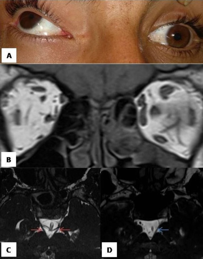
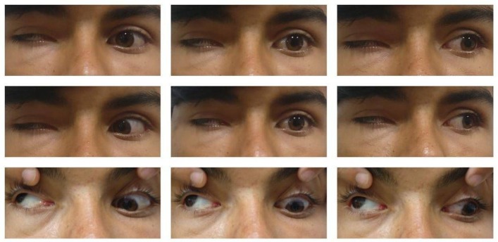
An 18-year-old Indian man was referred as Horner’s syndrome. He presented with severe ptosis in the right eye (RE) and limitation of movements in both eyes (BE) since birth. RE had low vision since early childhood. Family history was irrelevant. Best corrected visual acuity in RE was hand movement close to face and 20/20 in the left eye (LE). RE had severe ptosis with poor levator palpebrae superioris action; exotropia of >45◦ and hypertropia of 30◦, with total limitation of adduction and depression (figure 1). In LE, the patient had limitation in up gaze with poor bells phenomenon. No aberrant innervation was noted. The pupil in RE was severely miotic with no apparent reaction to light and accommodation, while it was normal in LE (figure 2). Anterior segment and fundus examination was within normal limits in BE. Systemic examination was also within normal limits. On MRI of brain and orbit, the right third cranial nerve could not be visualised along with significant atrophy of extraocular muscles (EOMs) supplied by it (figure 2). The patient was diagnosed to have congenital fibrosis of EOMs (CFEOM) in view of typical features of bilateral limitation of EOM movements with ptosis and severe atrophy of EOM supplied by the oculomotor nerve, as also confirmed on MRI. Poor vision in the RE was attributed to stimulus deprivation amblyopia caused by severe ptosis.
Figure 1.
Nine gaze photograph of the patient showing right eye complete ptosis with poor levator palpebrae superioris muscle action. Right eye is fixed in exotropia and supraduction, with limitation of all movements except abduction. Left eye shows limitation of elevation only.
Figure 2.

(A) Photograph showing miosis in right eye. (B–D) MRI of the patient. (B) Coronal T1 weighted section of orbit shows marked thinning of the extraocular muscles on the right side. (C) High resolution three-dimensional (3D) constructive interference in steady state (CISS) oblique axial section at the level of midbrain of a normal individual showing normal cisternal part of the third cranial nerves on both sides (red arrows). (D) High-resolution 3D CISS oblique axial section at the level of midbrain in our patient shows absence of right third cranial nerve while left side nerve appears thin (blue arrow).
CFEOM is a rare disorder that comes under the broad category of congenital cranial dysinnervation disorders. It is characterised by congenital non-progressive restrictive strabismus associated with ptosis. Due to extensive research using high-resolution MRI and genetic and experimental studies,1–3 it has been found that the primary defect is in innervations such as hypoplasia or absence of cranial nerve III which in turn leads to fibrosis, hypoplasia or aplasia of the EOM. Three phenotypes have been established based on the clinical features and the genes involved. CFEOM1 and CFEOM2 are fairly symmetric in BE.1 CFEOM3 can have variable ocular and neurological presentations. CFEOM3, usually the eye is fixed in infraduction or in primary gaze, while eyes are known to be fixed above the midline in CFEOM2. Pupil anomalies are much more common in CFEOM2, while they are very rare in CFEOM3.1 CFEOM2, in contrast to our Indian patient, has only been reported from Middle Eastern families with history of consanguineous marriages and involves a particular gene (PHOX2A).1 Our patient could not afford a genetic analysis, and seemed to have mixed features of CFEOM2 and CFEOM3.
Our patient was diagnosed with Horner’s syndrome initially by the primary physician due to the presence of ptosis and miosis. However, Horner’s syndrome does not include strabismus and typically the ptosis is mild as against findings of our case. The aetiology of pupil abnormalities in CFEOM is yet to be elucidated as patients do not appear to have structural atrophy of the iris. It has been postulated that the complete lack of pupillary reaction could imply a central pupillary abnormality of unknown aetiology or an atrophic superior cervical ganglion.2
Learning points.
Miosis in a case of congenital fibrosis of extraocular muscles (CFEOM) is a rare presentation, and may be a cause of misdiagnosis.
MRI can demonstrate hypoplasia of cranial nerves, supporting diagnosis of CFEOM in complicated strabismus.
Footnotes
Contributors: PG and BT worked up and diagnosed the patient. RG performed imaging and confirmed diagnosis. All authors wrote the manuscript, HO critically revised it. HO is the over all guarantor.
Funding: The authors have not declared a specific grant for this research from any funding agency in the public, commercial or not-for-profit sectors.
Competing interests: None declared.
Provenance and peer review: Not commissioned; externally peer reviewed.
Patient consent for publication: Obtained.
References
- 1. Whitman M, Hunter D, Engle E. Congenital fibrosis of the extraocular muscles. Gene reviews. Seattle: University of Washington, 2016:1993–2017. [PubMed] [Google Scholar]
- 2. Bosley TM, Oystreck DT, Robertson RL, et al. Neurological features of congenital fibrosis of the extraocular muscles type 2 with mutations in PHOX2A. Brain 2006;129(Pt 9):2363–74. 10.1093/brain/awl161 [DOI] [PubMed] [Google Scholar]
- 3. Merino P, Gómez de Liaño P, Fukumitsu H, et al. Congenital fibrosis of the extraocular muscles: magnetic resonance imaging findings and surgical treatment. Strabismus 2013;21:183–9. 10.3109/09273972.2013.811605 [DOI] [PubMed] [Google Scholar]


