Abstract
Carcinosarcoma of the uterine cervix is a very rare tumour that has been described in less than 70 cases in the literature. It is less common compared with carcinosarcoma of the uterine corpus and it can have two origins: the Müllerian ducts and the mesonephric duct remnants. The association of mesonephric carcinoma with a sarcomatous component was reported in only 11 cases, including the following. We describe a case of a 64-year-old woman, presenting with vaginal bleeding and a cervical lesion reported as a sarcoma of endometrial stroma in the first biopsy. After exclusion of distant disease, she was submitted to radical surgery and the final histopathological examination showed a carcinosarcoma of the cervix with mesonephric origin.
Keywords: obstetrics, gynaecology and fertility; cancer-see oncology; cervical cancer; pathology
Background
Malignant mesonephric tumours are a very rare malignancy of the female genital tract and arise from remnants of Wolffian (mesonephric) ducts.1–3 They account for less than 5% of uterine malignancies and most arise from the uterine body.4 Among these, cervical carcinosarcomas are exceedingly rare. In fact, cervical carcinosarcomas can have two origins: the paramesonephric (Müllerian) ducts and the mesonephric duct remnants.5 It is not uncommon to find mesonephric (Wolffian) remnants in the lateral wall of the vagina, in 20% of adult uterine cervix, broad ligament or mesosalpinx but sometimes they develop hyperplastic areas and rarely evolve to carcinoma.5 6
In the English literature there are 62 cases of cervical carcinosarcomas described, and among these, 42 of mesonephric origin. These neoplasms are often accompanied by a sarcomatous component, namely when there is a mesenchymal part along with the epithelial component: this is the case in 10 of the cases found in literature.5 The presence of a sarcomatous component associated with carcinomatous component has been explained by several theories but the most accepted is that carcinosarcomas are dedifferentiated (metaplastic) carcinomas with both elements, carcinomatous and sarcomatous.7 8 In immunohistochemical studies there are positivity for both cytokeratin (present in the carcinomatous element) and vimentin (positive in the mesenchymal component).2 5 9 Because they are extremely rare tumours, their biological behaviour is not well understood and there are not evidence-based guidelines for their treatment.9
Case presentation
A 64-year-old woman was sent to our gynaecological department because of abnormal vaginal bleeding in the past few months. She had no significant medical history. In her gynaecological history, she had two vaginal labours, menopause at age 55 years and no hormonal replacement therapy. We had no information about her most recent pap smear.
Previously, she had a polypoid structure being expelled by the uterine cervix and sent to histopathological evaluation. The lesion was classified as a sarcoma of the endometrial stroma and therefore she was referred to our department.
After exclusion of distant metastasis by CT scan and positron emission tomography (PET) scan, in our gynaecological exam she had a bleeding tumour of 6 cm arising from the cervix with apparent invasion of the contiguous vagina, but the parametriums were free, by rectal examination. The initial clinical impression was a cervical cancer.
It was decided in multidisciplinary reunion that she would be submitted to radical surgery.
Treatment
Radical hysterectomy, salpingo-oophorectomy, pelvic and paraaortic lymphadenectomy and omentectomy were performed.
Gross pathological examination showed a diffuse cervix enlargement. On cut section there was a fleshy white-red mass with 9.5×4×3 cm, partly exophytic and with diffuse infiltration of the cervix, the upper third of vagina, isthmus and lower third of uterus body.
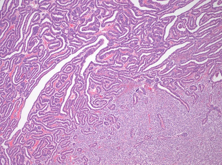
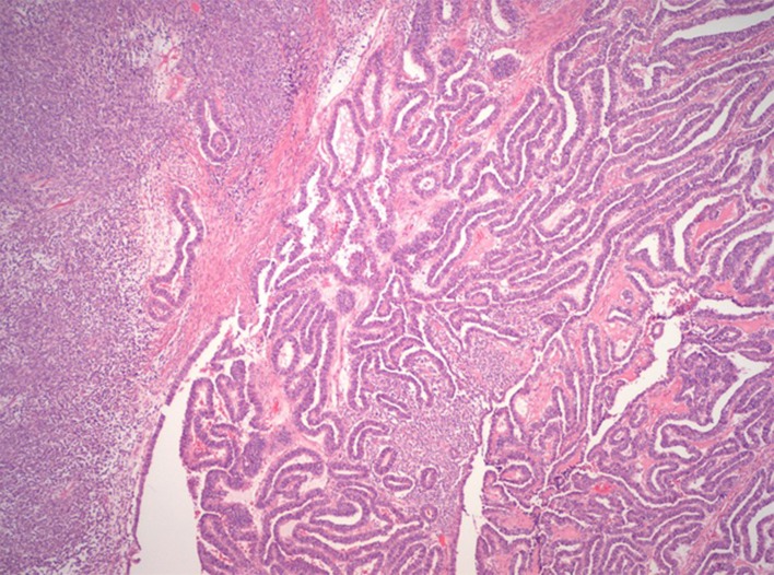
Microscopically, the neoplasm had two components, an epithelial and a mesenchymal component (figures 1 and 2).
Figure 1.
Left: endometrioid-like component. Right: sarcoma component.
Figure 2.
Sarcoma and carcinoma components.
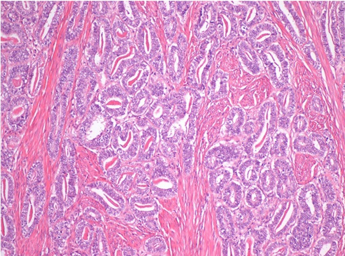
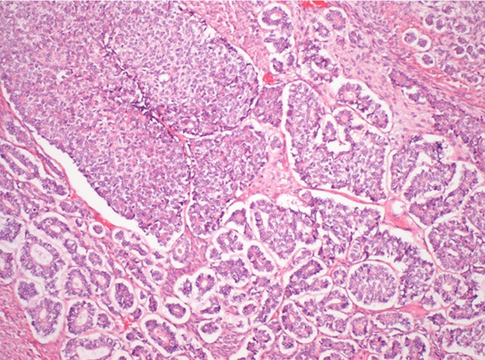
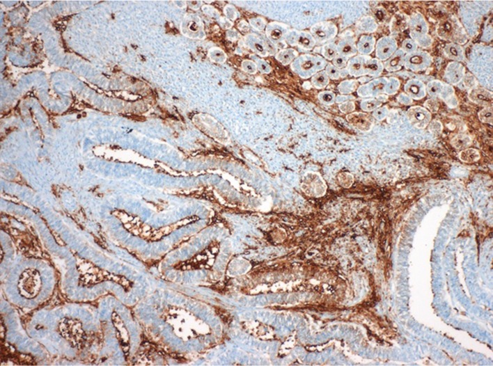
The epithelial component presented a varied appearance with elements resembling mesonephric origin (figure 3)—mesonephroid pattern (areas with small round tubules, a single row of cuboidal atypical cells with eosinophilic luminal content), retiform pattern (branching spaces with intraluminal papillae), endometrioid-like pattern (figure 1), sex-cord-like pattern (figure 4) and small areas resembling serous carcinoma. The immunohistochemical findings showed positive reaction in tumour cells for pancytokeratin, CD10 (apical and luminal) (figure 5), vimentin (focal) and p16 (focal). There was also mesonephric hyperplasia intimately merged with carcinoma (figure 6).
Figure 3.
Mesonephroid pattern of carcinoma.
Figure 4.
Sex-cord-like pattern of carcinoma.
Figure 5.
Immunohistochemical marking of CD 10.
Figure 6.
Mesonephric hyperplasia.
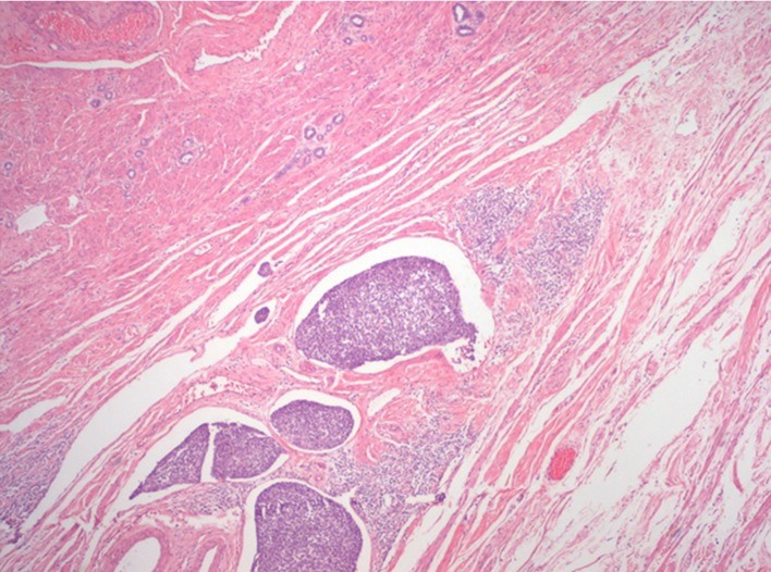
The mesenchymal component resembled a high grade endometrial stromal sarcoma, with closely juxtaposed high grade round cells, with confluent permeative growth. There was high mitotic activity (54 mitosis/10 HPF), and common lymphovascular invasion (figure 7). There were also two involved lymph nodes in the right parametrium by mesenchymal neoplasm component, the left parametrium was also invaded. The immunohistochemical findings showed positive reaction in tumour sarcoma cells for vimentin, and showed no reaction for cyclin D1, CD10, Estrogen Receptor (ER) and Progesterone Receptor (PR), thus not showing a high grade endometrial stromal sarcoma phenotype neither a low grade endometrial stromal sarcoma phenotype.
Figure 7.
Lymphovascular invasion by sarcoma.
The final diagnosis was of a mesonephric carcinosarcoma (malignant mixed mesonephric tumour), and the Pathological tumor-node-metastasis (pTNM) was pT2b N1 R0.
Outcome and follow-up
The postoperative period of 7 days was uneventful. Six weeks after the surgery, the patient was treated with adjuvant chemotherapy with carboplatin and paclitaxel and simultaneous external beam radiation.
In the follow-up the patient was admitted with asthenia and hypercalcaemia and the PET scan showed extensive bone metastasis. She died of progressive disease, 5 months after the diagnosis.
Discussion
Cervical carcinosarcomas are neoplasms that contain both epithelial and mesenchymal malignant elements. They can have origin in paramesonephric (Müllerian) ducts or mesonephric (Wolffian) duct remnants. At least 42 cases of mesonephric carcinomas have been described, corresponding to 16% of cervical carcinosarcomas, and a sarcomatous component was present in 11 cases (table 1), including the case we describe.5 The most common presentation in the literature is abnormal vaginal bleeding and most occur in postmenopausal women.6 7
Table 1.
Mesonephric carcinosarcomas in the literature and the present case
| Patient no | Age (years) | Clinical presentation | Figo stage | Sarcomatous component | Surgical treatment | Adjuvant therapy | Outcome | Follow-up/mean overall survival |
| 113 | 65 | PMB | Ib | Spindle cells; osteosarcoma | RAH, BSO | RT | NED | 23 months |
| 21 | 37 | Postcoital bleeding | Ib | Spindle cells | HT, BSO, LD | ChT | AWD | 11 months |
| 31 | 40 | AUB | Ib | Spindle cells | HT, BSO | RT | NED | 2.3 years |
| 41 | 73 | AUB+cervical polyp | Ib | Spindle cells | HT, BSO | RT | NED | 3 years |
| 51 2 | 39 | AUB | Ib | Spindle cells;osteosarcoma | HT, BSO | DOD | 6.2 years | |
| 69 | 54 | PMB | IIa | Spindle cells | HT, SBO, LD, omentectomy | DOD | 7 months | |
| 79 | 54 | NA | Ib | Spindle cells; chondrosarcoma | HT, BSO, LD, omentectomy | NED | 13 months | |
| 89 | 62 | NA | IVb | Spindle cells; rhabdomyosarcoma | HT, SOB, LD | RT | AWD | 36 months |
| 95 | 63 | PMB | IIa | Spindle cells | RAH, BSO, LD | NED | 10 months | |
| 106 | 59 | Tumour rupture | IIIb | Spindle cells | HT, SOB, LD | ChT, RT | NED | 4 months |
| 11(our case) | 64 | PMB + cervical polyp | IIa | Endometrial stroma sarcoma | RAH, BSO, LD omentectomy | ChT, RT | DOD | 7 months |
AUB, abnormal uterine bleeding; AWD, alive with disease; BSO, bilateral salpingo-oophorectomy; ChT, chemotherapy; DOD, died of disease; HT, hysterectomy; LD, lymphadenectomy; NA, not available; NED, no evidence of disease; PMB, postmenopausal bleeding; RAH, radical hysterectomy; RT, radiotherapy.
Clement et al reported a series of eight cases of mesonephric malignancies, four of them with a sarcomatous component.1 Laterza et al described the treatment and follow-up of 16 patients with cervical carcinosarcoma, 50% of them with early stage at diagnosis were free of disease at final follow-up.10 Sunwoo et al reported a case of carcinosarcoma treated with radical hysterectomy that at 14 months of follow-up remained free of malignancy.11 Kang et al performed radical hysterectomy and chemotherapy on a patient with stage IB cervical carcinosarcoma.12 Bloch et al described a unique case of osteosarcoma associated with atypical mesonephric rests occurring in the right lateral wall of the uterine cervix, treated with radical surgery and radiation.13
More recently, Kim et al described a case of carcinosarcoma of Müllerian duct origin treated with surgery and chemotherapy but the patient died at 7 months of follow-up.7 Another recent case was described by Meguro et al, reporting a postmenopausal woman with abnormal bleeding and a macroscopic cervical tumour that ended up being a mesonephric adenocarcinoma with a sarcomatous component.5
Despite abnormal bleeding being more frequent, Tseng et al reported an uncommon presentation of these neoplasms, describing a tumour rupture as the initial presentation of a mesonephric carcinosarcoma of the cervix.6
Unlike carcinosarcomas of the uterine body, cervical carcinosarcomas present at an earlier stage and have a better outcome although the relation of the sarcomatous component with the prognosis is not yet known.5
Although our case report refers to a carcinosarcoma of the uterine cervix and not uterine body, the diagnosis was made in an advanced stage and the prognosis was bad.
Despite the lack of guidelines and evidence-based recommendations regarding this kind of rare malignancies, surgery is the mainstay of treatment, complemented in some cases with adjuvant chemotherapy and radiation.7
Although these tumours are rare, they express classic morphological features, along with immunohistochemical markers, like CD 10 and vimentin, that can help to verify their origin. Suspicion of carcinosarcoma of the cervix should be raised in case of sarcomatous component, that can be found in 25% of tumour of mesonephric origin.2 5 7
Learning points.
Cervical carcinosarcomas are rare neoplasms that contain both epithelial and mesenchymal malignant elements and there are 62 cases reported in the English literature.
They arise from mesonephric or Müllerian duct remnants and are less frequent than carcinosarcomas of uterine origin.
Cervical mesonephric carcinosarcomas are extremely rare and there are 11 cases described in the literature, including this case.
Most of these tumours arise in postmenopausal women and the most frequent presentation is abnormal vaginal bleeding.
Guidelines regarding these malignancies are lacking, concerning their rarity, but the mainstay of treatment is surgery.
Footnotes
Contributors: BR: as main author contributed to the acquisition of data and interpretation; planning of the article (conception), writing of the manuscript, revision of contents. RS: contributed to the acquisition of data and figures, writing of the article and revision of contents. RD: contributed to the acquisition of data and figures, writing of the article and revision of contents. VP: contributed to the acquisition of data and interpretation; planning of the article (conception), writing of the manuscript, revision of contents.
Funding: The authors have not declared a specific grant for this research from any funding agency in the public, commercial or not-for-profit sectors.
Competing interests: None declared.
Provenance and peer review: Not commissioned; externally peer reviewed.
Patient consent for publication: Not required.
References
- 1. Clement PB, Young RH, Keh P, et al. Malignant mesonephric neoplasms of the uterine cervix. A report of eight cases, including four with a malignant spindle cell component. Am J Surg Pathol 1995;19:1158–71. [DOI] [PubMed] [Google Scholar]
- 2. Silver SA, Devouassoux-Shisheboran M, Mezzetti TP, et al. Mesonephric adenocarcinomas of the uterine cervix: a study of 11 cases with immunohistochemical findings. Am J Surg Pathol 2001;25:379–87. [DOI] [PubMed] [Google Scholar]
- 3. Clement PB, Zubovits JT, Young RH, et al. Malignant mullerian mixed tumors of the uterine cervix: a report of nine cases of a neoplasm with morphology often different from its counterpart in the corpus. Int J Gynecol Pathol 1998;17:211–22. [PubMed] [Google Scholar]
- 4. Arend R, Doneza JA, Wright JD. Uterine carcinosarcoma. Curr Opin Oncol 2011;23:1–6. 10.1097/CCO.0b013e328349a45b [DOI] [PubMed] [Google Scholar]
- 5. Meguro S, Yasuda M, Shimizu M, et al. Mesonephric adenocarcinoma with a sarcomatous component, a notable subtype of cervical carcinosarcoma: a case report and review of the literature. Diagn Pathol 2013;8:74 10.1186/1746-1596-8-74 [DOI] [PMC free article] [PubMed] [Google Scholar]
- 6. Tseng CE, Chen CH, Chen SJ, et al. Tumor rupture as an initial manifestation of malignant mesonephric mixed tumor: a case report and review of the literature. Int J Clin Exp Pathol 2014;7:1212–7. [PMC free article] [PubMed] [Google Scholar]
- 7. Kim M, Lee C, Choi H, et al. Carcinosarcoma of the uterine cervix arising from Müllerian ducts. Obstet Gynecol Sci 2015;58:251–5. 10.5468/ogs.2015.58.3.251 [DOI] [PMC free article] [PubMed] [Google Scholar]
- 8. Kadota K, Haba R, Ishikawa M, et al. Uterine cervical carcinosarcoma with heterologous mesenchymal component: a case report and review of the literature. Arch Gynecol Obstet 2009;280:839–43. 10.1007/s00404-009-1017-0 [DOI] [PubMed] [Google Scholar]
- 9. Bagué S, Rodríguez IM, Prat J. Malignant mesonephric tumors of the female genital tract: a clinicopathologic study of 9 cases. Am J Surg Pathol 2004;28:601–7. [DOI] [PubMed] [Google Scholar]
- 10. Laterza R, Seveso A, Zefiro F, et al. Carcinosarcoma of the uterine cervix: case report and discussion. Gynecol Oncol 2007;107:S98–S100. 10.1016/j.ygyno.2007.07.038 [DOI] [PubMed] [Google Scholar]
- 11. Sunwoo JG, Cho IS, Jeon S, et al. A case of malignant mixed Müllerian Tumor (MMMT) of the uterine cervix. Obstet Gynecol Sci 2008;51:350–4. [Google Scholar]
- 12. Kang JS, Kim T, Lee HK, et al. Primary carcinosarcoma of the uterine cervix. Obstet Gynecol Sci 1994;37:1872–9. [Google Scholar]
- 13. Bloch T, Roth LM, Stehman FB, et al. Osteosarcoma of the uterine cervix associated with hyperplastic and atypical mesonephric rests. Cancer 1988;62:1594–600. [DOI] [PubMed] [Google Scholar]


