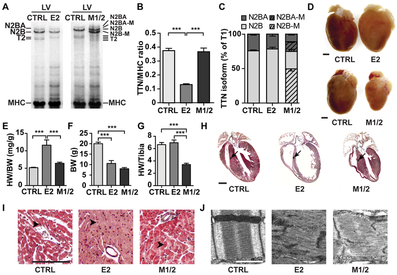
Figure 3. Complete titin loss leads to DCM while loss of M-band titin results in cardiac atrophy at 5 weeks.
A) Agarose protein gel electrophoresis of the left ventricle (LV) from control, E2- and M1/2-KO vs. control (E2, M1/2, CTRL) at 5-weeks. B) The total titin (TTN)/MHC protein ratio is reduced in the E2-KO; n = 6 control, n = 3 per KO. C) Titin N2BA and N2B isoform expression is unchanged in KO mice. In the M1/2-KO, the corresponding truncated isoforms amount to >50%; n = 6 for control n = 3 per KO. D) The heart of 5-weeks-old E2-KO mice is enlarged, while the deletion of titin’s M-band leads to cardiac atrophy (size bar = 1 mm). E, F) Heart weight/ body weight ratio (HW/BW) is increased in E2-KO, but body weight (BW) is reduced in both knockout strains. G) Normalization of the heart weight to the tibia length reflect the atrophy of the M1/2-KO; n = 13 control, n = 7 per KO. H) Masson trichrome staining of the heart from 5-weeks-old animals with DCM in the E2 knockout and atrophy of the M1/2 knockout heart (arrows: area of higher magnification in I; size bar = 1 mm). I) Higher magnification from H with increased number of nuclei (arrowheads indicate nuclei; size bar = 100 µm). J) Disassembly of the sarcomere in both strains. Electron microscopy of left ventricular tissue from 3-week-old animals (size bar = 1µm). Data in B, E, F and G were analyzed for statistical significance with one way ANOVA and Tukey post-test; *** p < 0.001

