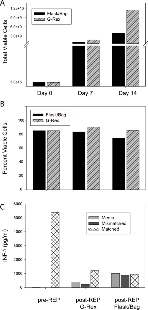
Figure 3.

REP TIL expansion in tissue culture flasks and bags compared to G-Rex®100MCS. 5 × 10⁶ pre-REP TIL isolated from melanoma tumors were cultured in T175 flasks or G-Rex®100MCS in the presence of IL-2 (3,000 IU/ml), normal donor mononuclear cells and agonistic anti-CD3 (30 ng/ml). On day 7 TIL cultured in T175 flasks were transferred to tissue culture bags. On day 14 TIL were harvested from culture vessels and cell counts and viability were determined. (A) 5 × 10⁶ TIL were set up in each culture system at day 0. Cell counts at day 7 and 14 were 5.7 × 10⁸ and 3.7 × 10⁹, respectively for T175 flask to bag cultures and 1.32 × 10⁹ and 1.16 × 10¹⁰, respectively for G-Rex®100MCS cultures. (B) Viability of pre-REP TIL were 85% at day 0, 83% and 74% at day 7 and 14, respectively, for T175 flask to bag cultures and 90% and 85%, respectively, for G-Rex®100MCS cultures. Data represent the mean of two independent experiments using expanded TIL from different patients. (C) TIL harvested at day 14 were cultured for 24 hours in complete media alone, or with melanoma cell lines mismatched or matched for MHC class I antigens. Culture supernatants were tested for IFN-γ levels by ELISA and compared to IFN-γ levels from the same pre-REP cells prior to REP expansion. Data denote results from a single representative experiment.
