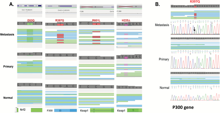
Figure 3.
Mutations in Keap1, Nrf2 and P300 detected by whole genome sequencing. (A) Visualisation of the mutations in four patients using the genomic browser IGV shows mutations in metastatic cells in Keap1, Nrf2 and P300. The mutated bases are flagged in green for glycine (G) or red (A) and the single-nucleotide deletion in pink. All mutations are supported by a large number of reads and were confirmed by Sanger sequencing. (B) Sanger sequencing confirming the WGS sequencing data. Sanger sequencing shows the presence of the G to A mutation in P300 gene.

