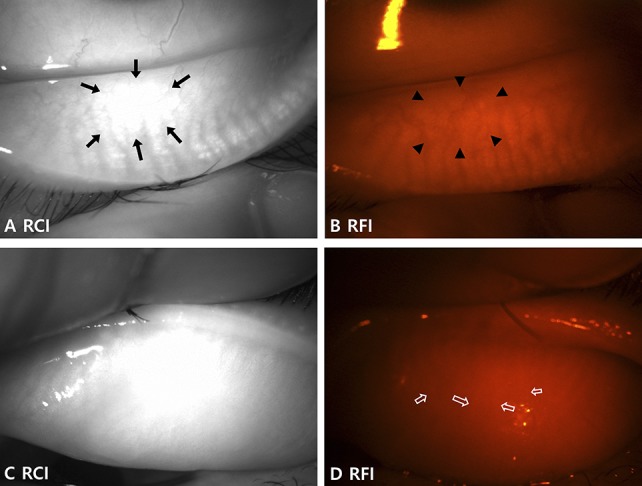
FIGURE 4.

Limitation of the RCIs in some cases. A and B, Images of the lower eyelid of the left eye of a 40-year-old female patient show (A) intact MGs with an indistinguishable area due to the bright reflection in the RCI (arrows). B, The outline of the MGs is more distinguishable in the same area of the RFI (arrowheads). C, The MGs are not quite distinguishable from the RCI of the upper eyelid of the left eye in a 75-year-old female patient. D, Although difficult, the MGs can be distinguished in the RFI of the same eyelid with careful evaluation (empty arrows) and may be easier when viewed live. Evaluation is usually more difficult in the upper eyelid of older patients.
