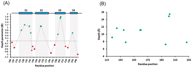
Figure 6.

(A) Membrane depth parameter values (Φ) for different spin-labeled Q1-VSD residues in POPC/POPG vesicles. Positive Φ values (green) indicate that the residue under investigation is embedded within the membrane, and negative Φ (red) values indicate that the residue is in the aqueous environment. (B) EPR depth measurement data for the residues residing in the lipid bilayer vesicles.
