Abstract
Importance
Primary resistance to immune checkpoint inhibitors is observed in 10% to 40% of patients with metastatic colorectal cancer (mCRC) displaying microsatellite instability (MSI) or defective mismatch repair (dMMR).
Objective
To investigate possible mechanisms underlying primary resistance to immune checkpoint inhibitors of mCRC displaying MSI or dMMR.
Design, Setting, and Participants
This post hoc analysis of a single-center, prospective cohort included 38 patients with mCRC diagnosed as MSI or dMMR by local laboratories and entered into trials of immune checkpoint inhibitors between January 1, 2015, and December 31, 2016. The accuracy of MSI or dMMR status was also assessed in a retrospective cohort comprising 93 cases of mCRC that were diagnosed as MSI or dMMR between January 1, 1998, and December 31, 2016, in 6 French hospitals. Primary resistance of mCRC was defined as progressive disease according to Response Evaluation Criteria in Solid Tumors criteria, 6 to 8 weeks after initiation of immune checkpoint inhibitors, without pseudo-progression. All tumor samples were reassessed for dMMR status using immunohistochemistry with antibodies directed against MLH1, MSH2, MSH6, and PMS2, and for MSI using polymerase chain reaction with pentaplex markers and with the HSP110 T17 (HT17) repeat.
Main Outcomes and Measures
The primary outcome was positive predictive value.
Results
Among the 38 patients (15 women and 23 men; mean [SD] age, 55.6 [13.7] years) in the study with mCRC displaying MSI or dMMR, primary resistance to immune checkpoint inhibitors was observed in 5 individuals (13%). Reassessment of the status of MSI or dMMR revealed that 3 (60%) of these 5 resistant tumors were microsatellite stable or displayed proficient mismatch repair. The positive predictive value of MSI or dMMR status assessed by local laboratories was therefore 92.1% (95% CI, 78.5%-98.0%). In the retrospective cohort of 93 patients (44 women and 49 men; mean [SD] age, 56.8 [18.3] years) without immune checkpoint inhibitor treatment, misdiagnosis of the MSI or dMMR status by local assessment was 10% (n = 9), with a positive predictive value of 90.3% (95% CI, 82.4%-95.0%). Testing for MSI with the HT17 assay confirmed the MSI or dMMR status in 2 of 4 cases showing discrepant results between immunohistochemistry and pentaplex polymerase chain reaction (ie, dMMR but microsatellite stable).
Conclusions and Relevance
Primary resistance of mCRC displaying MSI or dMMR to immune checkpoint inhibitors is due mainly to misdiagnosis of their MSI or dMMR status. Larger studies are required to confirm these findings. Microsatellite instability or dMMR status should be tested routinely using both immunohistochemistry and polymerase chain reaction methods prior to treatment with immune checkpoint inhibitors.
This cohort study investigates possible mechanisms underlying primary resistance to immune checkpoint inhibitors of metastatic colorectal cancers displaying microsatellite instability or mismatch repair deficiency.
Key Points
Question
What are the determinants of primary resistance to immune checkpoint inhibitors in metastatic colorectal cancers with microsatellite instability or mismatch repair deficiency?
Findings
In this cohort study of 38 patients with metastatic colorectal cancer, misdiagnosis of microsatellite instability or mismatch repair deficiency status was responsible for 3 of 5 cases of primary resistance to immune checkpoint inhibitors. These patients had been included in trials of immune checkpoint inhibitors based on positive microsatellite instability or mismatch repair deficiency tumor status, as determined by the institutes who referred them for enrollment in the trials.
Meaning
Microsatellite instability or mismatch repair deficiency status with both immunohistochemistry and polymerase chain reaction should be obtained for patients with metastatic colorectal cancer prior to treatment with immune checkpoint inhibitors.
Introduction
Microsatellite instability (MSI) is a molecular indicator of defective DNA mismatch repair (dMMR) and is observed in approximately 5% of metastatic colorectal cancers (mCRC). Microsatellite instability is a major predictive biomarker for the efficacy of immune checkpoint inhibitors. Despite high rates of response and durable clinical benefit with immune checkpoint inhibitors, 10% to 40% of mCRC displaying MSI or dMMR exhibit primary resistance to immunotherapy.1,2,3,4 A predictive biomarker for the efficacy of immune checkpoint inhibitors in MSI or dMMR tumors has yet to be found, however.5 The aim of this study was to start assessing molecular mechanisms that could underlie the primary resistance of mCRC displaying MSI or dMMR to immune checkpoint inhibitors. By reevaluating the MSI and dMMR status of tumors from patients who were entered into trials of immune checkpoint inhibitors, we found that misinterpretation of results for MSI and/or mismatch repair testing may explain most cases showing primary resistance to immune checkpoint inhibitors.
Methods
Study Populations
All patients with mCRC who were determined by local assessment as having MSI or dMMR and subsequently included in clinical trials for immune checkpoint inhibitors (NCT02460198 and NCT02060188) conducted at Saint-Antoine Hospital (Paris, France) were included in the study. Tumor samples from these patients were collected prospectively. A retrospective series comprising primary or metastatic tumor tissue from patients with mCRC who were previously diagnosed with MSI or dMMR in 6 French hospitals was also analyzed to assess the accuracy of current laboratory practice for the detection of MSI and dMMR. Ethical approval was provided the Comité de Protection des Personnes Ile de France IV Institutional Review Board (IRB No. 00003835). All patients provided written consent.
Central Analysis of Tumor Tissue
Tumor samples were tested by a central, specialized reference laboratory at Saint-Antoine Hospital for dMMR using immunohistochemistry (IHC) and for MSI using polymerase chain reaction (PCR) with the pentaplex mononucleotide repeat panel (eAppendix in Supplement 1). Microsatellite instability status was also assessed using the HSP110 (OMIM 610703) HT17 microsatellite marker (HT17 assay) previously shown to have better sensitivity and similar specificity to the pentaplex panel.6
End Points
Primary resistance to immune checkpoint inhibitors was defined according to Response Evaluation Criteria in Solid Tumors version 1.1 criteria7 as progressive radiographic disease on the first study scan (ie, 6 to 8 weeks after initiation of immune checkpoint inhibitors) with no pseudoprogression (confirmed progressive disease). False positives were defined as tumor samples initially diagnosed as MSI or dMMR by local assessment, but displaying both proficient mismatch repair (pMMR) using IHC and microsatellite stability (MSS) after reevaluation by the specialist central review laboratory. The HT17 assay was also incorporated into the definition of MSI and dMMR on account of its better sensitivity compared with the pentaplex panel. The positive predictive value of MSI and dMMR testing by local assessment was calculated as the proportion of false-positive cases among all positive cases.
Results
Primary Resistance to Immunotherapy
Forty-seven patients with mCRC initially diagnosed as MSI and/or dMMR in originating centers were included in immunotherapy trials evaluating anti–programmed cell death 1 with or without anti-CTL4 monoclonal antibodies between January 1, 2015, and December 31, 2016, at the Saint-Antoine Hospital. Nine patients were excluded owing to lack of suitable tumor tissue. Therefore, 38 patients with mCRC treated with immune checkpoint inhibitors who were assessed locally as having MSI and/or dMMR were included in this prospective cohort. Their histologic and molecular characteristics are shown in the eTable in Supplement 2. Primary resistance to immune checkpoint inhibitors was observed in 5 patients (13%). Of these, 3 tumors (60%) were found to display both pMMR and MSS after central review (Table, Figure 1, and Figure 2; eFigure in Supplement 1).
Table. Misdiagnosis of Microsatellite Instability and Mismatch Repair–Deficient Tumors by Local Assessment.
| Sample No.a | Local Assessment | Central Review | Best Response Under Immunotherapy | ||
|---|---|---|---|---|---|
| IHC | PCR | IHC | PCR | ||
| Patients included in immunotherapy trials (n = 38) | |||||
| 47 | pMMR | MSI | pMMR | MSS | Disease progression |
| 115 | NE | MSI | pMMR | MSS | Disease progression |
| 181 | dMMR | NE | pMMR | MSS | Disease progression |
| Retrospective historical cohort (n = 93) | |||||
| 29 | pMMR | MSI | pMMR | MSS | NAb |
| 41 | NE | MSI | pMMR | MSS | NA |
| 42 | NE | MSI | pMMR | MSS | NA |
| 43 | NE | MSI | pMMR | MSS | NA |
| 46 | NE | MSI | pMMR | MSS | NA |
| 56 | NE | MSI | pMMR | MSS | NA |
| 64 | pMMR | MSI | pMMR | MSS | NA |
| 94 | pMMR | MSI | pMMR | MSS | NA |
| 106 | NE | MSI | pMMR | MSS | NA |
Abbreviations: dMMR, mismatch repair deficient; IHC, immunohistochemistry; MSI, microsatellite instability; MSS, microsatellite stability; NA, not applicable; NE, not evaluated; PCR, polymerase chain reaction; pMMR, mismatch repair proficient.
The 12 misdiagnosed samples originate from 6 institutions, with a maximum of 3 errors in 1 single center (2 centers with 3 errors, 2 centers with 2 errors, and 2 centers with 1 error). Fixation, type of sample, IHC technique, antibodies, and PCR technique optimization did not differ between originating centers: all samples were formalin-fixed and paraffin-embedded, the PCR panel was the same for all centers (ie, pentaplex panel). No histopathologic or preanalytical variables (histologic type, differentiation grade, tissue origin, mucinous component, originating center) were associated with misdiagnosis.
Patients from the historical cohort did not receive immunotherapy.
Figure 1. False-Positive Tumor Due to Rare Microsatellite Polymorphisms: Immunohistochemistry.
Immunohistochemistry of tumor sections from patient 47 using antibodies targeting MLH1, PMS2, MSH2, and MSH6 showing the expression of all 4 mismatch repair proteins in tumor cells (ie, mismatch repair proficiency), and hematoxylin-eosin stain of a tumor section.
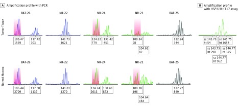
Figure 2. False-Positive Tumor Due to Rare Microsatellite Polymorphisms: Pentaplex Polymerase Chain Reaction (PCR) and HSP110 HT17 Assays.
A, Amplification profile from patient 47 using pentaplex PCR showing a microsatellite stable phenotype with 3 variant alleles mimicking unstable alleles that are outside the quasimonomorphic range (BAT-26, NR-24, and NR-21) in both normal mucosa and tumor tissue. B, Amplification profile using HSP110 HT17 assay confirming the microsatellite stable phenotype for tumor tissue. The colored areas indicate rare polymorphisms of 3 pentaplex markers that mimicked microsatellite instability in this patient.
Misdiagnosis of MSI and dMMR in Immunotherapy Clinical Trials
The positive predictive value of local testing for MSI and dMMR in the cohort treated with immune checkpoint inhibitors was 92.1% (3 patients misdiagnosed out of 38 patients; 95% CI, 78.5%-98.0%). Of the 3 misdiagnosed cases, 1 was owing to misinterpreted IHC results and 2 because of misinterpreted pentaplex PCR results. One of the 2 false-positive cases arising from pentaplex PCR occurred in an African man harboring 3 variant alleles that fell outside the quasimonomorphic variation range; his tumor was originally tested as MSI but pMMR (sample 47; Figure 1 and Figure 2).
Misdiagnosis of MSI and dMMR in Routine Laboratory Practice
A retrospective, multicenter cohort of 93 mCRC tumors diagnosed locally with MSI and/or dMMR between January 1, 1998, and December 31, 2016, was centrally reassessed for MSI and dMMR status. The pathologic information and molecular analyses are summarized in the eTable in Supplement 2. The positive predictive value of assessment of MSI and dMMR by local laboratories was 90.3% (95% CI, 82.4%-95.0%), with 9 of the 93 mCRC tumors showing both pMMR and MSS (Table).
HSP110 HT17 Assay for Discrepancies Between IHC and Pentaplex PCR
Four samples exhibited an MSS phenotype using pentaplex PCR but a dMMR status with IHC (immune checkpoint inhibitor–treated cohort, n = 1; retrospective cohort, n = 3). The HT17 assay confirmed the MSI phenotype for 2 cases (67%) from the retrospective cohort. The diagnostic performances of IHC, pentaplex PCR, and the HT17 assay are described in the eTable in Supplement 1.
Discussion
Microsatellite instability and dMMR have emerged as major predictive biomarkers for the efficacy of immune checkpoint inhibitors in mCRC.1,2,5 Several studies from expert centers, including ours, have aimed to standardize and validate the accepted reference methods for testing MSI and dMMR in colorectal cancer, mainly in patients whose disease is not metastatic.6,8,9,10,11 Information regarding the routine clinical use of these methods is lacking, notably in mCRC. This is despite the fact that novel immunotherapeutic strategies rely on accurate testing for MSI and/or dMMR, since these biomarkers now allow the use of immune checkpoint inhibitors in patients with mCRC after recent accelerated approvals by the US Food and Drug Administration.
A positive MSI or dMMR status by local assessment using only 1 diagnostic method (PCR or IHC) is currently sufficient for enrollment of patients with advanced cancer into most clinical trials of immune checkpoint inhibitors. Under these parameters, we have shown here that misdiagnosis of MSI and dMMR status may be responsible for most primary resistant cases observed in trials of immune checkpoint inhibitors trials for mCRC displaying MSI or dMMR. These results, confirmed with one of the largest retrospective collection of mCRC tumors with MSI, highlight the need for accurate diagnostic methods to assess MSI and dMMR to avoid patients being incorrectly treated with expensive and potentially harmful treatments. Similar studies in real-life routine practice should be provided for emerging diagnostic tools, such as next-generation sequencing, that are currently restricted to highly specialized laboratories but might spread throughout standard platforms in the near future. Importantly, no other predictive biomarker for response to immune checkpoint inhibitors has yet been found among patients with cancer displaying MSI, including KRAS (OMIM 190070), NRAS (OMIM 164790), or BRAF (OMIM 164757) mutational status; Lynch syndrome; tumor programmed death-ligand 1 expression; or tumor mutational burden.1,3,5
One of the 3 patients with mCRC whose tumor was mistakenly diagnosed as MSI had been screened with both IHC and PCR, whereas the other 2 misdiagnosed patients showing primary resistance to immune checkpoint inhibitors had been tested with only 1 method. We therefore recommend dual testing with both IHC and pentaplex PCR for all patients with mCRC before treatment with immune checkpoint inhibitors, and recommend that all samples showing discrepant results between PCR and IHC should be reassessed for MSI and dMMR status in specialized laboratories having this expertise.
Limitations
This study has some limitations. We investigated a prospective series of only 38 patients treated with immune checkpoint inhibitors for mCRC with MSI. Larger prospective studies are required to confirm our findings.
Conclusions
Misdiagnosis of MSI and dMMR status may explain most primary resistances to immunotherapy among patients with mCRC. Local assessment of MSI or dMMR status in patients with mCRC is associated with a false-positive rate of 9% in patients included in trials of immune checkpoint inhibitors in France. This error rate, which biases the results of clinical trials, should be evaluated in larger studies. We recommend dual testing using both IHC and PCR before treatment with immune checkpoint inhibitors, with referral to expert diagnostic centers in the case of discrepant results and consideration of HT17 as an additional marker because of its superior sensitivity.6
eAppendix. Methods
eFigure. CT Scan Evolution of a False-Positive Patient Treated with Immune Checkpoint Inhibitor (Patient #181)
eTable. Detection Rate and Sensitivity of Immunohistochemistry, Pentaplex and HSP110 T17 PCR
eTable
References
- 1.Overman MJ, McDermott R, Leach JL, et al. Nivolumab in patients with metastatic DNA mismatch repair-deficient or microsatellite instability-high colorectal cancer (CheckMate 142): an open-label, multicentre, phase 2 study. Lancet Oncol. 2017;18(9):551-555. doi: 10.1016/S1470-2045(17)30422-9 [DOI] [PMC free article] [PubMed] [Google Scholar]
- 2.Le DT, Uram JN, Wang H, et al. PD-1 blockade in tumors with mismatch-repair deficiency. N Engl J Med. 2015;372(26):2509-2520. doi: 10.1056/NEJMoa1500596 [DOI] [PMC free article] [PubMed] [Google Scholar]
- 3.Overman MJ, Lonardi S, Wong KYM, et al. Durable clinical benefit with nivolumab plus ipilimumab in DNA mismatch repair: deficient/microsatellite instability–high metastatic colorectal cancer. J Clin Oncol. 2018;36(8):773-779. doi: 10.1200/JCO.2017.76.9901 [DOI] [PubMed] [Google Scholar]
- 4.Le DT, Kavan P, Won Kim T, et al. KEYNOTE-164: pembrolizumab for patients with advanced microsatellite instability high (MSI-H) colorectal cancer. J Clin Oncol. 2018;36(suppl):abstract 3514. [Google Scholar]
- 5.Le DT, Durham JN, Smith KN, et al. Mismatch repair deficiency predicts response of solid tumors to PD-1 blockade. Science. 2017;357(6349):409-413. doi: 10.1126/science.aan6733 [DOI] [PMC free article] [PubMed] [Google Scholar]
- 6.Buhard O, Lagrange A, Guilloux A, et al. HSP110 T17 simplifies and improves the microsatellite instability testing in patients with colorectal cancer. J Med Genet. 2016;53(6):377-384. doi: 10.1136/jmedgenet-2015-103518 [DOI] [PubMed] [Google Scholar]
- 7.Eisenhauer EA, Therasse P, Bogaerts J, et al. New response evaluation criteria in solid tumours: revised RECIST guideline (version 1.1). Eur J Cancer. 2009;45(2):228-247. doi: 10.1016/j.ejca.2008.10.026 [DOI] [PubMed] [Google Scholar]
- 8.Umar A, Boland CR, Terdiman JP, et al. Revised Bethesda guidelines for hereditary nonpolyposis colorectal cancer (Lynch syndrome) and microsatellite instability. J Natl Cancer Inst. 2004;96(4):261-268. doi: 10.1093/jnci/djh034 [DOI] [PMC free article] [PubMed] [Google Scholar]
- 9.Buhard O, Cattaneo F, Wong YF, et al. Multipopulation analysis of polymorphisms in five mononucleotide repeats used to determine the microsatellite instability status of human tumors. J Clin Oncol. 2006;24(2):241-251. doi: 10.1200/JCO.2005.02.7227 [DOI] [PubMed] [Google Scholar]
- 10.Shia J. Immunohistochemistry versus microsatellite instability testing for screening colorectal cancer patients at risk for hereditary nonpolyposis colorectal cancer syndrome: part I, the utility of immunohistochemistry. J Mol Diagn. 2008;10(4):293-300. doi: 10.2353/jmoldx.2008.080031 [DOI] [PMC free article] [PubMed] [Google Scholar]
- 11.Zhang L. Immunohistochemistry versus microsatellite instability testing for screening colorectal cancer patients at risk for hereditary nonpolyposis colorectal cancer syndrome: part II, the utility of microsatellite instability testing. J Mol Diagn. 2008;10(4):301-307. doi: 10.2353/jmoldx.2008.080062 [DOI] [PMC free article] [PubMed] [Google Scholar]
Associated Data
This section collects any data citations, data availability statements, or supplementary materials included in this article.
Supplementary Materials
eAppendix. Methods
eFigure. CT Scan Evolution of a False-Positive Patient Treated with Immune Checkpoint Inhibitor (Patient #181)
eTable. Detection Rate and Sensitivity of Immunohistochemistry, Pentaplex and HSP110 T17 PCR
eTable


