Key Points
Question
How is spaceflight associated with human brain white matter and extracellular fluid distribution?
Findings
In this longitudinal analysis, increased extracellular fluid was observed in widespread areas at the base of the cerebrum and decreases along the posterior aspect of the vertex following spaceflight. After adjusting for extracellular fluid, there was altered white matter microstructure in areas encompassing the superior and inferior longitudinal fasciculi, the inferior fronto-occipital fasciculus, and the corticospinal tract following spaceflight.
Meaning
Spaceflight is associated with redistribution of brain extracellular fluids; white matter changes occur throughout the brain and, in some cases, are significantly associated with mission duration and postflight declines in balance.
Abstract
Importance
Spaceflight results in transient balance declines and brain morphologic changes; to our knowledge, the effect on brain white matter as measured by diffusion magnetic resonance imaging (dMRI), after correcting for extracellular fluid shifts, has not been examined.
Objective
To map spaceflight-induced intracranial extracellular free water (FW) shifts and to evaluate changes in brain white matter diffusion measures in astronauts.
Design, Setting and Participants
We performed retrospective, longitudinal analyses on dMRI data collected between 2010 and 2015. Of the 26 astronauts’ dMRI scans released by the National Aeronautics and Space Administration Lifetime Surveillance of Astronaut Health, 15 had both preflight and postflight dMRI scans and were included in the final analyses. Data were analyzed between 2015 and 2018.
Interventions or Exposures
Seven astronauts completed a space shuttle mission (≤30 days) and 8 completed a long-duration International Space Station mission (≤200 days).
Main Outcomes and Measures
The dMRI scans were acquired for clinical monitoring; in this retrospective analysis, we analyzed brain FW and white matter diffusion metrics corrected for FW. We also obtained scores from computerized dynamic posturography tests of balance to assess brain-behavior associations.
Results
Of the 15 astronauts included, the median (SD) age was 47.2 (1.5) years; 12 were men, and 3 were women. We found a significant, widespread increase in FW volume in the frontal, temporal, and occipital lobes from before spaceflight to after spaceflight. There was also a significant decrease in FW in the posterior aspect of the vertex. All FW changes were significant and ranged from approximately 2.5% to 4.0% across brain regions. We observed white matter changes in the right superior and inferior longitudinal fasciculi, the corticospinal tract, and cerebellar peduncles. All white matter changes were significant and ranged from approximately 0.75% to 1.25%. Spaceflight mission duration was associated with cerebellar white matter change, and white matter changes in the superior longitudinal fasciculus were associated with the balance changes seen in the astronauts from before spaceflight to after spaceflight.
Conclusions and Relevance
Free water redistribution with spaceflight likely reflects headward fluid shifts occurring in microgravity as well as an upward shift of the brain within the skull. White matter changes were of a greater magnitude than those typically seen during the same period with healthy aging. Future, prospective assessments are required to better understand the recovery time and behavioral consequences of these brain changes.
This study maps spaceflight-induced intracranial extracellular free water (FW) shifts and evaluates changes in brain white matter diffusion measures in astronauts following spaceflight.
Introduction
Understanding how spaceflight affects the human brain is essential to future space exploration. Neuroimaging studies have reported spaceflight-associated decreases in frontal and temporal gray matter volumes, increased somatosensory cortex,1 an upward displacement of the brain within the skull, and ventricular volume expansion.1,2 Increased periventricular white matter hyperintensities have also been reported in astronauts following flight.3 Further characterization of spaceflight-induced white matter changes is needed because variations in microstructural properties in normal-appearing brain white matter have been associated with cognitive4 and motor5 performance in healthy aging. This may provide insight into the neural mechanisms underlying the declines in balance, locomotion, and manual control6,7,8,9,10 that astronauts experience as a result of spaceflight.
Diffusion magnetic resonance imaging (dMRI) quantifies the diffusion of water molecules, which is affected by myelin density, membrane intactness, and organization of myelinated fibers.11 Thus, dMRI indices provide noninvasive measures of white matter components. In addition to the known microgravity-induced cephalad fluid shift,12 cerebrospinal fluid (CSF) redistribution following global and local gray matter tissue shifts in astronauts1,2 may compromise accurate estimation of dMRI indices. Therefore, we applied an established technique to estimate dMRI white matter metrics after correcting for free water (FW),13 enabling reliable white matter analyses.14 Based on our previous findings in bed-rest simulations of microgravity,15 we hypothesized that astronauts would have increased FW around the base of the brain and decreased FW in the superior and posterior portions, reflecting an upward shift of the brain within the skull after spaceflight. Further, we hypothesized that white matter microstructure would be altered following spaceflight, particularly in sensorimotor pathways. We hypothesized that the degree of white matter changes would be associated with mission duration, flight experience, and the magnitude of transient balance decrements that are evident postflight.
Methods
Study Design
We obtained whole-brain dMRI from 26 astronauts through the National Aeronautics and Space Administration (NASA) Lifetime Surveillance of Astronaut Health. Nine were excluded owing to missing preflight scans. Additionally, 1 data set with incomplete brain coverage and another with insufficient scan volumes were removed, leaving preflight and postflight scans from 15 unique individuals.
Participants
The astronauts were between ages 40 and 60 years; 7 (1 woman) completed a short-duration space shuttle mission (≤30 days) and 8 (2 women) completed a long-duration International Space Station mission (≤200 days). Prior spaceflight experience ranged from 0 (in 1 astronaut) to approximately 200 days in 0 to 3 prior missions (Table). Magnetic resonance imaging and posturography data were obtained for medical monitoring. Patients provided written informed consent and the study was approved by the NASA institutional review board. Preflight scans were collected at a median of 274 days before launch (range, 44-627 days) and postflight scans at a median of 6 days after return (range, 1-20 days).
Table. Demographics of Astronaut Cohort.
| Characteristic | Median (MAD) | |
|---|---|---|
| Shuttle | ISS | |
| Male | 6 | 6 |
| Female | 1 | 2 |
| Age, y | 47.2 (1.5) | 45.9 (2.5) |
| Mission duration, da | 13.0 (0.0) | 167.0 (1.0) |
| Previous spaceflight experience, d | 39.0 (26.0) | 16.0 (8.0) |
| Time elapsed between landing and postflight scan, da | 11.0 (5.0) | 3.5 (1.5) |
| Time elapsed between prior mission and preflight scan, db,c | 748.0 (118) | 1182.0 (340) |
Abbreviations: ISS, international space station; MAD, median absolute deviation.
Significantly different at P < .001.
Significantly different at P < .05.
One astronaut with no prior spaceflight experience not included in the report.
We also evaluated a control sample recruited by the NASA Johnson Space Center human test subject facility; these individuals were not astronauts in training. All the astronauts and control individuals passed an Air Force Class III equivalent physical examination; see the eMethods in the Supplement for analyses involving the control group.
Balance Control Assessments
Balance was evaluated preflight and postflight for 13 astronauts using the Sensory Organization Tests (SOTs) provided by the EquiTest System platform (NeuroCom). Participants performed three 20-second trials of SOT-5 (standing on a sway-referenced platform with eyes closed) and SOT-5M (added rhythmic head pitch motion ±20° synchronized to a 0.33-Hz auditory cue).9 Postflight testing was conducted within 2 days of landing.
Image Acquisition
Astronauts
Diffusion MRI scans were obtained on a 3-T Siemens Verio scanner using a diffusion-weighted 2-dimensional single-shot spin-echo prepared echo-planar imaging sequence with the following parameters: echo time, 95 milliseconds; repetition time (TR), 5800 milliseconds; flip angle, 90°; field of view, 230 × 230 mm; matrix size, 128 × 128; 40 axial slices of 3.9-mm slice thickness with zero gap; and 1.8 × 1.8 × 3.9-mm3 voxels. Twenty noncollinear gradient directions with diffusion weighting of b = 1000 s/mm2 were sampled 3 times. A volume with no diffusion weighting (b = 0 s/mm2) was acquired at the beginning of each sampling stream. In 10 astronauts, parameters were changed from preflight to postflight, resulting in a voxel resolution change to 1.95 × 1.95 × 3.9 mm3, with corresponding field of view changes. Shorter TRs were used in the preflight scans of 4 astronauts (4900, 5500, and two 5600), and in 1, the preflight and postflight scan TRs were 5900 and 5302 milliseconds, respectively.
Image Analysis
We analyzed the data using FMRIB Software Library (FSL), version 5.0; MATLAB R2015b; MathWorks advanced normalization tools (ANTs; 1.9.x16,17); and custom-written FW algorithms.13 Using a stepwise approach, the longitudinal nature of the data was taken into account when bringing the individual FW and FW-adjusted dMRI data into Montreal Neurological Institute standard space (MNI152). The processing pipeline in the following paragraphs is adapted from Schwarz et al.18
Preprocessing
Images were motion-corrected and eddy current–corrected in FSL by registering to the mean of the b = 0 images. Rotations that were applied to the dMRI volumes were also applied to the encoding b vectors. Motion correction plots and raw diffusion-weighted images were visually inspected for scan motion or artifacts. Artifact volumes were removed from the 4-dimensional image file and the b-value and b-vector matrices.
Tensor Fit
Diffusion tensor fitting was performed using an algorithm that fits a bi-tensor model.13 Scalar diffusion tensor imaging (DTI) indices corrected for FW and an FW image were produced for each time and individual. The FW images indicate the fractional volume of FW in a voxel, representing the proportion of water molecules that are not hindered or restricted by their surroundings.13 The following DTI indices were analyzed after FW correction: fractional anisotropy (FAT, reflecting diffusion anisotropy), axial diffusivity (ADT, reflecting water mobility parallel to a major fiber axis), and radial diffusivity (RDT, reflecting water mobility perpendicular to a major fiber axis). The subscript T indicates that the DTI measures are based on the tissue compartment.
Normalization
Patient template creation and normalization to standard space were carried out using FA images that were not corrected for FW generated using FSL’s diffusion tensor imaging fit. First, the outermost layer (1 voxel) of all FA images was eroded to remove noise and was zero-padded. Patient-specific templates were created using ANTs’ buildtemplateparallel.sh script in a fashion that was unbiased between the input images of any specific time. Next, we normalized these templates to MNI152 common space using ANTs’ SyN algorithm. Then, we combined the linear and nonlinear warp parameters from the individual patients’ FA image to the patient-specific FA template and the MNI152 standard space into 1 flow field. These flow fields were then used to transform and reslice the images to the same spatial resolution, bringing the zero-padded DTI scalar images that were adjusted for FW and the FW images into MNI152 common space. The normalized images were smoothed with a Gaussian kernel of 3.4-mm σ (approximately 8 mm full width at half maximum) to increase signal-to-noise ratio.
Voxelwise Data Analysis
Masking
Voxel-based analyses of diffusion indices were confined to a white matter mask constructed by eliminating the voxels where FA was less than 0.2. The final white matter mask consisted only of voxels that were nonzero in more than half of the sample.18 Free-water analyses were carried out within the whole-brain mask, made by binarizing the FMRIB58_FA_1mm standard template image.
Statistical Analysis
One-sample t tests on predifference to postdifference maps of FW and diffusion indices were conducted to test (1) whether there was any change as a function of spaceflight and (2) whether these changes were associated with mission duration, cumulative days in space, cumulative mission count, or the change in SOT-5 and SOT-5M scores. All tests were adjusted for the time elapsed between landing and the postflight MRI scan date. Tests of association were limited to brain areas where significant preflight to postflight changes in the DTI metrics and FW were detected; exploratory whole-brain analyses were also examined.
All tests were nonparametric permutation based using threshold-free cluster enhancement,19 with 15 000 random permutations implemented in FSL’s randomize,20 with 2.5-mm full width at half maximum variance smoothing. A voxel-level familywise error correction (significant at P < .05) was applied to adjust for multiple comparisons, and all P values were 2-sided. Anatomical labels of peak voxels showing significant white matter changes were defined using the Johns Hopkins University white matter tractography atlas. If no labels were detected, the International Consortium of Brain Mapping Diffusion Tensor Imaging 81 white matter labels atlas and the Harvard-Oxford Cortical Structural Atlas were consulted. The peak voxels within clusters showing FW changes were labeled using the Harvard-Oxford Cortical Structural Atlas.
Results
Balance Control
The astronauts showed reduced balance postflight on both SOT-5 and SOT-5M. Postflight scores (SOT-5 mean [SD], 79.5 [9.7]; SOT-5M mean [SD], 37.7 [32.0]) were significantly lower than preflight (SOT-5 mean [SD], 85.0 [8.3]; SOT-5M mean [SD], 70.6 [21.4]) as indicated by Wilcoxon signed rank tests (SOT-5 z = −2.9; P = .004; SOT-5M z = −3.1; P = .002). Although flight duration was not significantly associated with changes in SOT-5 (rs = 0.45, P = .12) or SOT-5M (rs = 0.19; P = .55), greater cumulative days in space were significantly associated with smaller preflight to postflight changes in SOT-5M (rs = 0.56; P = .045) but not in SOT-5 (rs = 0.51; P = .08).
Changes in FW and White Matter Microstructure in Astronauts
We observed significant widespread FW increases in the frontal, temporal, and occipital lobes. There was also a significant FW decrease in the superior and posterior portions of the brain from preflight to postflight (Figure 1).
Figure 1. Preflight to Postflight Change in Free Water (FW) in Astronauts (P < .05; Familywise Error Corrected).
Clusters in red indicate areas where FW significantly increased, and clusters in blue indicate where FW significantly decreased as a function of spaceflight. The results are overlaid on the Montreal Neurological Institute standard space brain template.
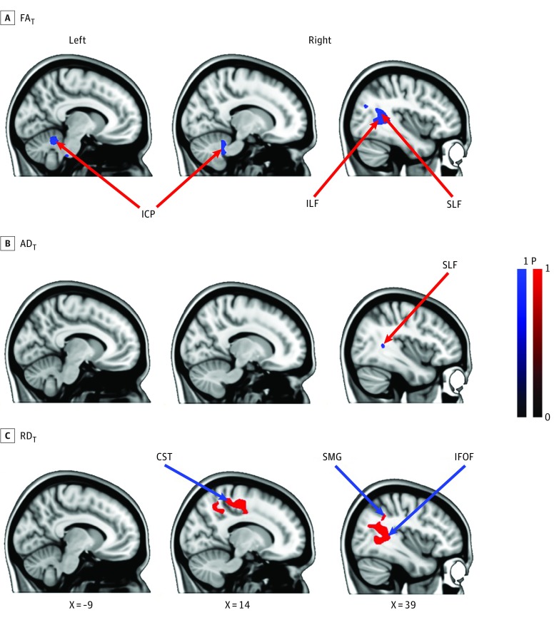
The FAT decreased in the right superior longitudinal fasciculus (SLF), inferior longitudinal fasciculus, inferior fronto-occipital fasciculus, corticospinal tract, and the inferior and middle cerebellar peduncles after spaceflight. Although to a lesser extent, significant increases in RDT and decreases in ADT values were detected in similar regions, with the exception of the cerebellar peduncles. Additionally, increased RDT was observed in white matter structures underlying the precentral and postcentral gyrus, the supramarginal gyrus, and angular gyrus (Figure 2).
Figure 2. Association of Spaceflight With White Matter Microstructure as Measured by Changes From Before Spaceflight to After Spaceflight for Free Water (FW)–Corrected Fractional Anisotropy (FAT), Axial Diffusivity (ADT), and Radial Diffusivity (RDT).
A and B, Blue gradients show regions in which the FAT and ADT values significantly decreased as a function of spaceflight (P < .05; familywise error corrected). The red gradient (C) shows regions in which the RDT significantly increased as a function of spaceflight (P < .05, familywise error corrected). The results are overlaid on the Montreal Neurological Institute standard space brain template. CST indicates corticospinal tract; ICP, inferior cerebellar peduncle; ILF, inferior longitudinal fasciculus; SLF, superior longitudinal fasciculus; SMG, supramarginal gyrus; IFOF, inferior-occipital fasciculus.
Association of FW and White Matter Changes with Mission Duration, Mission Count, and Balance Changes
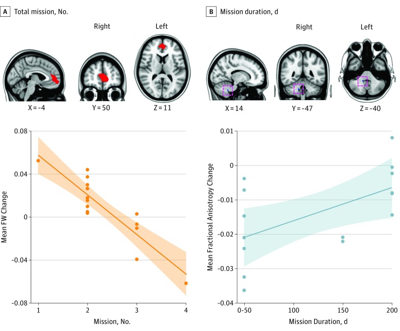
Only the exploratory whole-brain assessments revealed a significant association between preflight to postflight FW change and cumulative mission count. Astronauts who completed fewer missions showed greater FW increase in the anterior cingulate cortex, whereas those with more missions exhibited decreased FW postflight (Figure 3A).
Figure 3. Association of Spaceflight With Free Water (FW) Distribution and Fractional Anisotropy (FAT) Measures.
The clusters show brain areas where there was a significant association between the preflight to postflight change in FW and the total count of missions (A) and spaceflight-associated FA change and the mission duration (B) (P < .05; familywise error corrected). The results are overlaid on the Montreal Neurological Institute standard space brain template. The orange scatterplot depicts the mean change in FW of the cluster shown as a function of the number of missions. The blue scatterplot shows the mean change in FA of the cluster shown as a function of the current mission duration. The mission duration is expressed in bins of 50-day increments to prevent identification of individuals based on flight duration.
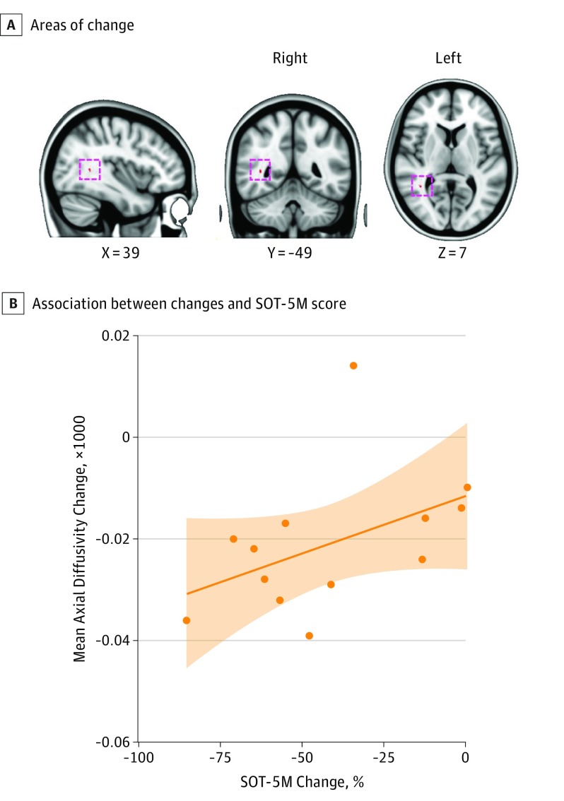
There was a significant association between mission duration and FAT changes with spaceflight in the right cerebellum lobule IX, where those with shorter mission durations showed greater decreases in FAT (Figure 3B). The ADT decreases with spaceflight in the SLF were greater in individuals whose SOT-5M performance declined more (Figure 4). These association results were confined to the area where significant spaceflight-associated changes were found in the respective indices.
Figure 4. Association Between Spaceflight-Induced Changes of Axial Diffusivity (ADT) and Sensory Organization Test (SOT) 5M Balance Score.
A, The red cluster (cluster size 13 mm3) shows the area in which the change in AD values was significantly associated with the change in SOT-5M performance (P < .05; familywise error corrected). B, The scatterplot depicts the association between the mean change of ADT value in the cluster shown in the upper panel and the difference in equilibrium score (expressed in percentage) of SOT-5M from preflight to postflight. The shaded area indicates the 95% confidence limits. The results are overlaid on the Montreal Neurological Institute standard space brain template.
Group comparisons between astronauts and the convenience control sample are reported in the eMethods in the Supplement. Results generally revealed preflight group differences in all brain measures, possibly owing to holdover from previous flight experiences in the astronauts (eFigure1 in the Supplement). The control group did not show significant changes in any of the brain metrics over time. Astronauts showed a greater degree of FW increase in the inferior portion of the cerebrum and a greater degree of RDT increase in the supramarginal gyrus.
Discussion
We observed widespread spaceflight-associated FW increases at the base of the cerebrum and decreases along the posterior vertex. After adjusting for FW, we observed altered white matter microstructural measures associated with spaceflight in the superior and inferior longitudinal fasciculi, the corticospinal tract, and cerebellar peduncles, regions involved in visuospatial processing, vestibular function, and movement control.
The co-occurrence of FW increases and decreases suggests an intracranial redistribution with spaceflight. This is similar to what we observed when participants spent 70 days in head-down bed rest to mimic microgravity effects,15 indicating that mechanical effects (ie, microgravity in space and reorientation of the head relative to the gravitational vector in bed rest)21 underlie these changes. In addition, the preflight to postflight FW shift we observed in this study complements our previous report of spaceflight-associated regional brain gray matter changes1 and the report of CSF compartment changes2 in astronauts, highlighting the link between fluid shifts and brain morphology alterations with spaceflight. Given the potential effect of brain positional alterations on the optic system, such as an upward repositioning of the optic nerves and globe flattening,2,22 fluid shifts and upward brain displacement with spaceflight are important to study. The mechanisms underlying spaceflight-associated neuro-ocular syndrome, a concern that has affected about one-third of long-duration astronauts,23 are not well understood. Thus, mapping of interstitial fluid shifts and associated brain changes may further our understanding of the development of spaceflight-associated neuro-ocular syndrome.
Interestingly, irrespective of the flight duration and cumulative days in space, astronauts with a greater number of missions showed FW decreases (as opposed to increases in more novice flyers) in the anterior, medial portion of the brain. This implies that the number of gravitational transitions experienced has an important effect on the FW compartment. The significant FW differences at baseline between astronauts and control individuals (eMethods in the Supplement) also support this notion. It is possible that repeated adaptation to multiple gravitational transitions may affect the gross morphology of the brain and its plasticity.
In this study, astronauts had significant spaceflight-associated decreases in FAT and ADT measures and increases in RDT values, indicative of disrupted white matter structural connectivity in multiple tracts. Disruption in these areas suggests that processes requiring prefrontal multimodal integration of sensory inputs24,25 may be at risk, such as visuomotor control and higher-order visuospatial processing.26,27,28,29 Particularly, the SLF is involved in vestibular processes by connecting the temporoparietal and prefrontal cortices, enabling coordination of body posture and spatially oriented actions.30 Indeed, astronauts who showed the largest spaceflight-associated balance disruptions had greater ADT decreases in the SLF following spaceflight, characteristic of less axonal density and organization.31,32
We also observed decreased FAT values in cerebellar afferent connections, including vestibular projections (ie, inferior cerebellar peduncle)33,34 and vertigo neural pathways (ie, middle cerebellar peduncle).35 Furthermore, increased RDT values in the right inferior and posterior parietal lobe were detected. These areas underlie body spatial representation with respect to the environment36 and central proprioceptive processing37,38 and are involved in vertical upright perception.39 These white matter alterations could reflect vestibular and proprioceptive processing changes that astronauts experience as a result of microgravity exposure.8,40
Spaceflight-associated decreases in FAT and increases in RDT values were detected in the corticospinal tract, the major motor pathway.41 Unlike the adaptive neuroplastic changes that manifest as increased gray matter volume in somatosensory regions in astronauts1 or increased corticospinal excitability in a spaceflight analog environment,42 the decreased corticospinal tract structural organization may reflect decreased lower limb muscle use.43 We also observed increased RDT values in white matter underlying the primary motor and sensory cortices. These findings suggest that dMRI may be effective for detecting adverse neuronal effects in astronauts.
Interestingly, long-duration fliers had less decline or even increased FAT values in the cerebellar white matter underlying lobule IX. This region participates in visually guided movement44 and the autonomic cardiovascular arousal that occurs during stressor tasks.45 Although it seems counterintuitive that white matter structural organization increases with longer mission durations, this may reflect adaptation over time. While cumulative days in space were not associated with any significant FW or diffusion changes, we found that astronauts with greater cumulative flight days had fewer balance declines immediately following spaceflight, supporting the presence of adaptive processes.
In our previous bed-rest spaceflight analog study,15 we observed significant FW redistribution but no changes in FW-corrected diffusion indices. Although we did not report the directional diffusivity measures from that study, a follow-up analysis confirmed no associations with ADT and RDT. The contrasting results between this spaceflight study and the analog study indicate a unique association of spaceflight with brain white matter microstructure. The absence of changes in diffusion indices in the presence of FW alterations in the bed rest study suggests that the white matter changes here are not entirely owing to intracranial fluid redistribution. In this study, the overlap of areas showing FAT and RDT changes suggests that white matter changes are driven largely by increased diffusion in the direction perpendicular to the main longitudinal axis (see eFigure 2 in the Supplement for RDT comparison with control individuals). In light of the reduced spinal myelin thickness in rodents after 30 days of spaceflight,46 the higher RDT value that is typically linked to demyelination47 may reflect a specific spaceflight effect on myelination.
White matter microstructure is relatively stable in adults; FA values in the SLF peak in the third decade of life then decrease moderately until the sixth decade, with a subsequent accelerating decrease.48 Given the astronauts’ age range, the time lapse between preflight and postflight scans is unlikely to explain the associations reported here. The spaceflight-associated white matter changes were approximately 1.5% to 2.5% per year. This is roughly 2-fold the reported longitudinal changes of less than 1.0% per year in a large sample with a mean age of 50 years.49
In comparison with the control individuals, astronauts had greater baseline RDT values. Given the spaceflight-associated RDT increase in this study, this may be a holdover from previous flights. Interestingly, the astronauts show greater ADT values at baseline than control individuals, specifically in the corticospinal tract and the cerebellar peduncles (eFigure 3 in the Supplement), which is indicative of greater coherence of fiber tracts that regulate sensorimotor and balance control in the astronauts. This may reflect white matter changes that occurred in response to preflight training or accumulated adaptive plasticity resulting from repeated sensorimotor adaptation to gravitational transitions.
Limitations
Limitations to working with this retrospective clinical data set include the varied timing of MRI acquisition and scan sequences across patients and the low spatial resolution of the scans. Although the changes in spatial resolution and TR did not exaggerate the preflight to postflight differences, our sensitivity to detect additional significant changes could have suffered as a result. In addition, future work with higher spatial resolution scans would increase localization confidence, especially for small structures that may be more affected by partial volume contamination. We are collecting astronaut dMRI scans in a longitudinal study with improved scanning parameters. Prospective studies with a controlled assessment schedule spanning multiple points before and after the mission will elucidate the enduring nature and behavioral consequences of FW and white matter changes associated with spaceflight.
Conclusions
In conclusion, analyses of this retrospective data set revealed significantly increased FW at the base of the cerebrum and decreases along the posterior vertex. After adjusting for these FW shifts, we found decreases in FAT and ADT measures and increases in RDT values, reflecting disrupted white matter structural connectivity in the cerebellum, corticospinal tract, and superior longitudinal fasiculus, among other regions. Some of these brain changes were associated with number of previous spaceflight missions, mission duration, and preflight to postflight balance declines. Ongoing, prospective studies will help to elucidate recovery time course and performance consequences. Future work should also address whether and how these brain changes are linked to spaceflight neuro-ocular syndrome.
eMethods.
eFigure 1. Comparison of Control and Astronauts’ Free Water (FW) Measure
eFigure 2. Comparison of Control and Astronauts’ FW-Corrected Radial Diffusivity (RDT) Measure
eFigure 3. Intercept Differences Between Crewmembers and Controls
eReferences
References
- 1.Koppelmans V, Bloomberg JJ, Mulavara AP, Seidler RD. Brain structural plasticity with spaceflight. NPJ Microgravity. 2016;2(1):2. [DOI] [PMC free article] [PubMed] [Google Scholar]
- 2.Roberts DR, Albrecht MH, Collins HR, et al. Effects of spaceflight on astronaut brain structure as indicated on MRI. N Engl J Med. 2017;377(18):1746-1753. doi: 10.1056/NEJMoa1705129 [DOI] [PubMed] [Google Scholar]
- 3.Alperin N, Bagci AM, Lee SH. Spaceflight-induced changes in white matter hyperintensity burden in astronauts. Neurology. 2017;89(21):2187-2191. doi: 10.1212/WNL.0000000000004475 [DOI] [PubMed] [Google Scholar]
- 4.Vernooij MW, Ikram MA, Vrooman HA, et al. White matter microstructural integrity and cognitive function in a general elderly population. Arch Gen Psychiatry. 2009;66(5):545-553. doi: 10.1001/archgenpsychiatry.2009.5 [DOI] [PubMed] [Google Scholar]
- 5.Seidler RD, Bernard JA, Burutolu TB, et al. Motor control and aging: links to age-related brain structural, functional, and biochemical effects. Neurosci Biobehav Rev. 2010;34(5):721-733. doi: 10.1016/j.neubiorev.2009.10.005 [DOI] [PMC free article] [PubMed] [Google Scholar]
- 6.Bock O, Weigelt C, Bloomberg JJ. Cognitive demand of human sensorimotor performance during an extended space mission: a dual-task study. Aviat Space Environ Med. 2010;81(9):819-824. doi: 10.3357/ASEM.2608.2010 [DOI] [PubMed] [Google Scholar]
- 7.Reschke MF, Bloomberg JJ, Harm DL, Paloski WH, Layne C, McDonald V. Posture, locomotion, spatial orientation, and motion sickness as a function of space flight. Brain Res Brain Res Rev. 1998;28(1-2):102-117. doi: 10.1016/S0165-0173(98)00031-9 [DOI] [PubMed] [Google Scholar]
- 8.Kornilova LN. Vestibular function and sensory interaction in altered gravity. Adv Space Biol Med. 1997;6:275-313. doi: 10.1016/S1569-2574(08)60087-8 [DOI] [PubMed] [Google Scholar]
- 9.Wood SJ, Paloski WH, Clark JB. Assessing sensorimotor function following ISS with computerized dynamic posturography. Aerosp Med Hum Perform. 2015;86(12)(suppl):A45-A53. doi: 10.3357/AMHP.EC07.2015 [DOI] [PubMed] [Google Scholar]
- 10.Mulavara AP, Feiveson AH, Fiedler J, et al. Locomotor function after long-duration space flight: effects and motor learning during recovery. Exp Brain Res. 2010;202(3):649-659. doi: 10.1007/s00221-010-2171-0 [DOI] [PubMed] [Google Scholar]
- 11.Beaulieu C. The basis of anisotropic water diffusion in the nervous system: a technical review. NMR Biomed. 2002;15(7-8):435-455. doi: 10.1002/nbm.782 [DOI] [PubMed] [Google Scholar]
- 12.Nelson ES, Mulugeta L, Myers JG. Microgravity-induced fluid shift and ophthalmic changes. Life (Basel). 2014;4(4):621-665. doi: 10.3390/life4040621 [DOI] [PMC free article] [PubMed] [Google Scholar]
- 13.Pasternak O, Sochen N, Gur Y, Intrator N, Assaf Y. Free water elimination and mapping from diffusion MRI. Magn Reson Med. 2009;62(3):717-730. doi: 10.1002/mrm.22055 [DOI] [PubMed] [Google Scholar]
- 14.Albi A, Pasternak O, Minati L, et al. ; PharmaCog Consortium . Free water elimination improves test-retest reproducibility of diffusion tensor imaging indices in the brain: A longitudinal multisite study of healthy elderly subjects. Hum Brain Mapp. 2017;38(1):12-26. doi: 10.1002/hbm.23350 [DOI] [PMC free article] [PubMed] [Google Scholar]
- 15.Koppelmans V, Pasternak O, Bloomberg JJ, et al. Intracranial fluid redistribution but no white matter microstructural changes during a spaceflight analog. Sci Rep. 2017;7(1):3154. doi: 10.1038/s41598-017-03311-w [DOI] [PMC free article] [PubMed] [Google Scholar]
- 16.Avants BB, Tustison NJ, Song G, Cook PA, Klein A, Gee JC. A reproducible evaluation of ANTs similarity metric performance in brain image registration. Neuroimage. 2011;54(3):2033-2044. doi: 10.1016/j.neuroimage.2010.09.025 [DOI] [PMC free article] [PubMed] [Google Scholar]
- 17.Avants BB, Yushkevich P, Pluta J, et al. The optimal template effect in hippocampus studies of diseased populations. Neuroimage. 2010;49(3):2457-2466. doi: 10.1016/j.neuroimage.2009.09.062 [DOI] [PMC free article] [PubMed] [Google Scholar]
- 18.Schwarz CG, Reid RI, Gunter JL, et al. ; Alzheimer’s Disease Neuroimaging Initiative . Improved DTI registration allows voxel-based analysis that outperforms tract-based spatial statistics. Neuroimage. 2014;94:65-78. doi: 10.1016/j.neuroimage.2014.03.026 [DOI] [PMC free article] [PubMed] [Google Scholar]
- 19.Smith SM, Nichols TE. Threshold-free cluster enhancement: addressing problems of smoothing, threshold dependence and localisation in cluster inference. Neuroimage. 2009;44(1):83-98. doi: 10.1016/j.neuroimage.2008.03.061 [DOI] [PubMed] [Google Scholar]
- 20.Winkler AM, Ridgway GR, Webster MA, Smith SM, Nichols TE. Permutation inference for the general linear model. Neuroimage. 2014;92:381-397. doi: 10.1016/j.neuroimage.2014.01.060 [DOI] [PMC free article] [PubMed] [Google Scholar]
- 21.Bijsterbosch JD, Lee KH, Hunter MD, et al. The effect of head orientation on subarachnoid cerebrospinal fluid distribution and its implications for neurophysiological modulation and recording techniques. Physiol Meas. 2013;34(3):N9-N14. doi: 10.1088/0967-3334/34/3/N9 [DOI] [PubMed] [Google Scholar]
- 22.Shinojima A, Kakeya I, Tada S. Association of space flight with problems of the brain and eyes. JAMA Ophthalmol. 2018;136(9):1075-1076. doi: 10.1001/jamaophthalmol.2018.2635 [DOI] [PMC free article] [PubMed] [Google Scholar]
- 23.Lee AG, Tarver WJ, Mader TH, Gibson CR, Hart SF, Otto CA. Neuro-ophthalmology of space flight. J Neuroophthalmol. 2016;36(1):85-91. doi: 10.1097/WNO.0000000000000334 [DOI] [PubMed] [Google Scholar]
- 24.Petrides M, Pandya DN. Efferent association pathways originating in the caudal prefrontal cortex in the macaque monkey. J Comp Neurol. 2006;498(2):227-251. doi: 10.1002/cne.21048 [DOI] [PubMed] [Google Scholar]
- 25.Rodríguez-Herreros B, Amengual JL, Gurtubay-Antolín A, et al. Microstructure of the superior longitudinal fasciculus predicts stimulation-induced interference with on-line motor control. Neuroimage. 2015;120:254-265. doi: 10.1016/j.neuroimage.2015.06.070 [DOI] [PubMed] [Google Scholar]
- 26.Aralasmak A, Ulmer JL, Kocak M, Salvan CV, Hillis AE, Yousem DM. Association, commissural, and projection pathways and their functional deficit reported in literature. J Comput Assist Tomogr. 2006;30(5):695-715. doi: 10.1097/01.rct.0000226397.43235.8b [DOI] [PubMed] [Google Scholar]
- 27.Cabeza R, Nyberg L. Imaging cognition II: an empirical review of 275 PET and fMRI studies. J Cogn Neurosci. 2000;12(1):1-47. doi: 10.1162/08989290051137585 [DOI] [PubMed] [Google Scholar]
- 28.Duffau H. Stimulation mapping of white matter tracts to study brain functional connectivity. Nat Rev Neurol. 2015;11(5):255-265. doi: 10.1038/nrneurol.2015.51 [DOI] [PubMed] [Google Scholar]
- 29.Fernández Coello A, Duvaux S, De Benedictis A, Matsuda R, Duffau H. Involvement of the right inferior longitudinal fascicle in visual hemiagnosia: a brain stimulation mapping study. J Neurosurg. 2013;118(1):202-205. doi: 10.3171/2012.10.JNS12527 [DOI] [PubMed] [Google Scholar]
- 30.Spena G, Gatignol P, Capelle L, Duffau H. Superior longitudinal fasciculus subserves vestibular network in humans. Neuroreport. 2006;17(13):1403-1406. doi: 10.1097/01.wnr.0000223385.49919.61 [DOI] [PubMed] [Google Scholar]
- 31.Arfanakis K, Haughton VM, Carew JD, Rogers BP, Dempsey RJ, Meyerand ME. Diffusion tensor MR imaging in diffuse axonal injury. AJNR Am J Neuroradiol. 2002;23(5):794-802. [PMC free article] [PubMed] [Google Scholar]
- 32.Pierpaoli C, Barnett A, Pajevic S, et al. Water diffusion changes in Wallerian degeneration and their dependence on white matter architecture. Neuroimage. 2001;13(6, pt 1):1174-1185. doi: 10.1006/nimg.2001.0765 [DOI] [PubMed] [Google Scholar]
- 33.Yamada K, Kizu O, Ito H, et al. Wallerian degeneration of the inferior cerebellar peduncle depicted by diffusion weighted imaging. J Neurol Neurosurg Psychiatry. 2003;74(7):977-978. doi: 10.1136/jnnp.74.7.977 [DOI] [PMC free article] [PubMed] [Google Scholar]
- 34.Mendoza J, Foundas A. Clinical Neuroanatomy: A Neurobehavioral Approach. New York, NY: Springer Science & Business Media; 2007. [Google Scholar]
- 35.Oas JG, Baloh RW. Vertigo and the anterior inferior cerebellar artery syndrome. Neurology. 1992;42(12):2274-2279. doi: 10.1212/WNL.42.12.2274 [DOI] [PubMed] [Google Scholar]
- 36.Andersen RA. Encoding of intention and spatial location in the posterior parietal cortex. Cereb Cortex. 1995;5(5):457-469. doi: 10.1093/cercor/5.5.457 [DOI] [PubMed] [Google Scholar]
- 37.Ben-Shabat E, Matyas TA, Pell GS, Brodtmann A, Carey LM. The right supramarginal gyrus is important for proprioception in healthy and stroke-affected participants: a functional MRI study. Front Neurol. 2015;6:248. doi: 10.3389/fneur.2015.00248 [DOI] [PMC free article] [PubMed] [Google Scholar]
- 38.Goble DJ, Coxon JP, Van Impe A, et al. Brain activity during ankle proprioceptive stimulation predicts balance performance in young and older adults. J Neurosci. 2011;31(45):16344-16352. doi: 10.1523/JNEUROSCI.4159-11.2011 [DOI] [PMC free article] [PubMed] [Google Scholar]
- 39.Kheradmand A, Lasker A, Zee DS. Transcranial magnetic stimulation (TMS) of the supramarginal gyrus: a window to perception of upright. Cereb Cortex. 2015;25(3):765-771. doi: 10.1093/cercor/bht267 [DOI] [PMC free article] [PubMed] [Google Scholar]
- 40.Roll R, Gilhodes JC, Roll JP, Popov K, Charade O, Gurfinkel V. Proprioceptive information processing in weightlessness. Exp Brain Res. 1998;122(4):393-402. doi: 10.1007/s002210050527 [DOI] [PubMed] [Google Scholar]
- 41.Davidoff RA. The pyramidal tract. Neurology. 1990;40(2):332-339. doi: 10.1212/WNL.40.2.332 [DOI] [PubMed] [Google Scholar]
- 42.Roberts DR, Zhu X, Tabesh A, Duffy EW, Ramsey DA, Brown TR. Structural brain changes following long-term 6° head-down tilt bed rest as an analog for spaceflight. AJNR Am J Neuroradiol. 2015;36(11):2048-2054. doi: 10.3174/ajnr.A4406 [DOI] [PMC free article] [PubMed] [Google Scholar]
- 43.Gopalakrishnan R, Genc KO, Rice AJ, et al. Muscle volume, strength, endurance, and exercise loads during 6-month missions in space. Aviat Space Environ Med. 2010;81(2):91-102. doi: 10.3357/ASEM.2583.2010 [DOI] [PubMed] [Google Scholar]
- 44.Stein JF, Glickstein M. Role of the cerebellum in visual guidance of movement. Physiol Rev. 1992;72(4):967-1017. doi: 10.1152/physrev.1992.72.4.967 [DOI] [PubMed] [Google Scholar]
- 45.Critchley HD, Corfield DR, Chandler MP, Mathias CJ, Dolan RJ. Cerebral correlates of autonomic cardiovascular arousal: a functional neuroimaging investigation in humans. J Physiol. 2000;523(pt 1):259-270. doi: 10.1111/j.1469-7793.2000.t01-1-00259.x [DOI] [PMC free article] [PubMed] [Google Scholar]
- 46.Povysheva TV, Rezvyakov PN, Shaimardanova GF, et al. Myelinated fibers of the mouse spinal cord after a 30-day space flight. Dokl Biol Sci. 2016;469(1):163-166. doi: 10.1134/S0012496616040153 [DOI] [PubMed] [Google Scholar]
- 47.Song S-K, Sun S-W, Ramsbottom MJ, Chang C, Russell J, Cross AH. Dysmyelination revealed through MRI as increased radial (but unchanged axial) diffusion of water. Neuroimage. 2002;17(3):1429-1436. doi: 10.1006/nimg.2002.1267 [DOI] [PubMed] [Google Scholar]
- 48.Westlye LT, Walhovd KB, Dale AM, et al. Life-span changes of the human brain white matter: diffusion tensor imaging (DTI) and volumetry. Cereb Cortex. 2010;20(9):2055-2068. doi: 10.1093/cercor/bhp280 [DOI] [PubMed] [Google Scholar]
- 49.Sexton CE, Walhovd KB, Storsve AB, et al. Accelerated changes in white matter microstructure during aging: a longitudinal diffusion tensor imaging study. J Neurosci. 2014;34(46):15425-15436. doi: 10.1523/JNEUROSCI.0203-14.2014 [DOI] [PMC free article] [PubMed] [Google Scholar]
Associated Data
This section collects any data citations, data availability statements, or supplementary materials included in this article.
Supplementary Materials
eMethods.
eFigure 1. Comparison of Control and Astronauts’ Free Water (FW) Measure
eFigure 2. Comparison of Control and Astronauts’ FW-Corrected Radial Diffusivity (RDT) Measure
eFigure 3. Intercept Differences Between Crewmembers and Controls
eReferences


