Abstract
Production of steroid hormones is complex and dependent upon steroidogenic enzymes, cofactors, receptors, and transporters expressed within a tissue. Collectively, these factors create an environment for tissue-specific steroid hormone profiles and potentially tissue-specific responses to drug administration. Our objective was to assess steroid production, including sulfated steroid metabolites in the boar testis, prostate, and liver following inhibition of aromatase, the enzyme that converts androgen precursors to estrogens. Boars were treated with the aromatase inhibitor, letrozole from 11 to 16 weeks of age and littermate boars received the canola oil vehicle. Steroid profiles were evaluated in testes, prostate, and livers of 16, 20, and 40 week old boars using liquid chromatography/mass spectrometry. Testis, prostate, and liver had unique steroid profiles in vehicle-treated animals. Only C18 steroid hormones were altered by treatment with the aromatase inhibitor, letrozole; no significant differences were detected in any of the C19 or C21 steroids evaluated. Testis was the only tissue with significantly decreased free estrogens following treatment with the aromatase inhibitor; estrone and estradiol concentrations were lower (p < 0.05) in testes from 16, 20, and 40 week letrozole-treated boars. However, concentrations of the sulfated conjugates, estrone-sulfate and estradiol-sulfate, were significantly decreased (p<0.05) in 16 and 20 week boar testes, prostates, and livers from letrozole-treated boars. Hence, the distribution of estrogens between the free and conjugated forms was altered in a tissue-specific manner following inhibition of aromatase. The results suggest sulfated testicular estrogens are important estrogen precursors for the prostate, potentially enabling peripheral target tissues to synthesize free estrogens in the male pig.
Introduction
To fully understand steroid hormone synthesis, regulation, and the mechanisms involved is challenging. Interplay and regulation of enzymes, cofactors, receptors, steroid hormones, and protein hormones within a single tissue are complex [1–10]. Recently, the role of steroid sulfates and the expression of sulfotransferase and sulfatase have been recognized as critical players in the regulation of steroid hormone production in a tissue [7, 11]. Quantitatively, the most important steroid hormone in the human circulation is dehydroepiandrostenedione-sulfate (DHEA-sulfate) with concentrations 100–10,000 times higher than circulating androgens and estrogens [12]. The classical theory was that sulfate conjugation was strictly for enhanced clearance of steroid hormones due to increased water solubility and inability to bind to steroid receptors [13]. More recently, increased biological half-life of sulfo-conjugates and desulfation as a path to free steroids are recognized as potential alternative roles [1, 2, 7, 14].
The boar is a distinctive model amongst male mammals because the testes produce high levels of circulating estrogens [15, 16]. The boar also possesses reproductive attributes such as large accessory sex glands and testes, large ejaculate volume, highly developed interstitial tissues in the testis, and the extensive steroid producing capabilities of the Leydig cells [15, 17]. To better understand the regulation and synthesis of steroid hormones, specifically estrogens, in the boar, we utilized an aromatase inhibitor, letrozole. Aromatase catalyzes the rate-limiting step in estrogen biosynthesis, the conversion of C19 androgens to C18 estrogens [18, 19]. Previously, our laboratory developed a letrozole-treated boar model to evaluate the influences of endogenous estrogens on the reproductive development of boars [20–25]. Although no effect of letrozole treatment on androgens had been detected in the prior work, effects of this inhibition of aromatase on less abundant androgens and other steroids in the testis and in other tissues remained unanswered. The questions of why the boar produces or requires the large amount of endogenous estrogens synthesized [26], and which mechanisms regulate this synthesis [27, 28] are not understood.
The potential roles of steroid sulfates, specifically DHEA-sulfate and estrogen sulfates, in steroid hormone production and distributions are intriguing, given the substantial amounts of sulfated steroid hormones in the boar [29, 30]. This study focuses on the interplay between free and conjugated steroid hormones in a tissue. Raeside et al. called for a more complete profile of steroid hormones in the boar testis, specifically estrogens [15] and the critical need to better understand local metabolism of steroid hormones within a tissue [31]. To address this critical gap, over 200 different C21, C19 and C18 steroids were assessed in testis, prostate, and liver at different developmental stages in boars treated with letrozole during the late juvenile interval. The multi-tissue approach in combination with a broad investigation of steroids and aromatase inhibition, revealed the complexity and unique role each organ has in steroid hormone synthesis. These data allow consideration of the roles of the testis, prostate, and liver in endogenous estrogen regulation and steroid hormone production.
Materials and methods
Animal treatment, tissue collection, and experimental design
Animals were treated humanely in strict accordance with protocols approved by the University of California, Davis Institutional Animal Care and Use Committee (#13398 and #16308). Avoidance of stress was a goal in administration of treatments and all efforts were made to eliminate suffering. Twenty-four boars were offspring from animals donated by PIC USA (Franklin, KY, USA) and semen donated by Genus plc (Hendersonville, TN, USA). Animal procedures have been detailed previously [22, 32]. Letrozole was a gift from Ciba-Geigy (Basel, Switzerland). Briefly, littermates (total of 24 boars) from five litters were randomly assigned to either vehicle treatment (n = 12) or letrozole treatment (n = 12). Between 11 to 16 weeks of age (prepubertal treatment), each boar received weekly oral administration of letrozole (0.1mg/kg body weight) or canola oil (vehicle treatment) mixed in 50 grams of feed. To collect tissue specimens, pigs were euthanized by electroshock and subsequent exsanguination at 16, 20, and 40 weeks of age. The 16 week age was selected since that is the time when sperm first appear in the epididymis, 20 weeks was selected since that age was associated with peak testicular testosterone [33] in this genetic background, and 40 weeks was considered a mature age to determine if prepubertal letrozole treatment reprogrammed testicular steroid concentrations. At each age, one boar treated with letrozole from four different litters and the corresponding littermate treated with vehicle provided tissue samples. Testis, prostate, and liver tissue samples were flash-frozen on dry ice and stored at -80°C until analysis. Only three liver samples were available to evaluate the 16 week letrozole-treated boars (n = 3).
Quantification of steroids
Samples were extracted using a previously described modification [34] of a published method [35]. Briefly, ~200 mg of tissue were homogenized and extracted twice with cold methanol 1:2, weight: volume. After centrifugation, the methanol supernatants were removed and stored in -20°C. The remaining pellet was extracted twice with 1ml chloroform, centrifuged, and the supernatant collected. Both the methanol and chloroform extracts were combined and dried with a centrifugal concentrator. The residue was reconstituted in 125 μl of methanol, filtered through a 0.2 μm ultracentrifuge filter (Millipore Inc., St. Louis, MO, USA), and 7 μl injected into the UPLC/MS-MS.
The ultra-performance liquid chromatography–electrospray mass spectrometry (UPLC-ESI/MS/MS) system consisted of a Waters Acquity UPLC system (Waters Inc., Milford, MA, USA) coupled to a Waters Xevo-TQ mass spectrometer (Waters Inc., Milford, MA, USA). A detailed descriptions of the method used to quantify steroids has been described previously [34, 36]. Analytical data were processed by TargetLynx 4.1 software (Waters Inc., Milford, MA, USA).
Tissue enzymes
Microsomes were prepared from tissue (from 16 week old animals) homogenized in 0.1M potassium phosphate buffer (pH 7.4) containing 20% glycerol as previously described [37]. Aromatase activity was determined using the tritiated water assay [38]. Steroid sulfatase (STS) gene expression was evaluated in tissues from 3 animals by qPCR using TCTGTCACGGGCATTCCATC as the forward primer and CGGAGTTAACGGGTCTGTCT as the reverse primer. Product size (89) of the PCR product was validated on an agarose gel. Efficiency was calculated at 98.6%, R2 for linearity was 0.99 and melt curves indicated a single product; these observations were validation of the qPCR. The reference gene was RARS [39].
Statistical analysis
Statistical analysis and principal component analysis of steroid concentrations were performed using Origin software (OriginLab, Northampton, MA, USA). Tissue steroid concentrations were analyzed using a fixed model and one-way analysis of variance (ANOVA). Differences between means from vehicle and letrozole treatments at the same age were evaluated with Tukey’s honestly significant difference. Effects were considered statistically significant at P < 0.05. Data were also transformed to improve normality (log, square root, log(square root) or reciprocal transformations) and ANOVA rerun. The absence of significant treatment effects for the C19 and C21 steroids was the same for transformed and nontransformed data. Detected treatment effects on the C18 steroids were similar for the transformed and nontransformed data although P values did vary slightly. Results are presented as means (of the initial data) ± standard deviation, as a percentage of summed total steroids and as a percentage of each class (C21, C19 and C18). Principal component analysis was used to visualize differences among tissue type and treatment. Logged transformed data were imported into Origin software with values below the limit of quantitation input as one half of the limit of detection. Aromatase activity and gene expression were analyzed using lmer (mixed model with litter as a random factor) and aov (fixed model) functions in R statistical programs [40].
Results
Letrozole effects on C18, C19, and C21 steroids
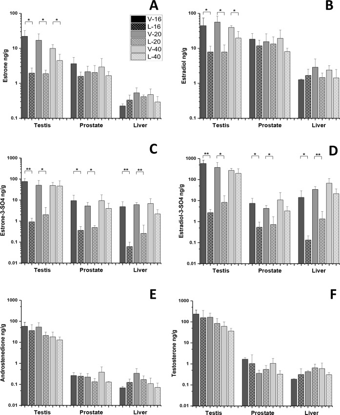
Letrozole administration resulted in statistically significant decreases in estradiol and estrone concentrations in testes at all ages, while no concentration changes in free estrogens were observed in prostate or liver (Fig 1A and 1B). Sulfated conjugates of estradiol and estrone were significantly decreased in the testis, prostate, and liver from the 16 and 20 week letrozole-treated boars but not in the 40 week letrozole-treated boars (Fig 1C and `D). Interestingly, estradiol sulfate and estrone sulfate concentrations were similar or lower than their free forms in the testis, prostate, and liver tissue extracts from 16 and 20 week letrozole-treated boars but not in their vehicle-treated littermates (Table 1). Hence, letrozole treatment alters testicular distribution of C18 steroid hormones in 16 and 20 week boars, but free C18 steroid distributions are less affected in the prostate and liver (Table 1). Letrozole treatment did not alter any androgen in any of the three tissues at any time point (Fig 1E and 1F, Table 1) compared with vehicle-treated littermates nor did it affect any C21 steroid.
Fig 1. Letrozole induced changes in tissue concentrations (pg/g) of estrogens and androgens in the testis, prostate, and liver.
Estrone (A) and estradiol (B) concentrations were decreased in the testis of 16, 20, and 40 week boars compared with vehicle-treated littermates. Estrone (A) and estradiol (B) concentrations in the prostate and liver were not affected by letrozole treatment. Estrone sulfate (C) and estradiol sulfate (D) concentrations were decreased compared with vehicle-treated littermates at week 16 and 20 in all tissues. The aromatase substrates, androstenedione (E) and testosterone (F), were not affected in any tissue by letrozole administration. * P < 0.05, ** P < 0.01.
Table 1. Steroid content of testis, prostate, and liver from puberal and postpuberal littermate boars treated with the aromatase inhibitor, letrozole, or with the canola oil vehicle1.
| Testis2 | ||||||
| C18 | V-16 | L-16 | V-20 | L-20 | V-40 | L-40 |
| Estrone | 22.07 (9.92) | 1.96 (0.78) | 17.03 (8.97) | 1.88 (0.49) | 9.96 (2.71) | 4.48 (2.28) |
| Estrone-3-SO4 | 76.11 (29.56) | 0.92 (0.45) | 50.75 (35.24) | 2.02 (2.44) | 49.52 (19.75) | 46.42 (35.49) |
| Estrone-3-GLU | ND | ND | ND | ND | ND | ND |
| 16α-Hydroxyestrone | 1.70 (0.68) | 0.04 (0.04) | 0.68 (0.08) | 0.13 (0.11) | 0.51 (0.31) | 0.33 (0.09) |
| Estradiol | 44.45 (29.11) | 7.88 (3.74) | 56.26 (27.04) | 7.86 (4.62) | 39.59 (4.83) | 19.68 (10.45) |
| Estradiol-3-SO4 | 586.5 (246.2) | 2.69 (1.14) | 381.6 (271.3) | 8.30 (8.91) | 273.9 (70.6) | 199.7 (129.9) |
| Estradiol-3-GLU | 0.27 (0.26) | 0.12 (0.18) | 0.31 (0.62) | 0.03 (0.06) | 0.02 (0.04) | 0.02 (0.03) |
| Estradiol-17-GLU | 3.64 (3.30) | 0.66 (0.77) | 2.94 (2.18) | 0.97 (1.08) | 2.13 (2.13) | 0.24 (0.38) |
| Estriol | 0.20 (0.29) | trace | 0.29 (0.41) | 0.01 (0.02) | 0.04 (0.08) | 0.04 (0.08) |
| C19 | V-16 | L-16 | V-20 | L-20 | V-40 | L-40 |
| Dehydroepiandrosterone | 85.96 (23.65) | 132.90 (138.80) | 98.91 (105.92) | 48.55 (45.99) | 52.26 (30.66) | 37.13 (32.51) |
| Dehydroepiandrosterone-SO4 | 237.8 (43.87) | 163.7 (179.6) | 193.7 (71.79) | 125.9 (70.36) | 98.37 (15.06) | 102.0 (55.65) |
| 5-Androstenediol | 75.40 (35.83) | 104.3 (107.7) | 86.29 (113.7) | 37.4 (30.84) | 84.27 (88.43) | 28.20 (22.01) |
| 4-Androstenediol | 5.58 (4.64) | 5.01 (4.81) | 5.35 (6.13) | 1.89 (1.21) | 4.77 (3.41) | 2.20 (1.87) |
| Androstenedione | 58.11 (31.33) | 36.20 (32.91) | 53.49 (32.36) | 21.24 (9.45) | 18.20 (12.55) | 13.08 (4.79) |
| 19-Hydroxyandrostendione | 12.80 (14.66) | 0.35 (0.34) | 10.60 (11.26) | 0.93 (0.99) | 2.61 (1.42) | 3.07 (2.78) |
| Testosterone | 236.2 (130.6) | 157.8 (192.4) | 166.5 (92.85) | 84.42 (59.00) | 61.86 (36.59) | 36.84 (12.43) |
| Epitestosterone | 4.50 (2.95) | 2.42 (3.65) | 4.81 (2.94) | 2.32 (0.76) | 1.01 (0.74) | 0.67 (0.49) |
| Epi/Testosterone-SO4 | 12.60 (6.59) | 11.32 (4.70) | 8.44 (6.36) | 4.27 (2.42) | 4.56 (3.92) | 7.53 (6.41) |
| 5α-Dihydrotestosterone | 1.87 (1.88) | 4.39 (6.76) | 2.42 (2.58) | 3.12 (3.31) | 0.54 (0.63) | 1.33 (1.09) |
| 6-Ketotestosterone | 59.83 (96.59) | 55.22 (75.38) | 45.08 (55.70) | 33.28 (27.30) | 7.44 (11.89) | 5.88 (5.80) |
| 6-Dehydrotestosterone | 0.86 (0.46) | 0.40 (0.25) | 0.29 (0.15) | 0.30 (0.09) | 0.21 (0.12) | 0.22 (0.13) |
| 17β-Dihydroepiandrosterone | 181.7 (162.6) | 174.8 (242.7) | 151.0 (170.3) | 98.98 (46.79) | 130.6 (93.27) | 82.13 (51.50) |
| 6α-Hydroxytestosterone | 12.16 (8.52) | 3.54 (6.53) | 9.13 (6.32) | 2.02 (3.49) | 1.49 (0.90) | 0.88 (0.79) |
| Adrenosterone | 2.74 (2.82) | 2.37 (2.47) | 1.40 (0.89) | 1.49 (1.15) | 0.39 (0.45) | 0.40 (0.29) |
| Epiandrosterone | 12.85 (7.10) | 15.66 (26.94) | 10.91 (7.72) | 10.13 (14.69) | 3.81 (4.12) | 6.51 (5.76) |
| C21 | ||||||
| Pregnenolone | 835.4 (357.8) | 822.5 (462.5) | 739.8 (594.0) | 806.3 (725.1) | 484.4 (107.0) | 397.8 (239.8) |
| Pregnenolone-3-SO4 | 59.3 (11.5) | 130.6 (177.8) | 66.7 (52.8) | 87.7 (77.3) | 39.8 (20.9) | 38.5 (33.6) |
| 17-Hydroxypregnenolone | 396.0 (265.0) | 406.5 (360.5) | 421.4 (507.5) | 395.9 (536.0) | 255.1 (208.4) | 214.1 (147.9) |
| Progesterone | 6.92 (2.26) | 10.30 (6.50) | 9.03 (9.34) | 16.86 (23.74) | 5.08 (4.11) | 4.14 (2.98) |
| 17-Hydroxyprogesterone | 2.52 (1.01) | 4.04 (2.70) | 3.48 (3.44) | 4.68 (4.61) | 2.89 (2.13) | 2.08 (1.08) |
| 11-Deoxycortisol | 2.88 (2.15) | 1.07 (0.72) | 2.25 (1.42) | 0.95 (0.71) | 0.97 (0.55) | 0.49 (0.30) |
| Cortisol | 4.14 (4.13) | 0.69 (0.79) | 3.27 (3.09) | 1.01 (0.99) | 2.10 (1.49) | 2.09 (1.39) |
| Deoxycorticosterone | 1.59 (1.22) | 0.50 (0.43) | 1.35 (1.02) | 0.44 (0.26) | 0.44 (0.35) | 0.27 (0.11) |
| Corticosterone | 0.27 (0.52) | 0.41 (0.43) | 1.02 (1.81) | 0.16 (0.12) | 0.37 (0.27) | 0.42 (0.27) |
| Epiallopregnanolone | 78.13 (62.52) | 120.8 (147.3) | 81.82 (97.05) | 162.6 (179.2) | 81.87 (95.14) | 47.55 (25.79) |
| Allo/Pregnanolone-SO4 | 1.59 (1.36) | 3.78 (3.38) | 0.93 (0.61) | 1.90 (1.84) | 0.42 (0.33) | 0.23 (0.18) |
| Epi/Epiallopregnanolone-SO4 | 2.91 (1.80) | 21.39 (35.92) | 7.94 (11.73) | 14.97 (18.26) | 4.37 (5.82) | 3.15 (3.72) |
| Prostate3 | ||||||
| C18 | V-16 | L-16 | V-20 | L-20 | V-40 | L-40 |
| Estrone | 3.64 (1.86) | 1.59 (0.51) | 2.14 (0.92) | 2.04 (1.18) | 2.94 (2.15) | 1.65 (0.49) |
| Estrone-3-SO4 | 9.37 (7.78) | 0.36 (0.19) | 5.27 (2.42) | 0.49 (0.12) | 9.32 (8.19) | 4.00 (3.01) |
| Estrone-3-GLU | ND | ND | ND | ND | ND | ND |
| 16α-Hydroxyestrone | ND | ND | ND | ND | ND | ND |
| Estradiol | 18.33 (8.28) | 11.80 (5.35) | 15.50 (10.56) | 13.34 (7.89) | 19.43 (14.06) | 8.04 (1.98) |
| Estradiol-3-SO4 | 7.44 (5.38) | 0.54 (0.42) | 4.28 (1.64) | 0.73 (0.99) | 10.86 (13.35) | 3.25 (1.92) |
| Estradiol-3-GLU | 0.35 (0.57) | 0.002 (0.002) | 0.05 (0.09) | 0.12 (0.17) | 1.31 (0.99) | 0.02 (0.03) |
| Estradiol-17-GLU | 0.42 (0.72) | 0.002 (0.001) | 0.20 (0.40) | 1.51 (2.00) | 1.40 (1.23) | 0.94 (0.86) |
| Estriol | ND | ND | ND | ND | ND | ND |
| C19 | ||||||
| Dehydroepiandrosterone | ND | ND | ND | ND | ND | ND |
| Dehydroepiandrosterone-SO4 | 29.29 (19.57) | 9.89 (7.27) | 15.36 (5.18) | 12.16 (7.53) | 31.03 (24.15) | 13.15 (10.51) |
| 5-Androstenediol | ND | ND | ND | ND | ND | ND |
| 4-Androstenediol | ND | ND | ND | ND | ND | ND |
| Androstenedione | 0.26 (0.10) | 0.24 (0.09) | 0.22 (0.10) | 0.13 (0.06) | 0.38 (0.29) | 0.13 (0.01) |
| 19-Hydroxyandrostendione | ND | ND | ND | ND | ND | ND |
| Testosterone | 1.67 (0.32) | 1.04 (1.67) | 0.36 (0.15) | 0.55 (0.29) | 1.07 (0.76) | 0.33 (0.15) |
| Epitestosterone | 0.01 (0.02) | 0.09 (0.07) | 0.02 (0.02) | 0.09 (0.10) | 0.27 (0.53) | 0.27 (0.28) |
| Epi/Testosterone-SO4 | ND | ND | ND | ND | ND | ND |
| 5α-Dihydrotestosterone | 2.51 (1.10) | 1.79 (1.17) | 1.29 (0.84) | 1.23 (0.39) | 0.93 (0.95) | 0.99 (0.49) |
| 6-Ketotestosterone | ND | ND | ND | ND | ND | ND |
| 6-Dehydrotestosterone | ND | ND | ND | ND | ND | ND |
| 17β-Dihydroepiandrosterone | trace | trace | trace | trace | trace | trace |
| 6α-Hydroxytestosterone | ND | ND | ND | ND | ND | ND |
| Adrenosterone | ND | ND | ND | ND | ND | ND |
| Epiandrosterone | 4.76 (2.82) | 6.24 (5.94) | 6.01 (4.52) | 7.01 (4.62) | 4.41 (3.25) | 3.27 (2.01) |
| C21 | ||||||
| Pregnenolone | ND | ND | ND | ND | ND | ND |
| Pregnenolone-3-SO4 | 0.49 (0.39) | 0.36 (0.29) | 0.42 (0.19) | 1.29 (0.88) | 0.98 (0.80) | 0.62 (0.55) |
| 17-Hydroxypregnenolone | ND | ND | ND | ND | ND | ND |
| Progesterone | 0.70 (0.34) | 0.31 (0.21) | 0.73 (0.86) | 1.64 (2.36) | 0.58 (0.50) | 0.25 (0.20) |
| 17-Hydroxyprogesterone | ND | ND | ND | ND | ND | ND |
| 11-Deoxycortisol | ND | ND | ND | ND | ND | ND |
| Cortisol | 2.85 (1.87) | 0.86 (0.72) | 3.13 (4.05) | 3.39 (3.70) | 3.38 (3.57) | 1.12 (0.69) |
| Deoxycorticosterone | 0.24 (0.36) | 0.11 (0.10) | 0.24 (0.26) | 0.24 (0.25) | 0.07 (0.11) | 0.08 (0.07) |
| Corticosterone | 1.84 (1.38) | 0.36 (0.50) | 0.82 (1.15) | 2.32 (3.12) | 0.26 (0.17) | 0.35 (0.28) |
| Epiallopregnanolone | ND | ND | ND | ND | ND | ND |
| Allo/Pregnanolone-SO4 | ND | ND | ND | ND | ND | ND |
| Epi/Epiallopregnanolone-SO4 | trace | trace | trace | trace | trace | trace |
| Liver4 | ||||||
| C18 | V-16 | L-16 | V-20 | L-20 | V-40 | L-40 |
| Estrone | 0.22 (0.03) | 0.33 (0.13) | 0.53 (0.20) | 0.41 (0.05) | 0.48 (0.21) | 0.29 (0.12) |
| Estrone-3-SO4 | 4.84 (3.40) | 0.06 (0.04) | 6.02 (1.51) | 0.26 (0.39) | 6.85 (4.79) | 2.18 (1.30) |
| Estrone-3-GLU | 1.06 (0.36) | 0.12 (0.22) | 0.14 (0.13) | 0.36 (0.44) | 1.40 (2.23) | 0.90 (1.01) |
| 16α-Hydroxyestrone | 0.01 (0.003) | 0.03 (0.01) | 0.02 (0.01) | 0.02 (0.01) | 0.02 (0.01) | 0.02 (0.004) |
| Estradiol | 1.27 (0.05) | 1.68 (0.84) | 2.89 (2.07) | 1.47 (0.50) | 2.40 (0.80) | 1.43 (1.06) |
| Estradiol-3-SO4 | 14.26 (15.44) | 0.13 (0.08) | 34.31 (12.07) | 1.35 (1.70) | 67.40 (43.92) | 21.61 (14.41) |
| Estradiol-3-GLU | 1.19 (1.33) | 0.004 (0.01) | 0.95 (0.88) | 0.02 (0.02) | 3.05 (3.03) | 2.18 (1.72) |
| Estradiol-17-GLU | 1.11 (1.57) | 0.32 (0.64) | 0.57 (0.47) | 0.13 (0.15) | 1.79 (0.29) | 0.62 (0.67) |
| Estriol | 0.07 (0.10) | trace | 0.16 (0.23) | trace | 0.01 (0.02) | 0.21 (0.34) |
| C19 | V-16 | L-16 | V-20 | L-20 | V-40 | L-40 |
| Dehydroepiandrosterone | ND | ND | ND | ND | ND | ND |
| Dehydroepiandrosterone-SO4 | 11.14 (4.38) | 6.96 (3.49) | 16.70 (4.33) | 11.66 (10.59) | 17.78 (14.61) | 9.23 (6.83) |
| 5-Androstenediol | ND | ND | ND | ND | ND | ND |
| 4-Androstenediol | ND | ND | ND | ND | ND | ND |
| Androstenedione | 0.07 (0.01) | 0.13 (0.05) | 0.34 (0.23) | 0.17 (0.08) | 0.11 (0.09) | 0.07 (0.04) |
| 19-Hydroxyandrostendione | ND | ND | ND | ND | ND | ND |
| Testosterone | 0.19 (0.01) | 0.31 (0.27) | 0.43 (0.05) | 0.65 (0.32) | 0.60 (0.49) | 0.31 (0.08) |
| Epitestosterone | 0.26 (0.08) | 0.18 (0.15) | 0.14 (0.11) | 0.25 (0.23) | 0.20 (0.21) | 0.19 (0.19) |
| Epi/Testosterone-SO4 | 2.35 (2.00) | 2.15 (2.10) | 2.87 (0.76) | 2.90 (3.31) | 4.93 (2.03) | 4.79 (4.31) |
| 5α-Dihydrotestosterone | 0.04 (0.06) | 0.09 (0.10) | 0.04 (0.07) | 0.01 (0.02) | 0.13 (0.15) | 0.05 (0.07) |
| 6-Ketotestosterone | ND | ND | ND | ND | ND | ND |
| 6-Dehydrotestosterone | ND | ND | ND | ND | ND | ND |
| 17β-Dihydroepiandrosterone | ND | ND | ND | ND | ND | ND |
| 6α-Hydroxytestosterone | ND | ND | ND | ND | ND | ND |
| Adrenosterone | ND | ND | ND | ND | ND | ND |
| Epiandrosterone | ND | ND | ND | ND | ND | ND |
| C21 | V-16 | L-16 | V-20 | L-20 | V-40 | L-40 |
| Pregnenolone | ND | ND | ND | ND | ND | ND |
| Pregnenolone-3-SO4 | 0.60 (0.02) | 0.71 (0.86) | 0.87 (0.54) | 1.07 (0.64) | 0.95 (0.39) | 0.96 (0.76) |
| 17-Hydroxypregnenolone | ND | ND | ND | ND | ND | ND |
| Progesterone | 0.06 (0.03) | 0.17 (0.11) | 1.09 (1.16) | 1.05 (1.09) | 0.30 (0.06) | 0.30 (0.14) |
| 17-Hydroxyprogesterone | ND | ND | ND | ND | ND | ND |
| 11-Deoxycortisol | 0.14 (0.08) | 0.28 (0.25) | 2.04 (2.09) | 1.87 (1.86) | 0.36 (0.27) | 0.30 (0.19) |
| Cortisol | 0.47 (0.06) | 1.64 (2.25) | 13.23 (12.51) | 7.73 (8.98) | 1.40 (1.14) | 1.70 (1.05) |
| Deoxycorticosterone | 0.03 (0.01) | 0.04 (0.04) | 0.38 (0.33) | 0.36 (0.45) | 0.11 (0.08) | 0.07 (0.05) |
| Corticosterone | 0.20 (0.21) | 0.30 (0.26) | 0.60 (0.54) | 0.86 (0.91) | 0.07 (0.06) | 0.12 (0.11) |
| Epiallopregnanolone | ND | ND | ND | ND | ND | ND |
| Allo/Pregnanolone-SO4 | ND | ND | ND | ND | ND | ND |
| Epi/Epiallopregnanolone-SO4 | trace | trace | trace | trace | trace | trace |
1Values represent means (SD) in ng/g. Values in bold indicate statistically significant differences between letrozole-treated boars and vehicle-treated littermates at the indicated age.
2Values for testis tissue at each age are based upon one boar from each of four litters treated with letrozole from 11 to 16 weeks of age and littermates treated with the canola oil vehicle. All boars came from a total of five litters.
3Values for prostate tissue at each age are based upon one boar from each of four litters treated with letrozole from 11 to 16 weeks of age (three boars treated with vehicle at 16 weeks of age and three boars treated with letrozole at 20 weeks of age) and littermates treated with the canola oil vehicle. Tissues came from same boars that donated testicular tissue.
4Values for liver tissue at each age are based upon one boar from each of four litters treated with letrozole from 11 to 16 weeks of age (three boars treated with vehicle at 16 weeks of age and at 20 weeks of age) and littermates treated with the canola oil vehicle. Tissues came from same boars that donated testicular tissue.
Testis, prostate, and liver have unique C18, C19, and C21 steroid profiles
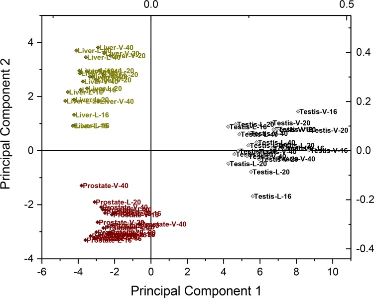
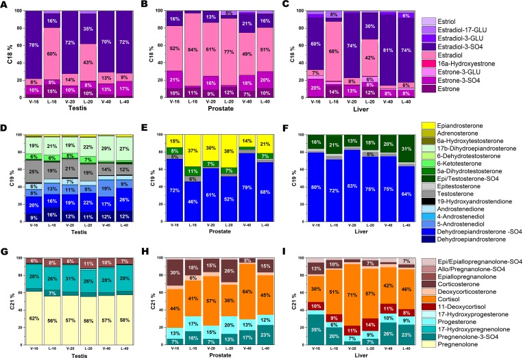
Each tissue had a unique steroid distribution, which drove the clustering of samples in principal component analysis regardless of treatment group (Figs 2 and 3), although DHEA-sulfate was the predominant C19 steroid hormone in all tissues. The testes had the most diverse steroid profile of the three tissues investigated, containing 40 different identified C18, C19, and C21 steroids (Table 1). This represents a relatively small proportion of the 92 estrogens (C18), 50 androgens (C19), and 60 progestogens and gluco/mineralocorticoids (C21) that might have been detected with this method (Table 2). Quantitatively, the C21 steroids had the largest overall contribution in the testes, followed by C19 and C18 steroids (Table 1). Pregnenolone was the most abundant steroid in boar testis followed by 17-hydroxypregnenolone or estradiol 3-SO4. Pregnenolone levels were twice the concentration of 17-hydroxypregnenolone and five or more fold higher than any other C21 steroid in the testis in all boars. Interestingly, neither pregnenolone nor 17-hydroxypregnenolone was detected in prostate or liver. Pregnenolone-sulfate was detected in all three tissues with concentrations in testis being 20 or more fold higher than concentrations in the prostate and liver.
Fig 2. Principal component analysis of steroids in the boar testis, prostate, and liver.
The source organ was the primary influence for clustering of steroid profiles rather than age or treatment. The first two of the principal components accounted for 68% of the total variance (54% and 14%) and remaining calculated components each explained 2–7% of the total variance and were not subsequently considered.
Fig 3. Steroid distributions of C18, C19, and C21 in the testis, prostate, and liver.
C18 steroids (A-C) consist of estrogen derivatives, C19 steroids (D-F) consist of androgen derivatives, and C21 steroids (G-I) consist of progestogens, gluco/mineralocorticoids, and derivatives. Each tissue had a distinct profile. C refers to vehicle-treated littermates and L to letrozole-treated littermates. The number following C or L refers to the age of the animal at tissue recovery. Data based on nmol/g tissue.
Table 2. List of steroids evaluated in profile.
| Steroid | LOD1 | Steroid | LOD1 |
|---|---|---|---|
| Pregnenolone | 1.58 | Estradiol-3-hemisuccinate | 0.001 |
| 17-Hydroxypregnenolone | 1.504 | Estrone-3-hemisuccinate | 0.007 |
| Dehydroepiandrosterone | 0.087 | Estradiol-17-hemisuccinate | 0.268 |
| 5-Androstenediol | 0.172 | Estradiol-3,17-di-hemisuccinate | 0.001 |
| Progesterone | 0.003 | Estriol-3-hemisuccinate | 0.064 |
| 17-Hydroxyprogesterone | 0.007 | Allopregnanolone | 0.785 |
| 11-Deoxycortisol | 0.007 | Alldihydroprogesterone | 1.58 |
| Cortisol | 0.068 | Allopregnanediol | 0.1 |
| 11-Deoxycorticosterone | 0.015 | 11α-Hydroxyprogesterone | 0.757 |
| Corticosterone | 0.072 | 11β-Hydroxyprogesterone | 0.076 |
| Aldosterone | 0.693 | 21-Hydroxypregnanolone | 3.008 |
| Androstenedione | 0.009 | 21-Hydroxypregnenolone | 3.008 |
| Testosterone | 0.009 | 7α-Hydroxypregnenolone | 3.008 |
| 5α-Dihydrotestosterone | 0.344 | 2α-Hydroxytestosterone | 0.81 |
| Estrone | 0.009 | 20α-Hydroxy-5a-pregnan-3-one | 0.785 |
| Estradiol | 0.009 | 20α-Hydroxyprogesterone | 0.521 |
| 11α-Hydroxyestrone | 0.087 | 17α-20a-Dihydroprogesterone | 0.923 |
| 11β-Hydroxyestrone | 0.873 | Pregnenolone-SO4 | 0.063 |
| 11α-Hydroxyestradiol | 0.173 | Pregnanolone | 0.785 |
| 11β-Hydroxyestradiol | 1.734 | Pregnanediol | 1.56 |
| 9(11)-Dehydroestradiol | 0.092 | Pregnanedione | 1.58 |
| 9(11)-Dehydroestrone | 0.093 | Epiallopregnanolone | 1.36 |
| 11-Ketoestrone | 0.088 | 17β-Dihydroandrosterone | 3.419 |
| Estradiol-3-SO4 | 0.025 | 17β-Dihydroepiandrosterone | 1.71 |
| Estrone-3-SO4 | 0.076 | 7α-Hydroxytestosterone | 0.821 |
| Estradiol-17-SO4 | 0.049 | 7α-Hydroxyandrostenedione | 0.083 |
| Estradiol-3,17-SO4 | 0.216 | 6α-Hydroxytestosterone | 0.019 |
| 3-o-methoxy-estrone | 0.009 | 6β-Hydroxytestosterone | . |
| Estradiol-3-GLU | 1.062 | Dehydroepiandrosterone-GLU | 3.46 |
| Estrone-3-GLU | 0.073 | 16α-Hydroxydehydroepiandrosterone | 8.55 |
| Estradiol-17-GLU | 1.062 | Testosterone-SO4 | 0.062 |
| Estrone-3,17-GLU | 0.327 | Desoxytestosterone | 0.544 |
| 4-Hydroxyestrone | 0.002 | Testosterone-GLU | 0.166 |
| 4-Hydroxyestradiol | 0.347 | 4-Androsten-3α-ol-17-one | 2.19 |
| 4-o-Methoxy-estrone | 0.083 | 2α-Hydroxyandrostenedione | 6.22 |
| 4-o-Methoxy-estradiol | 3.307 | 11α-Hydroxyandrostenedione | 1.84 |
| 4-o-Methoxy-estriol | 15.703 | 6-Ketotestosterone | 0.413 |
| 4-Hydroxy-estrone-2-SG | 0.042 | 11-Ketotestosterone | 0.928 |
| 4-Hydroxy-estradiol-2-SG | 0.169 | 9-Dehydrotestosterone | . |
| 4-Hydroxy-estrone-2-CYS | 0.025 | Allotetrahydrocortexone | . |
| 4-Hydroxy-estradiol-2-CYS | 0.614 | Adrenosterone | 0.227 |
| 4-Hydroxy-estrone-2-NAcCYS | 0.224 | Etiocholanolone | . |
| 4-Hydroxy-estradiol-2-NAcCYS | 0.223 | 4-Androstenediol | 0.826 |
| 4-Hydroxy-estrone-1-N3Ade | 0.012 | 16α-Ketotestosterone | 2.21 |
| 4-Hydroxy-estradiol-1- N3Ade | 0.024 | 19-Hydroxyandrostenedione | 0.235 |
| 4-Hydroxy-estrone-1-N7Gua | 0.23 | 4-Androsten-3,6-17-trione | 2.63 |
| 4-Hydroxy-estradiol-1- N7Gua | 0.229 | 7β-Hydroxydehydroepiandrosterone | 6.24 |
| 2-Hydroxyestrone | 0.349 | 7α-Hydroxydehydroepiandrosterone | 6.22 |
| 2-Hydroxyestradiol | 0.347 | 7-Ketodehydroepiandrosterone | 1.12 |
| 2-Hydroxy-estrone-1+4-SG | 0.423 | 9-Dehydroepiandrosterone | . |
| 2-Hydroxy-estradiol-1+4-SG | 0.421 | Cortisone | . |
| 2-Hydroxy-estrone-1+4-Cys | 0.247 | 9-Dehydroprogesterone | 5.29 |
| 2-Hydroxy-estradiol-1+4-Cys | 0.614 | 11-Dehydrotetrahydrocosticosterone | 0.814 |
| 2-Hydroxy-estrone-1+4-NAcCys | 0.112 | Tetrahydrocorticosterone | . |
| 2-Hydroxy-estradiol-1+4-S NAcCys | 0.557 | 6β-Hydroxycorticosterone | 0.618 |
| 2-Hydroxyestrone-6-N3Ade | 0.119 | 5α-Dihydrocorticosterone | 1.66 |
| 2-Hydroxyestradiol-6-N3Ade | 0.119 | 5α-Dihydropregnanolone | . |
| 2-Hydroxyestriol | 0.008 | Tetrahydrocortisol | . |
| 3-Methoxy-2-hydroxy-estrone | 0.166 | Pregnanetriol | 1.31 |
| 2-Methoxy-3-hydroxy-estrone | 0.008 | Allotetrahydrocortisol | . |
| 2-Methoxy-3-hydroxy-estradiol | 0.331 | 16α-Hydroxyprogesterone | 2.25 |
| 2,3 Dimethoxy-estrone | 0.032 | 6-Dehydrotestosterone | 3.27 |
| 2,3 Dimethoxy-estradiol | 0.079 | 9(11)-Didehydroepiandrosterone | 9.36 |
| 6α-Hydroxyestradiol | 0.173 | 11β-Hydroxyepiandrosterone | 1.28 |
| 6β-Hydroxyestradiol | 0.173 | 16α-Hydroxyepiandrosterone | 8.55 |
| 6-Ketoestrone | 0.088 | Androsterone | . |
| 6-Ketoestradiol | 0.087 | Epiandrosterone | 0.67 |
| 6-Dehydroestradiol | 0.092 | 7α-Hydroxyandrostenediol | 1.38 |
| 6-Dehydroestrone | 0.019 | 7β-Hydroxyandrostenediol | 1.29 |
| 16α-Hydroxyestrone | 0.175 | 5β-Dihydrocorticosterone | . |
| 17-Epiestriol | 0.173 | 4-Pregnen-3b-ol-20-one | 8.06 |
| Estriol | 0.347 | Urocortisone | 0.067 |
| 16,17-Epiestriol | 3.468 | 5-Androsten-3b,17-diol-16-one | 3.94 |
| 16-Epiestriol | 3.468 | 19-Hydroxydehydroepiandrosterone | 2.8 |
| Estriol-3-SO4 | 0.271 | 16α-Hydroxypregnenolone | 4.401 |
| Estriol-3-GLU | 2.153 | 5α-Dihydrocortexone | 1.62 |
| 3-Methoxy-estriol | 0.002 | Epitestosterone | 0.174 |
| 16-Keto-17β-estradiol | 3.492 | 5β-Androstanedione | . |
| 16-Ketoestriol | 0.331 | 11β-Hydroxyandrostenedione | . |
| 7-Dehydro-17β-estradiol | 3.699 | 11-Ketoetiocholanolone | . |
| Equilin | 0.019 | 5α-Pregnan-11a-ol-3,20-dione | 2.51 |
| Dihydroequilin-3-SO4 | 1.342 | 5α-Epoxypregnenolone | . |
| Equilin-3-SO4 | 6.749 | Allopregnanetrione | . |
| Estrone-9-N3-Ade | 0.124 | 11-Ketoprogesterone | 3.12 |
| Estradiol-9-N3-Ade | 0.247 | 11-Dehydrocorticosterone | . |
| Estradiol-3-acetate | 0.08 | 6β-Hydroxycortisol | . |
| Estrone-3-acetate | 0.16 | 20β-Dihydrocortisone | . |
| Estradiol-3,17α-di-acetate | 0.07 | 6β-Hydroxycortisone | . |
| Estradiol-3,17β-di-acetate | 1.402 | Etiocholanolone-GLU | 0.398 |
| 2-Hydroxy-estradiol-17-acetate | 0.003 | Dehydroepiandrosterone-SO4 | 0.198 |
| 6-Ketoestradiol-3,17-di-acetate | 0.006 | Epitestosterone-SO4 | 0.181 |
| 6-Ketoestradiol-tri-acetate | 0.023 | Epitestosterone-GLU | 0.126 |
| 6-Dehydroestradiol-di-acetate | 0.141 | Pregnanediol-GLU | 0.0183 |
| Estriol-3-acetate | 0.303 | 17α-Hydroxypregnanolone-GLU | 1.76 |
| Estriol-16-acetate | 0.303 | Pregnanolone-SO4 | 0.133 |
| Estriol-16,17-diacetate | 0.268 | 17α,20β-Dihydroxyprogesterone-GLU | 0.102 |
| 17-Epiestriol triacetate | 0.241 | Hydrocortisone-21-SO4 | . |
| Estriol-triacetate | 0.006 | Pregnenolone-GLU | . |
| 16,17-Epiestriol triacetate | 0.06 | Allopregnanolone-SO4 | 0.014 |
| 16-Epiestriol triacetate | 0.121 | Epiallopregnanolone-SO4 | 0.012 |
| Equilin acetate | 0.081 | Corticosterone-21-SO4 | . |
1LOD values are estimates of the limit of detection. Values in italics are based on a limited assessment and no estimates are available for compounds with a period.
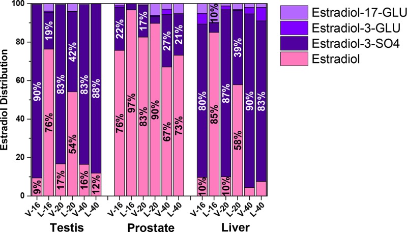
The prostate contained 20 different identified C18, C19, and C21 steroids (Table 1). Estradiol or DHEA-sulfate was the most abundant steroid in the prostate. Regardless of treatment, total C21,C19,C18 steroid concentrations in the prostate were ~50% C18, ~50% C19, and less than 10% C21 (Table 1). Total C18 steroids in letrozole-treated animals were less than half the concentration detected in the testis in vehicle-treated littermates. Most of the C18 steroids were present in an unconjugated form in the prostate in contrast to the conjugated form in the testis (Fig 4). Only six different C19 steroids were detected in the prostate compared with 17 detected in the testis. Although most C19 steroids were markedly lower in the prostate than the testis, 5α-dihydrotestosterone had a similar concentration in both tissues.
Fig 4. Distribution of estradiol and estradiol conjugates in testis, prostate, and liver.

Estradiol was the predominant C18 steroid in each of these tissues. The prominence of estradiol sulfate in testis and liver from vehicle-treated boars contrasts with the limited concentration of the sulfo-conjugate in the prostate, even in vehicle-treated boars. A shift in distribution between free estradiol and estradiol sulfate is apparent in the 16 and 20 week letrozole-treated littermates. C refers to vehicle-treated littermates and L to letrozole-treated littermates. The number following C or L refers to the age of the animal at tissue recovery.
The liver contained 23 different identified C18, C19, and C21 steroids. The C18 steroids quantitatively accounted for over 50% of total steroids in vehicle-treated boars and less than 20% of the total steroids in the liver from the 16 and 20 week letrozole-treated boars consistent with reduced production of C18 steroids in these boars (Table 1). Regardless of treatment, steroid levels in liver tissue were much, much lower than in the testis and total free steroid concentration was much lower than in the prostate.
Prostate enzymes
Aromatase activity in the prostate was detectable but almost 1000 fold lower than that in the testes of the same animals (0.08 vs 65.20 pmoles/mg protein/h, standard error = 0.27, P < 0.001). Concentrations of the aromatase precursors, testosterone and androstenedione were quite low in the prostate (less than 2% of testicular levels) and not affected by letrozole treatment. Measurable aromatase activity was not detectable in liver tissues from 16 week boars. In contrast, gene expression for steroid sulfatase (STS), the enzyme involved in removing sulfate, was detectable in prostate and testis and liver tissues with similar relative gene expression (ΔCt of 2.88, 1.86, and 2.0 respectively, P > 0.25, standard error = 0.92).
Discussion
To our knowledge, this study provides the most comprehensive assessment of steroid tissue concentrations in the boar to date. Although the steroids evaluated were extensive, the pheromone androstenone was not included in the panel. Testicular concentrations detected in this study were consistent with the testicular origin indicated for previously described steroid sulfates [41]. In contrast to a previous suggestion that dihydrotestosterone increased from puberty to maturity [42], our data do not suggest an increase in dihydrotestosterone in either the testis or the prostate although our data did not include comparisons with prepuberal timepoints. Apparent age-related decreases in testicular steroids among vehicle-treated boars may reflect increased testicular mass, due at least in part to increased spermatogenesis. Our multi-tissue approach in combination with a broad steroid profile provide critical information on the role of each of these tissues in steroidogenesis, steroid intracrinology, and steroid sulfate metabolism.
Endogenous estrogen synthesis and metabolism
Letrozole administration uniquely affected endogenous C18 steroids in the testis, prostate, and liver, revealing tissue-specific regulation of estrogen synthesis and synthetic pathways. Testicular estrogens in the boar are primarily synthesized locally via aromatization of androgen precursors [27, 41, 43]; testicular estrogen sulfates result from subsequent local conjugation by sulfotransferases [27, 29, 30]. Letrozole inhibited testicular aromatase, resulting in the direct decrease in estrone and estradiol concentrations and an indirect decrease in the corresponding testicular sulfo-conjugate concentrations. A decrease in 19-hydroxyandrostenedione, an intermediate of aromatase action on androstenedione, might be expected. Although values for 19-hydroxyandrostenedione were numerically much lower in letrozole-treated boars than in vehicle-treated littermates, values were not significantly different, perhaps due to high variability among boars. Letrozole administration during the prepuberal interval had a prolonged effect on estrogen production in this study similar to the reprogramming of aromatase including reduced protein and enzymatic activity observed long after letrozole administration ceased during the neonatal and juvenile intervals [44].
Estrogen sulfate and free estrogen concentrations were affected differently by letrozole administration in the prostate and liver; concentrations of free estrogens were unaffected by letrozole treatment while estrone-sulfate and estradiol-sulfate concentrations were significantly decreased in tissue from letrozole-treated boars. Consistent with the high levels of sulfatase present in the prostate [7, 45], in vitro evidence suggests estrone and estradiol are produced in greater quantities in the human prostate by sulfatase than by aromatase [46]. Similarly, the very, very low aromatase activity we detected in the prostate in conjunction with gene expression for steroid sulfatase, the enzyme that removes sulfate from sulfated steroids, suggests local transformation from estrogen sulfates may maintain free estrogen levels in the boar prostate as well. Higher estradiol concentrations in the prostates from 16 and 20 week letrozole-treated boars than concentrations in the testes from these same boars are consistent with this suggestion of local transformation in the prostate distinct from testicular regulation. The significant decrease in estrogen sulfates in the prostate may reflect desulfation of testicular-derived estrogen sulfates in order to maintain free estrogen concentrations in the prostate. Furthermore, prostate intracrine metabolism may enable independent regulation/synthesis of estrogens and androgens via separate testicular precursors, estrogen sulfate and DHEA-sulfate, respectively.
Dependence of boar prostate size on estrogens and androgens was previously addressed by Booth et al. [47–49]. Castrated boars were treated with dihydrotestosterone, DHEA, estrone, or estradiol and prostate size along with other secondary sex glands were monitored. Only males administered estrone or estradiol showed significantly increased mass of prostate, seminal vesicles, and bulbourethral glands [47, 48], confirming that these accessory sex glands are dependent on estrogens. Interestingly, castrated boars administered DHEA, did not have detectable levels of free estrogens and did not have an increased prostate mass [47]. This finding suggests that DHEA is unlikely to be a major estrogen precursor in the prostate consistent with primarily aromatase-independent synthesis of estrogens in the prostate. Our data extend these previous findings suggesting that the porcine prostate primarily synthesizes free estrogens from testicular estrogen sulfates, rather than from DHEA similar to the role of sulfatase proposed in the human prostate [9]. These results are also consistent with use of sulfatase inhibitors to inhibit local synthesis of active estrogens in prostate cancer therapy [46, 50, 51]. Hence, although free estrogen concentrations are significantly decreased in the primary steroidogenic tissue, testes, and systemic circulation [32], free estrogens in a peripheral tissue, the prostate, were not affected. The ability of the prostate to function independently of testicular and systemic influences strongly suggests that future steroid-hormone therapeutic approaches should account for the contribution of steroid sulfates and sulfatase in steroid hormone production in a tissue.
Historically, sulfated steroid hormones in the systemic system were considered metabolic end products, primarily synthesized in the liver, that aid in excretion [52]; although research has challenged and abandoned conceptions of this simple biological role and exclusive origin of steroid sulfates [3, 7, 53]. Based on the current study, the boar liver likely does not drive or significantly impact systemic estrogen sulfate concentrations but likely functions to aid in excretion of circulating estrogens. The expression of estrogen receptors [54] and estrogen-induced hepatocyte regeneration [55] suggest that liver can be a target for estrogens. Furthermore, the liver possesses metabolic machinery to both conjugate and de-conjugate estrogens [7, 27, 56], providing the ability to regulate estrogen concentrations and distribution of free estrogens independent of circulating concentrations. However, free estrogen levels were very low in the liver, suggesting minimal signaling.
Sulfated steroids in tissues can be altered after tissue collection by sulfatase enzymatic activity [30]. However, homogenization of the frozen tissue in ice-cold methanol in the present study would be expected to denature these enzymes prior to significant conversion. The ratios of sulfated to free steroids in the present study are consistent with postharvest inhibition of sulfatase activity in the testis [30].
Progestogen and gluco/mineralocorticoid synthesis and metabolism
The predominance of C21 steroids detected in the testis is consistent with the testis as a primary endocrine gland in the boar. Pregnenolone and 17-hydroxypregnenolone were prominent in the testis and most likely function as intermediary compounds in testicular steroidogenesis [57]; neither steroid is secreted in quantitatively significant amounts [26]. Endogenous estrogens and androgens can be synthesized metabolically from pregnenolone via either the Δ5- or Δ4- pathway in the boar [28, 58]. Metabolites in testis extract are consistent with predominance of the Δ5- pathway in the testis and support presence of both pathways. These results are consistent with the testis being the primary site for synthesis of systemic DHEA-sulfate [59, 60] as it is in most mammals other than primates [61].
Production of steroid hormones in the prostate likely result from precursor steroids provided systemically. The C21 steroid precursors, pregnenolone, 17-hydroxypregenenolone, and 17-hydroxyprogesterone, were not detectable suggesting they are not components of steroid synthesis within the prostate. Other steroid components of the Δ5- pathway were similarly not detectable in the prostate or liver and other components of the Δ4- pathway were not detectable or present in only minimal quantities. The lack of substantial free C21 steroids and paucity of the Δ5- or Δ4- pathway metabolites, support the hypothesis that the boar prostate and liver utilize testis and/or adrenal derived steroids for local steroid hormone synthesis [7, 27] and contrasts with the abundance of these C21 steroid precursors in primary endocrine glands like the testis.
Conclusion
A multi-tissue approach in combination with aromatase inhibition and investigation of a broad array of steroids, revealed complexity and unique roles for individual organs in steroid hormone synthesis. Results from animals treated with letrozole displayed the primary role of the testis in synthesis of estrogens and estrogen sulfates, and suggested the dependence of the prostate on sulfated testis-derived steroid hormone precursors. Furthermore, individual organs may maintain tissue-specific profiles of active steroid hormones independent of systemic influences. Our research sheds light on the critical regulatory role of steroid sulfates in a tissue and suggests future research on the interplay of free and sulfo-conjugated steroid hormones within a tissue.
Acknowledgments
The authors would like to thank Dr. Nilesh Gaikwad for his technical expertise.
Data Availability
An excel style file containing steroid concentrations for each tissue from each animal is available at https://doi.org/10.25338/B85P5T, a DASH site from the California Digital Library, University of California.
Funding Statement
Research was supported by the National Research Initiative Competitive Grant 2008-35203-19082 from USDA National Institute of Food and Agriculture (https://nifa.usda.gov/) to TB and AC, W3171 MSP (https://www.nimss.org/projects/view/mrp/outline/16596) to TB, a W.K. Kellogg Endowment to TB, and the infrastructure support of the Department of Animal Science, College of Agricultural and Environmental Sciences (https://animalscience.ucdavis.edu/), and the California Agricultural Experiment Station (https://caes.ucdavis.edu/research/aes of the University of California–Davis. The funders had no role in study design, data collection and analysis, decision to publish, or preparation of the manuscript.
References
- 1.Strott CA. Steroid sulfotransferases. Endocrine reviews. 1996;17(6):670–97. 10.1210/edrv-17-6-670 [DOI] [PubMed] [Google Scholar]
- 2.Strott CA. Sulfonation and molecular action. Endocrine reviews. 2002;23(5):703–32. 10.1210/er.2001-0040 [DOI] [PubMed] [Google Scholar]
- 3.Cameron CB. The liver and steroid hormone metabolism. British medical bulletin. 1957;13(2):119–25. [DOI] [PubMed] [Google Scholar]
- 4.Fluck CE, Pandey AV. Steroidogenesis of the testis—new genes and pathways. Annales d'endocrinologie. 2014;75(2):40–7. 10.1016/j.ando.2014.03.002 [DOI] [PubMed] [Google Scholar]
- 5.Gurpide E. Enzymatic modulation of hormonal action at the target tissue. Journal of toxicology and environmental health. 1978;4(2–3):249–68. 10.1080/15287397809529660 [DOI] [PubMed] [Google Scholar]
- 6.Labrie F, Luu-The V, Belanger A, Lin SX, Simard J, Pelletier G, et al. Is dehydroepiandrosterone a hormone? The Journal of endocrinology. 2005;187(2):169–96. 10.1677/joe.1.06264 [DOI] [PubMed] [Google Scholar]
- 7.Mueller JW, Gilligan LC, Idkowiak J, Arlt W, Foster PA. The regulation of steroid action by sulfation and desulfation. Endocrine reviews. 2015;36(5):526–63. 10.1210/er.2015-1036 [DOI] [PMC free article] [PubMed] [Google Scholar]
- 8.Payne AH, Hales DB. Overview of steroidogenic enzymes in the pathway from cholesterol to active steroid hormones. Endocrine reviews. 2004;25(6):947–70. 10.1210/er.2003-0030 [DOI] [PubMed] [Google Scholar]
- 9.Reed MJ, Purohit A, Woo LW, Newman SP, Potter BV. Steroid sulfatase: molecular biology, regulation, and inhibition. Endocrine reviews. 2005;26(2):171–202. 10.1210/er.2004-0003 [DOI] [PubMed] [Google Scholar]
- 10.Gower DB, Cooke GM. Regulation of steroid-transforming enzymes by endogenous steroids. Journal of steroid biochemistry. 1983;19(4):1527–56. [DOI] [PubMed] [Google Scholar]
- 11.Labrie F. Intracrinology. Molecular and cellular endocrinology. 1991;78(3):C113–8. [DOI] [PubMed] [Google Scholar]
- 12.Belanger A, Candas B, Dupont A, Cusan L, Diamond P, Gomez JL, et al. Changes in serum concentrations of conjugated and unconjugated steroids in 40- to 80-year-old men. The Journal of clinical endocrinology and metabolism. 1994;79(4):1086–90. 10.1210/jcem.79.4.7962278 [DOI] [PubMed] [Google Scholar]
- 13.Kuiper GG, Carlsson B, Grandien K, Enmark E, Haggblad J, Nilsson S, et al. Comparison of the ligand binding specificity and transcript tissue distribution of estrogen receptors alpha and beta. Endocrinology. 1997;138(3):863–70. 10.1210/endo.138.3.4979 [DOI] [PubMed] [Google Scholar]
- 14.Sinclair PA, Squires EJ, Raeside JI, Renaud R. Synthesis of free and sulphoconjugated 16-androstene steroids by the Leydig cells of the mature domestic boar. The Journal of steroid biochemistry and molecular biology. 2005;96(2):217–28. 10.1016/j.jsbmb.2005.02.017 [DOI] [PubMed] [Google Scholar]
- 15.Raeside JI, Renaud RL. Estrogen and androgen production by purified Leydig cells of mature boars. Biology of reproduction. 1983;28(3):727–33. [DOI] [PubMed] [Google Scholar]
- 16.Raeside JI. The formation of unconjugated and sulfoconjugated estrogens by Leydig cells of the boar testis. Canadian journal of biochemistry and cell biology = Revue canadienne de biochimie et biologie cellulaire. 1983;61(7):790–5. [DOI] [PubMed] [Google Scholar]
- 17.Claus R, Hoffmann B. Estrogens compared to other steroids of testicular origin in blood plasma of boars. Acta Endocrinologica. 1980;94(3):404–11. [DOI] [PubMed] [Google Scholar]
- 18.Ryan KJ. Biological aromatization of steroids. Journal of Biological Chemistry. 1959;234(2):268–72. [PubMed] [Google Scholar]
- 19.Conley A, Hinshelwood M. Mammalian aromatases. Reproduction. 2001;121(5):685–95. [DOI] [PubMed] [Google Scholar]
- 20.At-Taras EE, Berger T, McCarthy MJ, Conley AJ, Nitta-Oda BJ, Roser JF. Reducing estrogen synthesis in developing boars increases testis size and total sperm production. Journal of andrology. 2006;27(4):552–9. 10.2164/jandrol.05195 [DOI] [PubMed] [Google Scholar]
- 21.At-Taras EE, Conley AJ, Berger T, Roser JF. Reducing estrogen synthesis does not affect gonadotropin secretion in the developing boar. Biology of reproduction. 2006;74(1):58–66. 10.1095/biolreprod.105.043760 [DOI] [PubMed] [Google Scholar]
- 22.Berger T, Conley AJ, Van Klompenberg M, Roser JF, Hovey RC. Increased testicular Sertoli cell population induced by an estrogen receptor antagonist. Mol Cell Endocrinol. 2012. [DOI] [PubMed] [Google Scholar]
- 23.Katleba KD, Legacki EL, Conley AJ, Berger T. Steroid regulation of early postnatal development in the corpus epididymidis of pigs. The Journal of endocrinology. 2015;225(3):125–34. 10.1530/JOE-15-0001 [DOI] [PubMed] [Google Scholar]
- 24.Pearl CA, At-Taras E, Berger T, Roser JF. Reduced endogenous estrogen delays epididymal development but has no effect on efferent duct morphology in boars. Reproduction. 2007;134(4):593–604. 10.1530/REP-06-0239 [DOI] [PubMed] [Google Scholar]
- 25.Ramesh R, Pearl CA, At-Taras E, Roser JF, Berger T. Ontogeny of androgen and estrogen receptor expression in porcine testis: effect of reducing testicular estrogen synthesis. Animal reproduction science. 2007;102(3–4):286–99. 10.1016/j.anireprosci.2006.10.025 [DOI] [PubMed] [Google Scholar]
- 26.Raeside JI, Christie HL, Renaud RL, Sinclair PA. The boar testis: the most versatile steroid producing organ known. Society of Reproduction and Fertility supplement. 2006;62:85–97. [PubMed] [Google Scholar]
- 27.Robic A, Feve K, Louveau I, Riquet J, Prunier A. Exploration of steroidogenesis-related genes in testes, ovaries, adrenals, liver and adipose tissue in pigs. Animal science journal = Nihon chikusan Gakkaiho. 2016;87(8):1041–7. 10.1111/asj.12532 [DOI] [PubMed] [Google Scholar]
- 28.Robic A, Faraut T, Prunier A. Pathways and genes involved in steroid hormone metabolism in male pigs: A review and update. The Journal of steroid biochemistry and molecular biology. 2014;140:44–55. 10.1016/j.jsbmb.2013.11.001 [DOI] [PubMed] [Google Scholar]
- 29.Zimmer B, Tenbusch L, Klymiuk MC, Dezhkam Y, Schuler G. Sulfation pathways: Expression of SULT2A1, SULT2B1 and HSD3B1 in the porcine testis and epididymis. J Mol Endocrinol. 2018;61(2):M41–M55. 10.1530/JME-17-0277 [DOI] [PubMed] [Google Scholar]
- 30.Schuler G, Dezhkam Y, Tenbusch L, Klymiuk MC, Zimmer B, Hoffmann B. Sulfation pathways: Formation and hydrolysis of sulfonated estrogens in the porcine testis and epididymis. J Mol Endocrinol. 2018;61(2):M13–M25. 10.1530/JME-17-0245 [DOI] [PubMed] [Google Scholar]
- 31.Raeside JI, Christie HL, Renaud RL. Androgen and estrogen metabolism in the reproductive tract and accessory sex glands of the domestic boar (Sus scrofa). Biology of reproduction. 1999;61(5):1242–8. [DOI] [PubMed] [Google Scholar]
- 32.Berger T, Conley AJ. Reducing endogenous estrogen during prepuberal life does not affect boar libido or sperm fertilizing potential. Theriogenology. 2014;82(4):627–35. 10.1016/j.theriogenology.2014.05.021 [DOI] [PubMed] [Google Scholar]
- 33.At-Taras EE, Conley AJ, Berger T, Roser JF. Reducing estrogen synthesis does not affect gonadotropin secretion in the developing boar. Biology of reproduction. 2006;74(1):58–66. 10.1095/biolreprod.105.043760 [DOI] [PubMed] [Google Scholar]
- 34.Gaikwad NW. Ultra performance liquid chromatography-tandem mass spectrometry method for profiling of steroid metabolome in human tissue. Analytical chemistry. 2013;85(10):4951–60. 10.1021/ac400016e [DOI] [PubMed] [Google Scholar]
- 35.Nuttall J, Kucera HR, Supasai S, Gaikwad NW, Oteiza PI. Combined Effects of Gestational Phthalate Exposure and Zinc Deficiency on Steroid Metabolism and Growth. Toxicological sciences: an official journal of the Society of Toxicology. 2017. [DOI] [PMC free article] [PubMed] [Google Scholar]
- 36.Nguyen TV, Reuter JM, Gaikwad NW, Rotroff DM, Kucera HR, Motsinger-Reif A, et al. The steroid metabolome in women with premenstrual dysphoric disorder during GnRH agonist-induced ovarian suppression: effects of estradiol and progesterone addback. Translational psychiatry. 2017;7(8):e1193 10.1038/tp.2017.146 [DOI] [PMC free article] [PubMed] [Google Scholar]
- 37.Corbin CJ, Moran FM, Vidal JD, Ford JJ, Wise T, Mapes SM, et al. Biochemical assessment of limits to estrogen synthesis in porcine follicles. Biol Reprod. 2003;69(2):390–7. 10.1095/biolreprod.103.015578 [DOI] [PubMed] [Google Scholar]
- 38.Corbin CJ, Trant JM, Walters KW, Conley AJ. Changes in testosterone metabolism associated with the evolution of placental and gonadal isozymes of porcine aromatase cytochrome P450. Endocrinology. 1999;140(11):5202–10. 10.1210/endo.140.11.7140 [DOI] [PubMed] [Google Scholar]
- 39.Hughes JR, Berger T. Regulation of apical blebbing in the porcine epididymis. J Anat. 2018;232(3):515–22. 10.1111/joa.12755 [DOI] [PMC free article] [PubMed] [Google Scholar]
- 40.R_Core_Team. R: A language and environment for statistical computing. Vienna, Austria: R Foundation for Statistical Computing; 2014. [Google Scholar]
- 41.Schuler G, Dezhkam Y, Bingsohn L, Hoffmann B, Failing K, Galuska CE, et al. Free and sulfated steroids secretion in postpubertal boars (Sus scrofa domestica). Reproduction. 2014;148(3):303–14. 10.1530/REP-14-0193 [DOI] [PubMed] [Google Scholar]
- 42.Cooke GM, Pothier F, Murphy BD. The effects of progesterone, 4,16-androstadien-3-one and MK-434 on the kinetics of pig testis microsomal testosterone-4-ene-5alpha-reductase activity. J Steroid Biochem Mol Biol. 1997;60(5–6):353–9. [DOI] [PubMed] [Google Scholar]
- 43.Moran FM, Ford JJ, Corbin CJ, Mapes SM, Njar VC, Brodie AM, et al. Regulation of microsomal P450, redox partner proteins, and steroidogenesis in the developing testes of the neonatal pig. Endocrinology. 2002;143(9):3361–9. 10.1210/en.2002-220329 [DOI] [PubMed] [Google Scholar]
- 44.Berger T, McCarthy M, Pearl CA, At-Taras E, Roser JF, Conley A. Reducing endogenous estrogens during the neonatal and juvenile periods affects reproductive tract development and sperm production in postpuberal boars. Anim Reprod Sci. 2008;109(1–4):218–35. 10.1016/j.anireprosci.2007.10.003 [DOI] [PubMed] [Google Scholar]
- 45.Klein H, Molwitz T, Bartsch W. Steroid sulfate sulfatase in human benign prostatic hyperplasia: characterization and quantification of the enzyme in epithelium and stroma. Journal of steroid biochemistry. 1989;33(2):195–200. [DOI] [PubMed] [Google Scholar]
- 46.Nakamura Y, Suzuki T, Fukuda T, Ito A, Endo M, Moriya T, et al. Steroid sulfatase and estrogen sulfotransferase in human prostate cancer. The Prostate. 2006;66(9):1005–12. 10.1002/pros.20426 [DOI] [PubMed] [Google Scholar]
- 47.Booth WD. Development of some male characteristics supported by oestrone but not dehydroepiandrosterone in the boar. Journal of reproduction and fertility. 1983;68(1):9–16. [DOI] [PubMed] [Google Scholar]
- 48.Parrott RF, Booth WD. Behavioural and morphological effects of 5 alpha-dihydrotestosterone and oestradiol-17 beta in the prepubertally castrated boar. Journal of Reproduction and Fertility Supplement. 1984;71(2):453–61. [DOI] [PubMed] [Google Scholar]
- 49.Booth WD. In-vitro metabolism of unconjugated androgens, oestrogens and the sulphate conjugates of androgens and oestrone by accessory sex organs of the mature domestic boar. The Journal of endocrinology. 1983;96(3):457–64. [DOI] [PubMed] [Google Scholar]
- 50.Giton F, Sirab N, Franck G, Gervais M, Schmidlin F, Ali T, et al. Evidence of estrone-sulfate uptake modification in young and middle-aged rat prostate. The Journal of steroid biochemistry and molecular biology. 2015;152:89–100. 10.1016/j.jsbmb.2015.05.002 [DOI] [PubMed] [Google Scholar]
- 51.Purohit A, Foster PA. Steroid sulfatase inhibitors for estrogen- and androgen-dependent cancers. The Journal of endocrinology. 2012;212(2):99–110. 10.1530/JOE-11-0266 [DOI] [PubMed] [Google Scholar]
- 52.Heftmann E. Androgens Steroid biochemistry: Academic Press; 1970. [Google Scholar]
- 53.Neunzig J, Sanchez-Guijo A, Mosa A, Hartmann MF, Geyer J, Wudy SA, et al. A steroidogenic pathway for sulfonated steroids: the metabolism of pregnenolone sulfate. The Journal of steroid biochemistry and molecular biology. 2014;144 Pt B:324–33. [DOI] [PubMed] [Google Scholar]
- 54.Pinzone JJ, Stevenson H, Strobl JS, Berg PE. Molecular and cellular determinants of estrogen receptor alpha expression. Molecular and cellular biology. 2004;24(11):4605–12. 10.1128/MCB.24.11.4605-4612.2004 [DOI] [PMC free article] [PubMed] [Google Scholar]
- 55.Uebi T, Umeda M, Imai T. Estrogen induces estrogen receptor alpha expression and hepatocyte proliferation in the livers of male mice. Genes to cells: devoted to molecular & cellular mechanisms. 2015;20(3):217–23. [DOI] [PubMed] [Google Scholar]
- 56.Hobkirk R. Steroid sulfotransferases and steroid sulfate sulfatases: characteristics and biological roles Canadian journal of biochemistry and cell biology = Revue canadienne de biochimie et biologie cellulaire. 1985;63(11):1127–44. [DOI] [PubMed] [Google Scholar]
- 57.Brooks RI, Pearson AM. Steroid hormone pathways in the pig, with special emphasis on boar odor: a review. Journal of animal science. 1986;62(3):632–45. [DOI] [PubMed] [Google Scholar]
- 58.Shet MS, Fisher CW, Tremblay Y, Belanger A, Conley AJ, Mason JI, et al. Comparison of the 17 alpha-hydroxylase/C17,20-lyase activities of porcine, guinea pig and bovine P450c17 using purified recombinant fusion proteins containing P450c17 linked to NADPH-P450 reductase. Drug metabolism reviews. 2007;39(2–3):289–307. 10.1080/03602530701468391 [DOI] [PubMed] [Google Scholar]
- 59.Tan HS, Raeside JI. Developmental patterns of plasma dehydroepiandrosterone sulfate and testosterone in male pigs. Animal reproduction science. 1980;3(1):73–81. [Google Scholar]
- 60.Clark AF, Raeside JI, Solomon S. Studies on the urinary exretion of 17-ketosteroids by the domestic pig. Endocrinology. 1965;76:427–33. 10.1210/endo-76-3-427 [DOI] [PubMed] [Google Scholar]
- 61.Conley AJ, Bernstein RM, Nguyen AD. Adrenarche in nonhuman primates: the evidence for it and the need to redefine it. The Journal of endocrinology. 2012;214(2):121–31. 10.1530/JOE-11-0467 [DOI] [PubMed] [Google Scholar]
Associated Data
This section collects any data citations, data availability statements, or supplementary materials included in this article.
Data Availability Statement
An excel style file containing steroid concentrations for each tissue from each animal is available at https://doi.org/10.25338/B85P5T, a DASH site from the California Digital Library, University of California.


