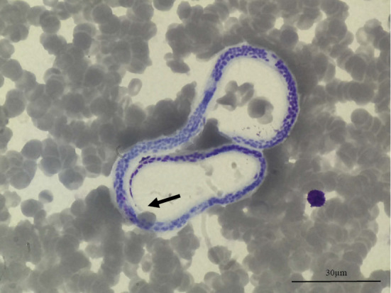
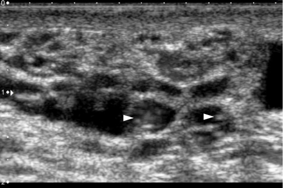
A 32-year-old Nepalese man had lived in Japan for 2 years before presenting at our hospital with an 8-month history of left hydrocele. He was born and raised in a lymphatic filariasis-endemic area of Nepal. He had had no respiratory symptoms or eosinophilia. A Giemsa-stained smear (Picture 1; 1,000x) of the peripheral blood collected at night showed morphological characteristics of microfilariae that were compatible with Wuchereria bancrofti: A stained sheath and no nuclei in the tail (Picture 1, arrow). This was confirmed to be W. bancrofti by polymerase chain reaction-restriction fragment length polymorphism. Ultrasonography of the hydrocele (Picture 2) revealed dilated lymphatic vessels (echo-free area) and adult worms (Picture 2, arrowheads) showing the typical filarial “dance sign” (1). Treatment with diethylcarbamazine was successful, and a remarkable decrease in the microfilarial count in the peripheral blood was noted. The patient returned to Nepal where he planned to undergo surgical repair of the hydrocele.
Picture 1.

Picture 2.

The authors state that they have no Conflict of Interest (COI).
Acknowledgement
This report is supported by the AMED.
References
- 1.Mand S, Marfo-Debrekyei Y, Dittrich M, Fischer K, Adjei O, Hoerauf A. Animated documentation of the filaria dance sign (FDS) in bancroftian filariasis. Filaria J 2: 3, 2003. [DOI] [PMC free article] [PubMed] [Google Scholar]


