

Abstract
Background
During childbirth, many women sustain trauma to the perineum, which is the area between the vaginal opening and the anus. These tears can involve the perineal skin, the pelvic floor muscles, the external and internal anal sphincter muscles as well as the rectal mucosa (lining of the bowel). When these tears extend beyond the external anal sphincter they are called 'obstetric anal sphincter injuries' (OASIS). When women sustain an OASIS, they are at increased risk of developing anal incontinence either immediately following birth or later in life. Anal incontinence is associated with significant medical, hygiene and social problems. Endoanal ultrasound (EAUS) can be performed with a bedside scanner by inserting a small probe into the anus and the structures of the anal canal and perineum can be reviewed in real‐time. We proposed that by examining the perineum with EAUS after the birth of the baby and before the tear has been repaired, there would be an increase in detection of OASIS. This increased detection could lead to improved primary repair of the external and internal anal sphincter resulting in reduced rates of anal incontinence and improved quality of life for women. EAUS may also have a role after perineal repair in the evaluation of residual injury and may help guide a woman's management in subsequent pregnancies and allow for early referral to specialised units, minimising long‐term complications.
Objectives
To evaluate the effectiveness of EAUS in the detection of OASIS following vaginal birth and in reducing the risk of anal sphincter complications related to OASIS.
Search methods
We searched the Cochrane Pregnancy and Childbirth Group's Trials Register (31 August 2015) and reference list of the one retrieved study.
Selection criteria
Randomised control trials (RCTs) comparing EAUS versus no ultrasound in women prior to repair of perineal trauma and EAUS performed after perineal repair. RCTs published in abstract form only and trials using a cluster‐randomised design were eligible for inclusion, but none were identified.
Trials using a cross‐over design and quasi‐RCTs were not eligible for inclusion.
Data collection and analysis
The two review authors independently assessed the single trial for inclusion and assessed trial quality. Both review authors independently extracted data. Data were checked for accuracy.
Main results
We included one trial that randomised 752 primiparous women with clinically detectable second‐degree perineal tears to either further assessment with EAUS prior to perineal repair or standard care. We assessed this trial as being at a low risk of bias. The trial reported women's anal incontinence at three and 12 months as well as their pain scores and quality of life assessment. The trial authors reported outcomes at three months for 719 women (364 in the experimental group, 355 in the control group, 4% loss to follow‐up), and an outcome at 12 months for 684 women (342 in the experimental group, 342 in the control group, 9% loss to follow‐up).
Primary outcome
Compared with clinical examination (routine care), the use of EAUS prior to perineal repair was associated with a reduction in the rate of severe anal incontinence (defined as involuntary loss of faeces or flatus that constitutes social and/or hygiene problems, or as defined by authors), at greater than six months postpartum (risk ratio (RR) 0.48, 95% confidence interval (CI) 0.24 to 0.97, 684 women at the 12‐month time point).
Secondary outcomes
Severe anal incontinence at less than six months was reduced with the use of EAUS prior to repair when compared with clinical examination (routine care) (RR 0.38, 95% CI 0.20 to 0.72, 719 women). However, increased perineal pain at three months was associated with the use of EAUS prior to perineal repair when compared with routine care (RR 5.86, 95% CI 1.74 to 19.72, 684 women). There was no clear difference in the number of women who reported any anal incontinence at either less than six months or equal to or greater than six months (outcomes not prespecified in our published protocol). Similarly, there was no clear difference between groups in terms of faecal incontinence, flatal incontinence, faecal urgency, or maternal quality of life. The study did not report any data on the need for secondary repair of external anal sphincter, dyspareunia, women's satisfaction with care or the planned or actual mode of birth in any subsequent pregnancy. We were unable to assess the detection rates of OASIS with EAUS from the included study because women with clinically‐detected OASIS were excluded from randomisation.
Authors' conclusions
There is some evidence to suggest that EAUS prior to perineal repair is associated with reduced risk of severe anal incontinence but an increase in the incidence of perineal pain at three months postpartum. However, these results are based on one small study involving 752 women. The study took place in a large teaching hospital with an average to busy labour ward. The trial participants were similar to those found in most large obstetric units in developed countries, thus increasing applicability of the evidence, but were restricted to primiparous women.
More research is needed to further evaluate the effectiveness of EAUS in the detection of OASIS following vaginal birth and in reducing the risk of anal sphincter complications related to OASIS. More high‐quality RCTs are needed to fully evaluate the intervention before the routine use of EAUS on the labour ward could be supported. It would be particularly useful if future trials could assess detection rates of OASIS with EAUS versus clinical examination alone as this is the basis of the theory for improved outcomes with this intervention. Cost and the training required to implement EAUS should be considered, along with maternal quality of life and individual symptoms experienced by postnatal women . It would also be useful to follow up women after their subsequent vaginal births to determine if subsequent mode of delivery affects long‐term outcomes. Future studies in multiparous women may also be useful.
Plain language summary
To investigate if using ultrasound to look at anal muscles after childbirth reduces women's suffering from gas, liquid or solid stool leakage
Many women have a tear to the skin between the vagina and anus (the perineum) following childbirth. These tears can sometimes involve the muscles that control the function of the anus, and women can suffer for the rest of their lives with difficulty controlling gas, liquid and solid stool. Evidence shows that by properly repairing these tears, complications can be reduced. The muscles can often be difficult to see with the naked eye due to a number of factors including reduced lighting, swelling of the tissues and poor pain relief.
In this review, we investigated whether using an ultrasound probe that is inserted into the anus before repairing the tear could help to identify undetected tears, delineate the extent of the muscle tear and allow for better repair and subsequently reduce long‐term problems. We also looked at the use of anal ultrasound in women who had an anal muscle tear repaired and whether this improved these outcomes by influencing their subsequent management.
We found one randomised controlled trial that met our inclusion criteria. This trial included 752 women who, following vaginal birth, had a tear to the skin between the vagina and anus that did not include the anal muscles on clinical examination. The women were allocated to receive either routine care (clinical examination) or anal ultrasound prior to their tear being sutured (stitched). We found that women who had anal ultrasound before undergoing perineal repair were about half as likely to suffer from severe anal symptoms. This difference was clear at less than six months and greater than or equal to six months after giving birth. Women were, however, more likely to have significant perineal pain at three months after birth if they underwent ultrasound examination. Solid stool incontinence and involuntary loss of gas (flatulence) were not clearly different between the two groups of women. There was also no difference in terms of maternal quality of life.
The study did not report on the need for secondary repair of external anal sphincter, the number of women who reported pain during sexual intercourse, women's satisfaction with care or details relating to mode of birth in any subsequent pregnancy. It was not possible to look at how effective anal ultrasound examination was at detecting anal sphincter injuries because women with clinically‐detected obstetric‐related anal sphincter injury were excluded from the study.
We assessed the included trial as being at low risk of bias as it was a well designed trial, but it involved a small number of women. More trials are needed in this area to further evaluate this intervention and inform future practice. We did not find any studies that used ultrasound after the tear had been sutured. It would be helpful if future studies could evaluate how effective ultrasound is at detecting obstetric anal sphincter injury. The cost of the intervention and any training needs should be considered, along with maternal quality of life and individual symptoms experienced by postnatal women. It would also be useful to examine how women give birth with subsequent pregnancies and longer‐term outcomes.
Background
Description of the condition
Trauma to the perineum (area between vagina and anus) is a common consequence of childbirth with 71.7% of Australian women (Li 2012) and 85% of British women (McCandlish 1998) sustaining some degree of perineal trauma during childbirth (Li 2012). Perineal trauma can occur via a spontaneous tear or an episiotomy (cut made to assist childbirth), and is classified by the degree of trauma sustained. The following classification, described by Sultan (Sultan 1999) has been adopted by the international consultation on incontinence (Norton 2002) and will be used in this review (RCOG 2007).
First degree – injury to the perineal skin only.
Second degree – injury to perineum involving perineal muscles but not involving the anal sphincter.
-
Third degree – injury to perineum involving the anal sphincter complex (external and internal anal sphincter):
3a – less than 50% of the external anal sphincter (EAS);
3b – more than 50% of the EAS;
3c – both the EAS and internal anal sphincter (IAS).
Fourth degree – injury to perineum involving the anal sphincter complex (EAS and IAS) and anal epithelium.
Obstetric anal sphincter injuries (OASIS) is a term used to define trauma that includes third‐ and fourth‐degree tears. These are of particular concern, as they are associated with high rates of short‐ and long‐term consequences for women. In the USA in 2003, 4.4% of deliveries had a documented anal sphincter injury whilst in 2010, 1.8% of Australian women sustained an OASIS (Li 2012).
Risk factors for obstetric anal sphincter injuries (OASIS)
Women are at a higher risk of sustaining an OASIS if they have a forceps birth, are having their first baby, are in the second stage of labour for more than one hour, deliver a baby weighing more than 4 kg, have a midline episiotomy or if the baby is in a persistent occipitoposterior position (RCOG 2007).
Diagnosis of obstetric anal sphincter injuries (OASIS)
Following vaginal birth all women are examined by the accoucher (person delivering baby) to assess the presence and degree of perineal trauma. This examination should include a PR (per rectum) examination to assess the extent of any sphincter/anal trauma. However, assessing these tears can often be difficult due to multiple factors: poor lighting, difficulty identifying anatomy due to significant oedema and bruising and maternal discomfort during examination. This can lead to a potential classification of an OASIS as second‐degree perineal trauma. There is also a disincentive on behalf of accouchers to report such injuries as they are often the subject of clinical audits and peer review meetings within obstetric units (Andrews 2006).
A prospective cohort study (Andrews 2006) of 254 primiparous women showed that 25% sustained OASIS of which 87% were missed by the midwives, 14% missed by specialist registrars and 67% missed by senior house officers. In this particular study, 98.8% of these OASIS were subsequently detected on clinical examination by a trained research fellow prior to endoanal ultrasound (EAUS). Another prospective cohort study showed that 28% of the second‐degree tears actually had an OASIS (clinically undetected at the time of injury). When a woman has an occult injury (the OASIS is missed), the rest of the perineum is repaired using a rapidly dissolving suture which provides only minimal support for the anal sphincter, leaving a long‐term weakness in the anal sphincter complex. This may result in immediate anal incontinence or delayed symptoms that may only manifest after subsequent deliveries or with increasing age. Women with occult injuries which are not repaired have an odds ratio of 8.8 of having anal incontinence at three months when compared with women who did not have an OASIS on EAUS (Faltin 2000). Under‐reporting and under‐identification of OASIS may lead to a sub‐optimal primary repair and increased rates of maternal morbidity longer term.
The magnitude and impact of OASIS is even more serious in the developing world where inadequate staffing, insufficient health‐care facilities and ineffective reporting systems make detection and primary repair of OASIS more difficult (WHO 1999).
Outcomes and complications of anal sphincter injury
OASIS is the most important aetiological factor in the pathogenesis of anal incontinence in women (Swash 1993). Anal incontinence is defined by the International Continence Society as involuntary loss of flatus or faeces (Haylen 2010) and affects up to 40% of women following childbirth (Faltin 2000). Of women who have an OASIS visible on EAUS, which was not detected at the time of repair, 37% (95% confidence interval (CI) 22% to 51%) will have anal incontinence at three months (Faltin 2000) whilst, if no OASIS injury was seen on EAUS, they have a 93% (95% CI 88% to 98%) likelihood of being continent (Faltin 2000).
The embarrassing nature of this problem leads to social isolation and many women not seeking medical treatment for their symptoms (Fernando 2013). With primary detection and appropriate repair, women can be reassured that the prognosis is good with 60% to 80% being asymptomatic at 12 months (RCOG 2007). Possible complications after anal sphincter injury range from faecal urgency, anal incontinence of gas, liquid or solid stool to perineal pain, difficulty with emptying the bladder or bowels, long‐term dyspareunia (painful sexual intercourse) and more rarely, rectovaginal fistulas (abnormal connection between vagina and rectum). However, there are also rates varying from 20% to 40% of occult OASIS injuries.
Description of the intervention
EAUS is a simple procedure that does not constitute any known risk to the mother in itself and involves minimal discomfort. A probe inserted into the anus allows the practitioner to view the structures of the anal canal. A study of 150 women showed that anal endosonography after vaginal delivery was well tolerated, even when women had no epidural analgesia (Faltin 2000). EAUS has been shown to be equivalent to MRI (magnetic resonance imaging) and superior to electromyographic sphincter mapping in detecting sphincter defects (Felt‐Bersma 2006). It is also much cheaper and more easily accessible with bedside units suitable for use on the labour ward. All women currently have a digital rectal examination as part of the assessment of perineal trauma and the EAUS could be performed at the same time as this. Obstetricians and pregnant women are also familiar with ultrasound techniques as they are commonly used throughout pregnancy via transabdominal and transvaginal approaches, making the intervention potentially more acceptable to women at this point in their lives.
How the intervention might work
EAUS performed prior to repair of visible obstetric trauma could improve detection rates of OASIS and allow for improved surgical repair and, by so doing, reduce the morbidity suffered by women undergoing vaginal birth. Perineal tears are routinely assessed and repaired immediately following birth, which makes identification of anal sphincter tears difficult and subsequently they are often missed (Andrews 2006). We propose that the routine use of EAUS would allow for a higher proportion of OASIS to be detected and subsequently repaired leading to reduced long‐term complications. Primary repair is associated with a 63% likelihood of a good outcome versus secondary repair where the result can be less than 50% continence (Oliver 2008).
EAUS could also be used as part of the early follow‐up of patients with OASIS to identify patients at higher risk of long‐term complications. This would allow for early referral to specialist centres or to colorectal surgeons and intervention with either physiotherapy and dietary manipulation or, if needed, secondary sphincter repair (Fowler 2009).
EAUS may also be useful in pregnant women who have had a prior OASIS in planning the mode of subsequent deliveries. From 17% to 24% of women who underwent a vaginal delivery after OASIS had worsening of their faecal symptoms (RCOG 2007). However, a large prospective cohort study found that recurrence of OASIS could not be predicted and that 95% of women with a prior injury did not sustain further overt sphincter damage during a subsequent vaginal delivery (Harkin 2003). This raises the question as to whether antenatal evaluation with EAUS may help identify those patients at high risk of worsening symptoms so they could be offered an elective lower segment caesarean section (LSCS) to reduce these risks.
Why it is important to do this review
It is important to systematically review the evidence for using EAUS in the detection and management of primary OASIS. Such an intervention may be useful in decreasing complications by directing more suitable treatment/surgical repairs, leading to improved long‐term physical symptoms and overall quality of life for women worldwide.
Objectives
To evaluate the effectiveness of EAUS in:
the detection of OASIS in the immediate postpartum period following vaginal birth;
reducing the rates of anal sphincter complications related to OASIS.
Methods
Criteria for considering studies for this review
Types of studies
We planned to include published and unpublished randomised controlled trials, cluster‐randomised trials and studies presented only as abstracts. Trials using quasi‐randomised methodology and cross‐over design were not eligible for inclusion.
Types of participants
All women experiencing, or who have previously had a vaginal birth at any gestation, including spontaneous and assisted vaginal births.
Types of interventions
EAUS performed following vaginal birth and prior to repair of any perineal trauma.
EAUS performed following vaginal birth after repair of any perineal trauma, including women undergoing EAUS during subsequent pregnancies.
Types of outcome measures
Primary outcomes
Severe anal incontinence, at >/= six months (defined as involuntary loss of faeces or flatus that constitutes social and/or hygiene problems, or as defined by authors).
Secondary outcomes
Severe anal incontinence measured at < six months (defined as involuntary loss of faeces or flatus that constitutes social and/or hygiene problems, or as defined by authors).
Any anal incontinence at < six months (outcome not prespecified).
Any anal incontinence at >/= six months (outcome not prespecified).
Detection of OASIS (defined as interruption of the external anal sphincter, with or without damage to the internal anal sphincter or anal mucosa).
Faecal incontinence (involuntary loss of solid stool) as specified by trialist.
Flatal incontinence (involuntary loss of flatus) as specified by trialist.
Faecal urgency as specified by trialist.
Perineal pain, as assessed by a visual analogue scale at three months postpartum or as specified by trialist.
Dyspareunia assessed by a visual analogue scale as specified by trialist.
Need for secondary repair of external anal sphincter.
Maternal quality of life as specified by trialist.
Planned mode of birth in subsequent pregnancy.
Actual mode of birth in subsequent pregnancy.
Women's satisfaction with care.
Search methods for identification of studies
The following methods section of this review is based on a standard template used by the Cochrane Pregnancy and Childbirth Group.
Electronic searches
We searched the Cochrane Pregnancy and Childbirth Group’s Trials Register by contacting the Trials Search Co‐ordinator (31 August 2015).
The Cochrane Pregnancy and Childbirth Group’s Trials Register is maintained by the Trials Search Co‐ordinator and contains trials identified from:
monthly searches of the Cochrane Central Register of Controlled Trials (CENTRAL);
weekly searches of MEDLINE (Ovid);
weekly searches of Embase (Ovid);
monthly searches of CINAHL (EBSCO);
handsearches of 30 journals and the proceedings of major conferences;
weekly current awareness alerts for a further 44 journals plus monthly BioMed Central email alerts.
Details of the search strategies for CENTRAL, MEDLINE, Embase and CINAHL, the list of handsearched journals and conference proceedings, and the list of journals reviewed via the current awareness service can be found in the ‘Specialized Register’ section within the editorial information about the Cochrane Pregnancy and Childbirth Group.
Trials identified through the searching activities described above are each assigned to a review topic (or topics). The Trials Search Co‐ordinator searches the register for each review using the topic list rather than keywords.
Searching other resources
We searched the reference list of the one retrieved study.
We did not apply any language or date restrictions.
Data collection and analysis
Selection of studies
Both review authors independently assessed for inclusion the potential study that was identified as a result of the search strategy. Any discrepancies were resolved through discussion.
Data extraction and management
We designed a form to extract data. For the eligible study, both review authors extracted the data using the agreed form. We resolved discrepancies through discussion. We entered data into Review Manager software (RevMan 2014) and checked for accuracy.
When information regarding any of the above was unclear, we attempted to contact authors of the original report to provide further details.
Assessment of risk of bias in included studies
Both review authors independently assessed risk of bias for the study using the criteria outlined in the Cochrane Handbook for Systematic Reviews of Interventions (Higgins 2011).
(1) Random sequence generation (checking for possible selection bias)
We described for the included study the method used to generate the allocation sequence in sufficient detail to allow an assessment of whether it should produce comparable groups.
We assessed the method as:
low risk of bias (any truly random process, e.g. random number table; computer random number generator);
high risk of bias (any non‐random process, e.g. odd or even date of birth; hospital or clinic record number);
unclear risk of bias.
(2) Allocation concealment (checking for possible selection bias)
We described for the included study the method used to conceal allocation to interventions prior to assignment and assessed whether intervention allocation could have been foreseen in advance of, or during recruitment, or changed after assignment.
We assessed the methods as:
low risk of bias (e.g. telephone or central randomisation; consecutively numbered sealed opaque envelopes);
high risk of bias (open random allocation; unsealed or non‐opaque envelopes, alternation; date of birth);
unclear risk of bias.
(3.1) Blinding of participants and personnel (checking for possible performance bias)
We described for the included study the methods used, if any, to blind women and personnel from knowledge of which intervention a participant received. We considered that the study was at low risk of bias if it was blinded, or if we judged that the lack of blinding would have been unlikely to affect results. We assessed blinding separately for different outcomes or classes of outcomes.
We assessed the methods as:
low, high or unclear risk of bias for women;
low, high or unclear risk of bias for personnel.
(3.2) Blinding of outcome assessment (checking for possible detection bias)
We described for the included study the methods used, if any, to blind outcome assessors from knowledge of which intervention a participant received. We assessed blinding separately for different outcomes or classes of outcomes.
We assessed methods used to blind outcome assessment as:
low, high or unclear risk of bias.
(4) Incomplete outcome data (checking for possible attrition bias due to the amount, nature and handling of incomplete outcome data)
We described for the included study, and for each outcome or class of outcomes, the completeness of data including attrition and exclusions from the analysis. We stated whether attrition and exclusions were reported and the numbers included in the analysis at each stage (compared with the total randomised participants), reasons for attrition or exclusion where reported, and whether missing data were balanced across groups or were related to outcomes. Where sufficient information was reported, or could be supplied by the trial authors, we planned to re‐include missing data in the analyses which we undertook.
We assessed methods as:
low risk of bias (e.g. no missing outcome data; missing outcome data balanced across groups);
high risk of bias (e.g. numbers or reasons for missing data imbalanced across groups; ‘as treated’ analysis done with substantial departure of intervention received from that assigned at randomisation);
unclear risk of bias.
(5) Selective reporting (checking for reporting bias)
We described for the included study how we investigated the possibility of selective outcome reporting bias and what we found.
We assessed the methods as:
low risk of bias (where it is clear that all of the study’s prespecified outcomes and all expected outcomes of interest to the review have been reported);
high risk of bias (where not all the study’s prespecified outcomes have been reported; one or more reported primary outcomes were not prespecified; outcomes of interest are reported incompletely and so cannot be used; study fails to include results of a key outcome that would have been expected to have been reported);
unclear risk of bias.
(6) Other bias (checking for bias due to problems not covered by (1) to (5) above)
We described for the included study any important concerns we had about other possible sources of bias.
We assessed whether the study was free of other problems that could put it at risk of bias:
low risk of other bias;
high risk of other bias;
unclear whether there is risk of other bias.
(7) Overall risk of bias
We made explicit judgements about whether the study was at high risk of bias, according to the criteria given in the Cochrane Handbook (Higgins 2011). With reference to (1) to (6) above, we assessed the likely magnitude and direction of the bias and whether we considered it is likely to impact on the findings. We explored the impact of the level of bias through undertaking sensitivity analyses ‐ seeSensitivity analysis.
Measures of treatment effect
Dichotomous data
For dichotomous data, we presented results as summary risk ratio with 95% confidence intervals.
Continuous data
For continuous data, we planned to use the mean difference if outcomes were measured in the same way between trials. We planned to use the standardised mean difference to combine trials that measure the same outcome, but used different methods. We did not identify continuous data for inclusion in this version of the review.
Unit of analysis issues
Cluster‐randomised trials
In future updates of this review, if identified, we will include cluster‐randomised trials in the analyses along with individually‐randomised trials. We will adjust their standard errors using the methods described in the Handbook using an estimate of the intracluster correlation co‐efficient (ICC) derived from the trial (if possible), from a similar trial or from a study of a similar population. If we use ICCs from other sources, we will report this and conduct sensitivity analyses to investigate the effect of variation in the ICC. If we identify both cluster‐randomised trials and individually‐randomised trials, we plan to synthesise the relevant information. We will consider it reasonable to combine the results from both if there is little heterogeneity between the study designs and the interaction between the effect of intervention and the choice of randomisation unit is considered to be unlikely.
We will also acknowledge heterogeneity in the randomisation unit and perform a sensitivity analysis to investigate the effects of the randomisation unit.
Other unit of analysis issues
In future updates, if we include studies with multiple arms, we will combine groups to create a single pair‐wise comparison.
Dealing with missing data
For the included study, we noted levels of attrition. We planned to explore the impact of including studies with high levels of missing data in the overall assessment of treatment effect by using sensitivity analysis.
For all outcomes, we carried out analyses, as far as possible, on an intention‐to‐treat basis, i.e. we attempted to include all participants randomised to each group in the analyses, and all participants were analysed in the group to which they were allocated, regardless of whether or not they received the allocated intervention. The denominator for each outcome in each trial was the number randomised minus any participants whose outcomes were known to be missing.
We attempted to contact the authors to clarify additional information not available in the paper, however, we were unsuccessful.
Assessment of heterogeneity
In future updates of this review we will assess statistical heterogeneity in each meta‐analysis using the T², I² and Chi² statistics. We will regard heterogeneity as substantial if the I² is greater than 30% and either the T² is greater than zero, or there is a low P value (less than 0.10) in the Chi² test for heterogeneity.
Assessment of reporting biases
As there was only one included trial, assessment of reporting bias was not undertaken in this version of the review, however it may be implemented in future reviews.
Had there been 10 or more studies in the meta‐analysis, we planned to investigate reporting biases (such as publication bias) using funnel plots by assessing funnel plot asymmetry visually. If asymmetry was suggested by a visual assessment, we planned to perform exploratory analyses to investigate it.
Data synthesis
We carried out statistical analysis using the Review Manager software (RevMan 2014). As there was only one included study we were unable to combine data, however in future versions we will use fixed‐effect meta‐analysis to combine data where it is reasonable to assume that studies are estimating the same underlying treatment effect: i.e. where trials are examining the same intervention, and the trials’ populations and methods are judged sufficiently similar. If there is clinical heterogeneity sufficient to expect that the underlying treatment effects differ between trials, or if substantial statistical heterogeneity is detected, we will use random‐effects meta‐analysis to produce an overall summary, if an average treatment effect across trials is considered clinically meaningful. The random‐effects summary will be treated as the average range of possible treatment effects and we will discuss the clinical implications of treatment effects differing between trials. If the average treatment effect is not clinically meaningful, we will not combine trials.
If we use random‐effects analyses, the results will be presented as the average treatment effect with 95% confidence intervals, and the estimates of T² and I².
Subgroup analysis and investigation of heterogeneity
This review contains a single included study. In future updates, if we identify substantial heterogeneity, we will investigate it using subgroup analyses and sensitivity analyses. We will consider whether an overall summary is meaningful, and if so, we will use a random‐effects analysis to produce it.
We plan to carry out the following subgroup analysis.
Women following their first vaginal birth versus women who have had multiple vaginal births.
Assisted versus spontaneous vaginal birth.
Subgroup analysis will be restricted to this review's primary outcomes.
We will assess subgroup differences by interaction tests available within RevMan (RevMan 2014) and we will report the results of subgroup analyses quoting the χ² statistic and P value, and the interaction test I² value.
Sensitivity analysis
In future updates we will carry out sensitivity analyses to explore the effects of trial quality as assessed by concealment of allocation or missing data for a particular outcome on the summary statistic. We will excluded trials with greater than 20% missing data. We will exclude studies of poor quality from the analysis in order to assess for any substantive difference to the overall result. If no substantive difference exists, we will leave the studies in the analysis. Where we include data from cluster‐randomised trials, we will carry out sensitivity analysis in order to investigate the effect of the randomisation unit. All sensitivity analysis will be restricted to this review's primary outcomes.
Results
Description of studies
See: Included studies.
Results of the search
The search of the Cochrane Pregnancy and Childbirth Group's Trials Register retrieved one trial report (see: Figure 1).
1.

Study flow diagram.
Included studies
There was only one trial that met the inclusion criteria for our review. This study (Faltin 2005) was a prospective randomised controlled trial investigating the use of endoanal ultrasound (EAUS) after birth but prior to perineal repair, in addition to standard clinical examination in primiparous women with clinical second‐degree perineal tears. The trial took place in a large university hospital in Geneva that delivered approximately 3500 women each year over a two‐year period between 1999 and 2001. All women were nulliparous over the age of 18 years and able to read French. All women with an intact perineum, or clinically‐detected third‐ or fourth‐degree tears on examination and per rectum digital examination were excluded. This means that it is not possible to comment on detection rates of obstetric anal sphincter injuries (OASIS) from this trial. If an anal sphincter tear was identified on EAUS, the perineum was surgically explored in theatre and the sphincter repaired. There were 752 women in this study (Faltin 2005) with 376 women randomised to each arm. As part of this trial, outcomes were assessed at three and 12 months with a questionnaire to assess the women's symptoms of perineal pain, incontinence of flatus, liquid and solid stool as well as their need to wear a pad and any lifestyle alterations they had made due to their anal incontinence. Women who reported fecal incontinence were also asked to answer an extra quality of life questionnaire. By the three‐month time point, 30 women had discontinued the trial (10 in the experimental group and 20 in the control group), and three were lost to follow‐up (two in the experimental group and one in the control group); this meant that the three‐month outcomes were for 719 women (364 in the experimental group and 355 in the control group with 4% overall loss to follow‐up). By 12 months, 65 women had discontinued (33 in the experimental group and 32 in the control group), and three were lost to follow‐up (one in the experimental group and two in the control group); resulting in reported outcomes for 684 women (342 in the experimental group and 342 in the control group with 9% loss to follow‐up).
Excluded studies
There are no excluded studies.
Risk of bias in included studies
We assessed the included trial and found it to have a low risk of bias overall.
Allocation
The single included study (Faltin 2005) has been assessed as low risk for allocation (selection) bias. A central computer‐generated random sequences of numbers in blocks of varying sizes (four, six, eight women) arranged in random order was used. The study allocations were then placed in opaque sealed envelopes which were opened by a supervising midwife not directly involved in the delivery.
Blinding
In the Faltin 2005 study, due to the design and nature of the trial, it was not possible to blind the women or the clinician to the allocation, the trial has therefore been assessed as being high risk for performance bias. The outcome assessors, however, were blinded to the allocation and thus the trial is at low risk of detection bias.
Incomplete outcome data
In Faltin 2005, 12 women were reported as having "protocol violations" from their allocated group (in the control group two women underwent EAUS prior to the repair and in the intervention group, 10 women did not have an ultrasound. However, the results from all these women were included in their intention‐to‐treat group. At the three‐month questionnaire, 33 patients were excluded from analysis due to being lost to follow‐up or discontinuing involvement in the study. At 12 months postpartum a total of 34 women (9%) were excluded from analysis from both groups. We have therefore assessed this study as being low risk due to attrition bias.
Selective reporting
All the prespecified outcomes were reported on in the trial and no extra outcomes were added after the trial was completed. In particular the trial authors prespecified the cut‐offs they would use to differentiate between mild and severe incontinence.
The trial authors collected mode of delivery data, but did not report on this, which prevented us from performing our subgroup analysis. We have attempted to contact the authors for these data and should it become available we will include them in future reviews.
We have therefore assessed the trial as being at low risk of reporting bias.
Other potential sources of bias
No other potential sources of bias were identified.
Effects of interventions
Anal endosonography versus routine care
Primary outcome
Severe anal incontinence, at >/= six months (defined as involuntary loss of faeces or flatus that constitutes social and/or hygiene problems, or as defined by authors)
Anal endosonography resulted in a reduction in severe anal incontinence (risk ratio (RR) 0.48, 95% confidence interval (CI) 0.24 to 0.97, one study, 684 women (Analysis 1.1)). This outcome was based on the 12‐month time point in the included trial (Faltin 2005).
1.1. Analysis.

Comparison 1 Anal endosonography versus routine care, Outcome 1 Severe anal incontinence at >/= 6 months, severity as defined by trial authors.
Secondary outcomes
The study we included (Faltin 2005), reported on some (but not all) of the secondary outcomes of this review.
Severe anal incontinence < six months, (defined as involuntary loss of faeces or flatus that constitutes social and/or hygiene problems, or as defined by authors)
Anal endosonography resulted in a reduction in severe anal incontinence at less than six months (RR 0.38, 95% CI 0.20 to 0.72, 719 women (Analysis 1.2)) .
1.2. Analysis.

Comparison 1 Anal endosonography versus routine care, Outcome 2 Severe anal incontinence <6 months, severity as defined by trial authors.
Any anal incontinence defined as involuntary loss of flatus or faeces, >/= six months (outcome not prespecified).
Anal endosonography resulting in no clear difference in anal incontinence at more than six months postpartum (RR 0.95, 95% CI 0.73 to 1.22, 684 women (Analysis 1.3)).
1.3. Analysis.

Comparison 1 Anal endosonography versus routine care, Outcome 3 any anal Incontinence >/= 6 months (outcome not prespecified).
Any anal incontinence defined as involuntary loss of flatus or faeces, < six months (outcome not prespecified).
Anal endosonography resulted in no clear difference in anal incontinence at less than six months (RR 1.03, 95% CI 0.83 to 1.27, 719 women (Analysis 1.4)).
1.4. Analysis.

Comparison 1 Anal endosonography versus routine care, Outcome 4 any anal incontinence <6 months (outcome not prespecified).
Detection of obstetric anal sphincter Injuries (OASIS)
This outcome was not reported in Faltin 2005 as all clinically‐detected OASIS were excluded from randomisation. However, following randomisation a further two women were found to have an OASIS on clinical examination and in the experimental group 21 of 376 (5.6%) women were found to have a previously undiagnosed OASIS. This would suggest the use of EAUS increased the detection of OASIS by a further 5.6 % (absolute risk).
Faecal incontinence (involuntary loss of solid stool)
Anal endosonography did not reduce the rate of faecal incontinence (RR 1.00, 95% CI 0.20 to 4.92, 684 women (Analysis 1.7)). This was assessed at the 12‐month time point in the included trial (Faltin 2005).
1.7. Analysis.

Comparison 1 Anal endosonography versus routine care, Outcome 7 Faecal incontinence.
Flatal incontinence (involuntary loss of flatus)
There was also no clear difference in flatal incontinence between the anal endosonography and standard care groups (RR 1.05, 95% CI 0.80 to 1.37, 684 women (Analysis 1.6)).
1.6. Analysis.

Comparison 1 Anal endosonography versus routine care, Outcome 6 Flatal incontinence.
Faecal urgency
Anal endosonography resulted in a non significant reduction in faecal urgency (RR 0.75, 95% CI 0.32 to 1.76, 684 women (Analysis 1.8)).
1.8. Analysis.

Comparison 1 Anal endosonography versus routine care, Outcome 8 Faecal urgency.
Perineal pain at three months postpartum as assessed by a visual analogue scale or as specified by trialist
Anal endosonography was associated with an increase in perineal pain at three months postpartum when compared with routine care (RR 5.86, 95% CI 1.74 to 19.72, 684 women (Analysis 1.9)).
1.9. Analysis.

Comparison 1 Anal endosonography versus routine care, Outcome 9 Perineal pain at 3 months.
Dyspareunia assessed by a visual analogue scale, or as specified by trialist
This outcome was not reported in Faltin 2005.
Need for secondary repair of external anal sphincter
This outcome was not reported in Faltin 2005.
Maternal quality of life as specified by trialist
Maternal quality of life was reported by Faltin 2005 who reported on lifestyle impact at 12 months ‐ there was no clear difference between groups (RR 0.57, 95% CI 0.17 to 1.93, 684 women (Analysis 1.12)).
1.12. Analysis.
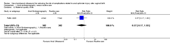
Comparison 1 Anal endosonography versus routine care, Outcome 12 Maternal quality of life.
Planned mode of birth in subsequent pregnancy
This outcome was not reported in Faltin 2005.
Actual mode of birth in subsequent pregnancy
This outcome was not reported in Faltin 2005.
Women's Satisfaction with care
This outcome was not reported in Faltin 2005.
Subgroup analysis
We were unable to perform any subgroup analysis as the one included trial did not investigate women following multiple vaginal births and although Faltin 2005 collected data on assisted and spontaneous vaginal birth, the authors did not report on these data. There were no trials identified that investigated the use of endoanal ultrasound (EAUS) following repair of the perineum.
Discussion
We included one trial that randomised 752 primiparous women with clinically detectable second‐degree perineal tears to either further assessment with endoanal ultrasound (EAUS) prior to perineal repair or standard care. We assessed this trial as being at a low risk of bias.
Summary of main results
The use of EAUS prior to repair was shown to reduce the rate of severe anal incontinence at both time points less than and greater than or equal to six months. This is in keeping with the theory that the improved detection leads to improved primary repair of obstetric anal sphincter injuries (OASIS), reducing the number of unrepaired anal sphincter injuries in the postnatal population. Such unrepaired anal sphincter injuries are associated with high rates of anal incontinence.
Use of EAUS, however, was associated with increased perineal pain at three months. A possible underlying mechanism for this increase in pain is that when more OASIS are identified, there is an increase in surgical exploration and a more extensive repair with sutures which take longer to dissolve.
There was no clear difference between EAUS and standard care in relation to the following outcomes: flatal incontinence, faecal incontinence, faecal urgency or maternal quality of life.
Overall completeness and applicability of evidence
The Faltin 2005 trial took place in a large teaching hospital with an average to busy labour ward using their regular staff, who underwent some training on the use of EAUS. The women in the trial were similar to those found in most large obstetric units in developed countries, thus increasing applicability of the evidence. This review only includes primiparous women who did not have a clinically‐detected OASIS injury, however, we would not expect multiparous women to have a different outcome. Future studies in multiparous women may be useful as there is a risk that women with a previously undetected OASIS injury may be misdiagnosed in this delivery as having sustained an OASIS as EAUS is unlikely to be able to discriminate between an acute or a long‐term anal sphincter injury.
No data were reported for some of this review's secondary outcomes: detection of OASIS (defined as interruption of the external anal sphincter, with or without damage to the internal anal sphincter, or anal mucosa, or both); dyspareunia assessed by visual analogue scale as specified by the trialist; need for secondary repair of external anal sphincter; planned mode of birth in subsequent pregnancy; actual mode of birth in subsequent pregnancy; or women's satisfaction with care.
Quality of the evidence
As this version of the review is limited to one trial it has little power to suggest a change in practice. The quality of Faltin 2005 was high with a well designed trial. However, the validity of a questionnaire as its sole outcome assessment could be better established. Further studies are needed in this area.
Potential biases in the review process
There is a low risk that the review process introduced bias.
Agreements and disagreements with other studies or reviews
Although more evidence is required to support the recommendation for the use of EAUS in the prevention of anal incontinence, the findings of this review are in keeping with the studies identified in the literature search indicating that currently a substantial number of OASIS are missed at time of delivery and these injuries have a high association with anal incontinence later in life (Faltin 2000; Johnson 2007; Norton 2002).
EAUS as a tool to detect anal sphincter trauma has been assessed in multiple trials, all of which found EAUS an accurate way of assessing the perineum for anal sphincter for trauma (Faltin 2000; Felt‐Bersma 2006; Liverpool 2008; Martinez 2003). However, EAUS is not the only option available to increase detection. A study by Andrews et al. showed that a second examination by a focused trained clinician increased the detection rate of OASIS from 11% to 24.5% and the additional use of EAUS after this expert re‐examination did not significantly improve rates further (Andrews 2006). This leads to the suggestion that improved training and re‐examination by a second clinician would be an equally effective approach to improving outcomes from OASIS. Sultan would agree that although EAUS is an accurate means of diagnosing OASIS, is it no better that a well trained clinician and is expensive and invasive (Sultan 1999).
EAUS has also been shown to be useful in identifying anal sphincter trauma in other clinical scenarios . For example, a cohort study of 22 women aged 29 to 68 years who had severe anal incontinence and previously had a vaginal birth underwent EAUS to identify OASIS. Following surgical repair of the tears that had been identified, 86% of these women had a significant improvement in their continence (Martinez 2003). This supports the hypothesis that OASIS identified on EAUS and subsequently repaired improves continence, even when women are already symptomatic.
The increase in perineal pain found in our review is most likely due to more OASIS being identified and subsequently repaired in the intervention group. There were 16 women in the experimental group who had an EAUS diagnosed OASIS and 18 women who reported increased rates of perineal pain at three months postnatal. Third‐degree tears are associated with increased perineal pain when compared to second‐degree tears at three months, however, their was no difference noted at 12 months (Faltin 2005).
Authors' conclusions
Implications for practice.
From the available data, results are consistent with improvement in severe anal incontinence with the use of endoanal ultrasound (EAUS) as an adjunct to clinical examination prior to perineal repair in primiparous women at both >/= six months and > six months postpartum. In contrast, EAUS was shown to be associated with an increase in women's perineal pain at the three‐month time point.
However, since this evidence is based on only one trial, which only followed women for 12 months, more research evidence is needed in order to confirm or refute these findings.
Implications for research.
Future high‐quality randomised controlled trials are needed before the routine use of EAUS on the labour ward could be supported. It would be particularly useful if future trials could assess detection rates of obstetric anal sphincter injuries (OASIS) with EAUS versus clinical examination alone as this is the basis of the theory for improved outcomes with this intervention. The cost and training required to implement EAUS would also need to be analysed. It would also be useful to investigate women after their subsequent vaginal births to determine if the subsequent mode of delivery affects long‐term outcomes. Further trials are also needed to investigate effects on maternal quality of life, individual symptoms experienced by postnatal women and to investigate its use in multiparous women.
Acknowledgements
As part of the pre‐publication editorial process, this review has been commented on by two peers (an editor and referee who is external to the editorial team), a member of the Pregnancy and Childbirth Group's international panel of consumers and the Group's Statistical Adviser.
This project was supported by the National Institute for Health Research, via Cochrane Infrastructure funding to Cochrane Pregnancy and Childbirth. The views and opinions expressed therein are those of the authors and do not necessarily reflect those of the Systematic Reviews Programme, NIHR, NHS or the Department of Health.
Data and analyses
Comparison 1. Anal endosonography versus routine care.
| Outcome or subgroup title | No. of studies | No. of participants | Statistical method | Effect size |
|---|---|---|---|---|
| 1 Severe anal incontinence at >/= 6 months, severity as defined by trial authors | 1 | 684 | Risk Ratio (M‐H, Fixed, 95% CI) | 0.48 [0.24, 0.97] |
| 2 Severe anal incontinence <6 months, severity as defined by trial authors | 1 | 719 | Risk Ratio (M‐H, Fixed, 95% CI) | 0.38 [0.20, 0.72] |
| 3 any anal Incontinence >/= 6 months (outcome not prespecified) | 1 | 684 | Risk Ratio (M‐H, Fixed, 95% CI) | 0.95 [0.73, 1.22] |
| 4 any anal incontinence <6 months (outcome not prespecified) | 1 | 719 | Risk Ratio (M‐H, Fixed, 95% CI) | 1.03 [0.83, 1.27] |
| 5 Detection of obstetric anal sphincter injuries (defined as interruption of the external anal sphincter, with or without damage to the internal anal sphincter or anal mucosa) | 0 | 0 | Risk Ratio (M‐H, Fixed, 95% CI) | 0.0 [0.0, 0.0] |
| 6 Flatal incontinence | 1 | 684 | Risk Ratio (M‐H, Fixed, 95% CI) | 1.05 [0.80, 1.37] |
| 7 Faecal incontinence | 1 | 684 | Risk Ratio (M‐H, Fixed, 95% CI) | 1.0 [0.20, 4.92] |
| 8 Faecal urgency | 1 | 684 | Risk Ratio (M‐H, Fixed, 95% CI) | 0.75 [0.32, 1.76] |
| 9 Perineal pain at 3 months | 1 | 684 | Risk Ratio (M‐H, Fixed, 95% CI) | 5.86 [1.74, 19.72] |
| 10 Dyspareunia assessed by a visual analogue scale as specified by trialists | 0 | 0 | Mean Difference (IV, Fixed, 95% CI) | 0.0 [0.0, 0.0] |
| 11 Need for secondary repair of external anal sphincter | 0 | 0 | Risk Ratio (M‐H, Fixed, 95% CI) | 0.0 [0.0, 0.0] |
| 12 Maternal quality of life | 1 | 684 | Risk Ratio (M‐H, Fixed, 95% CI) | 0.57 [0.17, 1.93] |
| 13 Planned mode of birth in subsequent pregnancy | 0 | 0 | Risk Ratio (M‐H, Fixed, 95% CI) | 0.0 [0.0, 0.0] |
| 14 Actual mode of birth in subsequent pregnancy | 0 | 0 | Risk Ratio (M‐H, Fixed, 95% CI) | 0.0 [0.0, 0.0] |
| 15 Women's satisfaction with care | 0 | 0 | Risk Ratio (M‐H, Fixed, 95% CI) | 0.0 [0.0, 0.0] |
Characteristics of studies
Characteristics of included studies [ordered by study ID]
Faltin 2005.
| Methods | Computer‐generated randomised controlled trial. | |
| Participants | Nulliparous women aged over 18 years with no planned caesarean who were able to read French, and sustained a clinically detectable second‐degree tear. Women with an intact perineum and women with a clinically detectable anal sphincter tears were excluded. 752 women were randomised to the trial. However, there were only 719 women for whom outcomes were reported at 3 months and only 684 women at 12 months. | |
| Interventions | Prior to repair of the perineal trauma, the women underwent anal endosonography using a 7.5 MHZ Endo‐P II sound system in addition to clinical examination by the Obstetrician. All the Obstetricians working in the unit received formal training on endoanal ultrasound as well as their images reviewed and feedback provided. The Obstetrician was not blinded to the findings and all detected sphincter tears were then repaired in theatre, with surgical exploration and end‐to‐end anastomosis with 2‐0 monofilament polyglyconate sutures (Maxon) post operatively women were advised to avoid constipation and use occasional stool softeners. In the control group the Obstetrician sutured the perineum after clinical examination only. |
|
| Outcomes | Questionaire at 3 and 12 months. Main outcome: fecal incontinence using Wexner scale grading at 3 months: no incontinence (0); mild incontinence (1‐4); severe incontinence (5‐20). Secondary outcomes: fecal incontinence quality of life scale questionnaire, flatus, liquid, stool incontinence, need to wear a pad, lifestyle alterations due to fecal incontinence. |
|
| Notes | ||
| Risk of bias | ||
| Bias | Authors' judgement | Support for judgement |
| Random sequence generation (selection bias) | Low risk | Computer‐generated random sequences of numbers in blocks of varying sizes (4, 6, 8 participants) arranged in random order. |
| Allocation concealment (selection bias) | Low risk | A supervising midwife not directly involved in the delivery opened consecutively‐numbered, opaque sealed envelopes. |
| Blinding of participants and personnel (performance bias) All outcomes | High risk | The participants and personal examining patients and performing the repair were not blinded to the allocation group. |
| Blinding of outcome assessment (detection bias) All outcomes | Low risk | The outcome assessors who interpreted the questionnaires and the midwives who performed the phone interviews when no response had been received were blinded to the allocation group. |
| Incomplete outcome data (attrition bias) All outcomes | Low risk | By the 12‐month time point 9% of participants were lost from both arms of the trial, however, it was balanced between both treatment groups. |
| Selective reporting (reporting bias) | Low risk | All outcomes were reported on and all results were prespecified. |
| Other bias | Low risk | The baseline characteristics of patients was similar in both arms. |
Differences between protocol and review
There are some differences between our published protocol (Walsh 2013) and this version of the full review, these are listed below.
Objectives
We have changed this section to further clarify the objectives of this review.
Protocol To evaluate the role of EAUS in:
the detection of OASIS in the immediate postpartum period following vaginal birth;
reducing the risk of associated maternal complications related to OASIS.
Review
To evaluate the effectiveness of EAUS in:
the detection of OASIS in the immediate postpartum period following vaginal birth;
reducing the rates of anal sphincter complications related to OASIS.
Methods/Electronic searches
The Pregnancy and Childbirth Group's standard methods have been updated to include monthly searches of CINAHL (EBSCO).
Methods/types of studies
We decided to consider cluster‐randomised trials although there were none identified for inclusion in this version of the review.
Methods/types of outcomes/primary outcome
We clarified the definition of our primary outcome. Originally we had "anal incontinence, defined as involuntary loss or flatus or faeces, which is a social or hygiene problem" but based on editorial feedback we changed it to "severe anal incontinence (defined as involuntary loss of faeces or flatus that constitutes social and/or hygiene problems, or as defined by authors). Whilst there is no agreed definition of severity, both severe and social and hygiene problem are frequently used interchangeably. We also added the >/= six months time point into our primary outcome to help in clarification as we felt longer‐term outcomes were more clinically relevant to patients.
Methods/types of outcomes/secondary outcomes
The outcome was changed from 'Anal incontinence, defined as involuntary loss of flatus or faeces, which is a social or hygiene problem measured at < three months, three to 12 months and > 12 months' was changed to 'severe anal incontinence measured at < six months (defined as involuntary loss or faeces or flatus that constitutes social and/or hygiene problems, or as defined by the trial authors). We felt this simplified the review and minimised the number of time points assessed.
We have added two new secondary outcomes that were not prespecified in our published protocol.
Any anal incontinence at < six months.
Any anal incontinence at >/= six months.
We felt these outcomes were relevant and were inadvertently missed in our protocol.
Contributions of authors
Kate Walsh is the guarantor for the review and co‐ordinated the preparation of the protocol and review. Rosalie Grivell provided advice on clinical and methodological aspects of the protocol and review. Both review authors independently assessed the single study for inclusion, risk of bias and extracted data.
Declarations of interest
Kate A Walsh ‐ none known.
Rosalie M Grivell ‐ none known.
New
References
References to studies included in this review
Faltin 2005 {published data only}
- Faltin DL, Boulvain M, Floria LA, Irion O. Diagnosis of anal sphincter tears to prevent fecal incontinence. Obstetrics & Gynecology 2005;106(1):6‐13. [DOI] [PubMed] [Google Scholar]
Additional references
Andrews 2006
- Andrews V, Sultan AH, Thakar R, Jones PW. Occult anal sphincter injuries ‐ myth or reality?. BJOG: an international journal of obstetrics and gynaecology 2006;113(2):195‐200. [DOI] [PubMed] [Google Scholar]
Faltin 2000
- Faltin DL, Boulvain M, Irion O, Bretones S, Stan C, Weil A. Diagnosis of anal sphincter tears by postpartum endosonography to predict fecal incontinence. Obstetrics and Gynecology 2000;95(5 Pt 1):643‐7. [DOI] [PubMed] [Google Scholar]
Felt‐Bersma 2006
- Felt‐Bersma RJ, Cazemier M. Endosonography in anorectal disease; an overview. Scandinavian Journal of Gastroenterology. Supplement 2006;243:165‐74. [DOI] [PubMed] [Google Scholar]
Fernando 2013
- Fernando RJ, Sultan AH, Kettle C, Thakar R. Methods of repair for obstetric anal sphincter injury. Cochrane Database of Systematic Reviews 2013, Issue 12. [DOI: 10.1002/14651858.CD002866.pub3] [DOI] [PMC free article] [PubMed] [Google Scholar]
Fowler 2009
- Fowler GE. Obstetric anal sphincter injury. Journal of the Association of Chartered Physiotherapists in Women's Health 2009;104:12‐9. [Google Scholar]
Harkin 2003
- Harkin R, Fitzpatrick M, O'Connell PR, O'Herlihy C. Anal sphincter disruption at vaginal delivery: is recurrence predictable?. European Journal of Obstetrics, Gynecology, and Reproductive Biology 2003;109(2):149‐52. [DOI] [PubMed] [Google Scholar]
Haylen 2010
- Haylen BT, Ridder D, Freeman RM, Swift SE, Berghmans B, Lee J, et al. An International Urogynecological Association (IUGA)/International Continence Society (ICS) joint report on the terminology for female pelvic floor dysfunction. Neurourology and Urodynamics 2010;29(1):4‐20. [DOI] [PubMed] [Google Scholar]
Higgins 2011
- Higgins JPT, Green S, editors. Cochrane Handbook for Systematic Reviews of Interventions Version 5.1.0 [updated March 2011]. The Cochrane Collaboration, 2011. Available from www.cochrane‐handbook.org.
Johnson 2007
- Johnson JK, Lindow SW, Duthie GS. The prevalence of occult obstetric anal sphincter injury following childbirth ‐ literature review. Journal of Maternal‐Fetal and Neonatal Medicine 2007;20(7):547‐54. [DOI] [PubMed] [Google Scholar]
Li 2012
- Li Z, Zeki R, Hilder L, Sullivan EA. Perineal status after vaginal birth. Australia’s Mothers and Babies 2010. Perinatal Statistics Series no. 27. Cat. no. PER 57. Canberra: AIHW National Perinatal Epidemiology and Statistics Unit, 2012:52. [Google Scholar]
Liverpool 2008
- Fowler G, Adams E, Bolderson J, Hosker G, Lowe D, Richmond D, et al. Liverpool Ultrasound Pictoria Chart: The development of a new method of documenting anal sphincter injury diagnosed by endoanal ultrasound. BJOG: an international journal of obstetrics and gynaecology 2008;115:767‐72. [DOI] [PubMed] [Google Scholar]
Martinez 2003
- Martínez Hernández Magro P, Villanueva Sáenz E, Jaime Zavala M, Sandoval Munro RD, Rocha Ramírez J. Endoanal sonography in assessment of fecal incontinence following obstetric trauma. Ultrasound in Obstetrics and Gynecology 2003;22(6):616‐21. [DOI] [PubMed] [Google Scholar]
McCandlish 1998
- McCandlish R, Bowler U, Asten H, Berridge G, Winter C, Sames L, et al. A randomised controlled trial of care of the perineum during the second stage of normal labour. BJOG: an international journal of obstetrics and gynaecology 1998;105:1262‐72. [DOI] [PubMed] [Google Scholar]
Norton 2002
- Norton C, Christiansen J (editors). Anal incontinence. In: Abrams P, Cardozo L, Khoury S, Wein A editor(s). Incontinence: 2nd International Consultation on Incontinence. 2nd Edition. Plymouth: Health Books, 2002:985‐1043. [Google Scholar]
Oliver 2008
- Oliver R, Thakar R, Sultan AH. Undiagnosed acute obstetric anal sphincter injuries (OAISIS)‐ an opportunity lost?. http://www.ics.org/Abstracts/Publish/46/000444.pdf (accessed 2015) 2008.
RCOG 2007
- Royal College of Obstetrician and Gynaecologists. The Management of Third and Fourth Degree Perineal Tears: a Greentop Guideline. London: RCOG, 2007. [Google Scholar]
RevMan 2014 [Computer program]
- The Nordic Cochrane Centre, The Cochrane Collaboration. Review Manager (RevMan). Version 5.3. Copenhagen: The Nordic Cochrane Centre, The Cochrane Collaboration, 2014.
Sultan 1999
- Sultan AH. Editorial: obstetric perineal injury and anal incontinence. Clinical Risk 1999;5:193‐6. [Google Scholar]
Swash 1993
- Swash M. Faecal incontinence: childbirth is responsible for most cases. BMJ 1993;307:636‐7. [DOI] [PMC free article] [PubMed] [Google Scholar]
WHO 1999
- Villar J, Gulmezoglu AM, Khanna J, Carroli G, Hofmeyr GJ, Schulz K, et al. Evidence‐based reproductive health in developing countries. The WHO Reproductive Health Library No 2;Geneva: Worold Health Organization 1999.
References to other published versions of this review
Walsh 2013
- Walsh KA, Grivell RM. Use of endoanal ultrasound for reducing the risk of complications related to anal sphincter injury after vaginal birth. Cochrane Database of Systematic Reviews 2013, Issue 11. [DOI: 10.1002/14651858.CD010826] [DOI] [PMC free article] [PubMed] [Google Scholar]